[Preprint]. 2024 May 13:2024.05.10.593630.
doi: 10.1101/2024.05.10.593630.

Structural insights into GrpEL1-mediated nucleotide and substrate release of human mitochondrial Hsp70

Affiliations

Structural insights into GrpEL1-mediated nucleotide and substrate release of human mitochondrial Hsp70

Marc A Morizono et al. bioRxiv. .

Update in

Abstract

Maintenance of protein homeostasis is necessary for cell viability and depends on a complex network of chaperones and co-chaperones, including the heat-shock protein 70 (Hsp70) system. In human mitochondria, mitochondrial Hsp70 (mortalin) and the nucleotide exchange factor (GrpEL1) work synergistically to stabilize proteins, assemble protein complexes, and facilitate protein import. However, our understanding of the molecular mechanisms guiding these processes is hampered by limited structural information. To elucidate these mechanistic details, we used cryoEM to determine the first structures of full-length human mortalin-GrpEL1 complexes in previously unobserved states. Our structures and molecular dynamics simulations allow us to delineate specific roles for mortalin-GrpEL1 interfaces and to identify steps in GrpEL1-mediated nucleotide and substrate release by mortalin. Subsequent analyses reveal conserved mechanisms across bacteria and mammals and facilitate a complete understanding of sequential nucleotide and substrate release for the Hsp70 chaperone system.


Conflict of interest statement

Ethics Declaration The authors declare no competing interests.

Figures
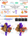
Figure 1:
Figure 1:. Structural determination of the Hsmortalin-GrpEL1 complex.
A. Canonical Hsp70 substrate capture and release cycle. B. Domain topology of Hsmortalin and HsGrpEL1. MTS: mitochondrial targeting signal. IDL: interdomain linker. C. Representative 2-D classes of the Hsmortalin-GrpEL1 complex. Colored arrows correspond to the densities observed in D. D. DeepEMhancer-sharpened map of the Hsmortalin-GrpEL1 complex colored by subunits and subdomains. The structure represents the exchange intermediate depicted in A.
Figure 2:
Figure 2:. The Hsmortalin NBD is fully expanded upon interaction with GrpEL1.
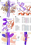
A. Interface mapping between the mortalin IIB-NBD lobe and the GrpEL1-B short α-helix, and the mortalin IB-NBD lobe and the GrpEL1-A β-wing domain. Lack of EM density within the NBD suggests an apo-nucleotide NBD. B. Structural comparison between the NBDs of Hsmortalin-GrpEL1, MtDnaK-GrpE (PDB: 8GB3), and ADP-PO4-bound MgDnaK (PDB: 5OBW). Compared to ADP-PO4-bound MgDnaK, the IIB-NBD lobe of Hsmortalin expands ~15° upon interaction with GrpEL1. C-E. Comparison of the ATP binding residues in the IIB-NBD lobe. ATP-stabilizing interactions in the IIB-NBD lobe of Hsmortalin-GrpEL1 are removed by movement of the IIB-NBD lobe. ADP-PO4 is modeled from MgDnaK-ADP-PO4.
Figure 3:
Figure 3:. The mortalin interdomain linker is stabilized by interaction with the GrpEL1 long α-helix.
A. Interaction mapping between the Hsmortalin linker and the GrpEL1 long α-helix. B. Multiple sequence alignment between Hsp70 and GrpE-like species. The interdomain linker of Hsp70 is highly conserved whereas the interacting GrpE-like region is variable. Hs: homo sapiens, Mg: mycoplasma genitalium, Mt: mycobacterium tuberculosis, Gk: geobacillus kaustophilus, Ec: eschericia coli. C. Superposition of GrpE-like species from our Hsmortalin-GrpEL1 structure, MtDnaK-GrpE (PDB: 8GB3), and the AlphaFold247 prediction of HsGrpEL1.
Figure 4:
Figure 4:. Hsmortalin in complex with GrpEL1 is substrate-bound to a mortalin truncation product.
A. Model of the Hsmortalin substrate binding domain (SBD) bound to a substrate fit into the DeepEMhancer-sharpened cryoEM map. B. Rigid-body docking of the mortalin SBD into the posterior substrate EM density (low-pass filtered to 5Å). C. Residue mapping of the substrate within the mortalin substrate binding site. D. Comparison of the modeled Hsmortalin substrate with the crystal structure of EcDnaK bound to the NRLLLTG peptide (PDB: 4EZW).
Figure 5:
Figure 5:. GrpEL1 forms unique interactions with the Hsmortalin NBD and SBD.
A. Structural mapping of the Hsmortalin SBD with the GrpEL1-B long α-helix and β-wing domain. B. Residue mapping of the interactions between the GrpEL1-B long α-helix and Hsmortalin SBDβ cleft. C. Residue mapping of the interactions between the GrpEL1-B β-wing domain and SBDα helical domain. D. Multiple sequence alignment of the GrpEL1-B β-wing-SBDα interacting regions across GrpE-like and Hsp70 species. E. Designation of the GrpEL1 β-wing face that interacts with the Hsmortalin NBD (β-wing face-N) and the face that interacts with the SBD (β-wing face-S).
Figure 6:
Figure 6:. Mutation of Y173A in GrpEL1 results in a shift of the Hsmortalin SBD.
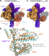
A. DeepEMhancer-sharpened map of mortalin-GrpEL1Y173A. Shadow represents the silhouette of the mortalin-GrpEL1WT SBD position. B. Superposition of mortalin-GrpEL1WT and mortalin-GrpEL1Y173A. In the mortalin-GrpEL1Y173A structure, the SBD is translated ~6 Å away from GrpEL1-B compared to WT. C. DeepEMhancer-sharpened map of mortalin-GrpEL1Y173A-lid. Shadow represents the silhouette of the mortalin-GrpEL1WT SBD position.
Figure 7:
Figure 7:. Flexibility analysis of mortalin-GrpEL1WT and mortalin-GrpEL1Y173A.
A. Anisotropic network modeling (ANM) modes of the SBDα lateral motion observed in both mortalin-GrpEL1WT and mortalin-GrpEL1Y173A. SBD plane is colored in beige. B. ANM mode of the lateral and medial SBDα motion found exclusively in the mortalin-GrpEL1Y173A ANM analysis. SBD plane is colored in beige. C. All-atom MD simulation of mortalin-GrpEL1WT. The position of the SBDα lid lateral motion is visualized across a representative 150ns simulation. Timepoints at 0, 70, and 130ns are represented as the start, middle, and end of the simulation. D. All-atom MD simulation of mortalin-GrpEL1Y173A. The medial motion of the SBDα lid is visualized across the 150ns simulation. Timepoints at 0, 70, and 130ns are represented as the start, middle, and end of the simulation.
Figure 8:
Figure 8:. GrpEL1-mediated nucleotide and substrate release mechanism of human mortalin.
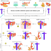
Upon interaction with a substrate and J-protein, mortalin hydrolyzes ATP and stably interacts with substrate. GrpEL1 interacts asymmetrically with mortalin and facilitates ADP release via interactions at the NBD. Flexibility within the SBD loosens the SBDα subdomain. Following ATP binding in a step-wise mechanism, the NBD dissociates first which enables opening of the SBD and subsequent substrate release. Alternatively, ATP binding induces concerted dissociation of mortalin whereby decoupling of SBDα and GrpEL1-B enables substrate release. The intermediate representing the mortalin-GrpEL1WT structure is highlighted in orange.

References

    1. Kim Y. E.; Hipp M. S.; Bracher A.; Hayer-Hartl M.; Ul-rich Hartl F. Annu. Rev. Biochem. 2013, 82, 323–355. - PubMed
    1. Voos W. Biochimica et Biophysica Acta (BBA) - Molecular Cell Research 2013, 1833, 388–399. - PubMed
    1. Genest O.; Wickner S.; Doyle S. M. Journal of Biological Chemistry 2019, 294, 2109–2120. - PMC - PubMed
    1. Morán Luengo T.; Mayer M. P.; Rüdiger S. G. Trends in Cell Biology 2019, 29, 164–177. - PubMed
    1. Clerico E. M.; Tilitsky J. M.; Meng W.; Gierasch L. M. Journal of Molecular Biology 2015, 427, 1575–1588. - PMC - PubMed

Publication types