[Preprint]. 2024 May 14:rs.3.rs-4384494.
doi: 10.21203/rs.3.rs-4384494/v1.

Sleep-wake body temperature regulates tau secretion in mice and correlates with CSF and plasma tau in humans

Affiliations

Sleep-wake body temperature regulates tau secretion in mice and correlates with CSF and plasma tau in humans

Geoffrey Canet et al. Res Sq. .

Update in

  • Sleep-wake variation in body temperature regulates tau secretion and correlates with CSF and plasma tau.
    Canet G, Da Gama Monteiro F, Rocaboy E, Diego-Diaz S, Khelaifia B, Godbout K, Lachhab A, Kim J, Valencia DI, Yin A, Wu HT, Howell J, Blank E, Laliberté F, Fortin N, Boscher E, Fereydouni-Forouzandeh P, Champagne S, Guisle I, Hébert SS, Pernet V, Liu H, Lu W, Debure L, Rapoport DM, Ayappa I, Varga AW, Parekh A, Osorio RS, Lacroix S, Burns MP, Lucey BP, Blessing EM, Planel E. Canet G, et al. J Clin Invest. 2025 Feb 4;135(7):e182931. doi: 10.1172/JCI182931. J Clin Invest. 2025. PMID: 39903530 Free PMC article.

Abstract

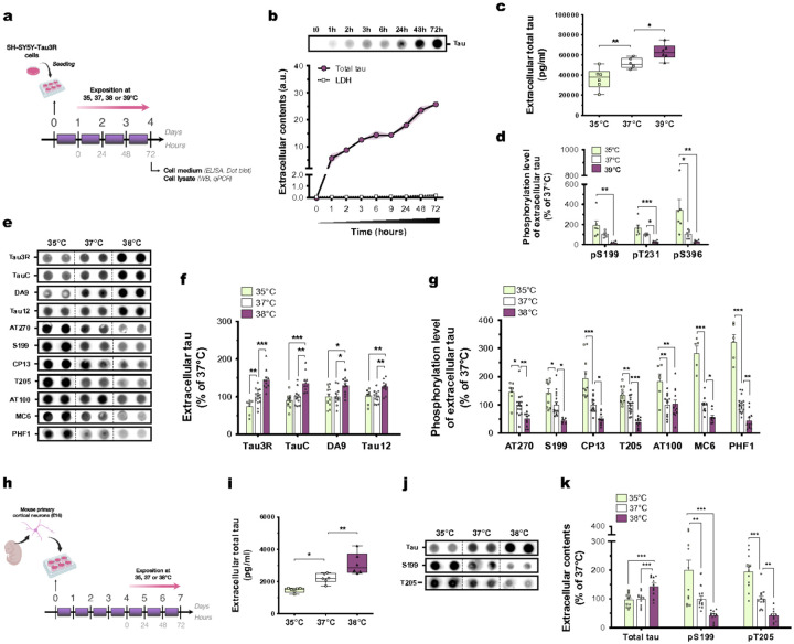
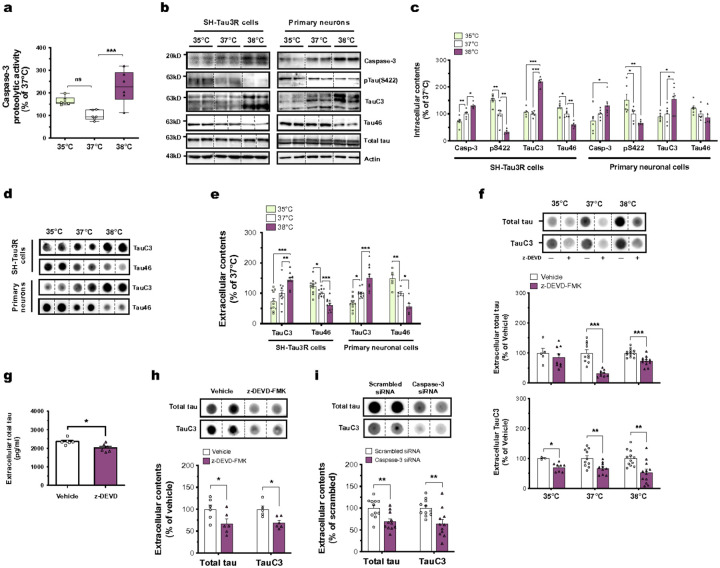
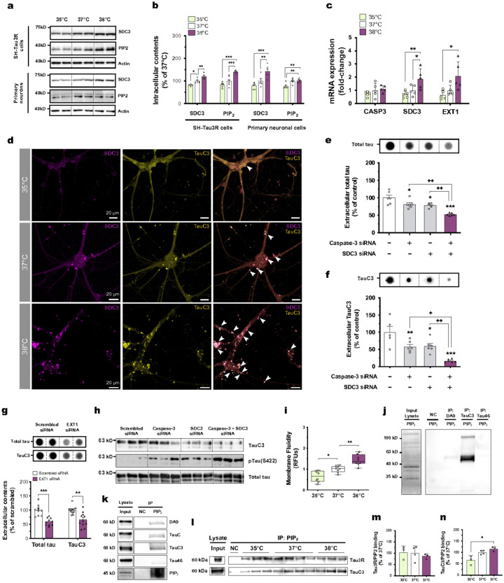
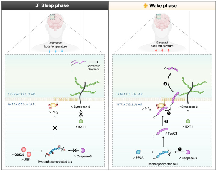
The sleep-wake cycle regulates interstitial fluid and cerebrospinal fluid (CSF) tau levels in both mouse and human by mechanisms that remain unestablished. Here, we reveal a novel pathway by which wakefulness increases extracellular tau levels in mouse and humans. In mice, higher body temperature (BT) associated with wakefulness and sleep deprivation increased CSF tau. In vitro, wakefulness temperatures upregulated tau secretion via a temperature-dependent increase in activity and expression of unconventional protein secretion pathway-1 components, namely caspase-3-mediated C-terminal cleavage of tau (TauC3), and membrane expression of PIP2 and syndecan-3. In humans, the increase in both CSF and plasma tau levels observed post-wakefulness correlated with BT increase during wakefulness. Our findings suggest sleep-wake variation in BT may contribute to regulating extracellular tau levels, highlighting the importance of thermoregulation in pathways linking sleep disturbance to neurodegeneration, and the potential for thermal intervention to prevent or delay tau-mediated neurodegeneration.

Keywords: Alzheimer’s disease; body temperature; sleep-wake cycle; tau; unconventional protein secretion.


Conflict of interest statement

Competing interests: All authors declare no competing interests.

Figures

Figure 1
Figure 1. Tau secretion is temperature-dependent in neuronal cells.
(a) 24 hours after seeding, SH-Tau3R cells were exposed to 35, 37, 38 or 39°C for 72 hours. (b) Extracellular accumulation of tau protein (Tau3R; n = 6–12; mean ± s.e.m (error envelopes in light purple)) and LDH (n= 4–9; mean ± s.e.m) over-time in cell medium of neurons cultured at 37°C. (c) The increase in extracellular tau levels is temperature-dependent in SH-Tau3R cells exposed to 35, 37 or 39°C (n = 6; Dunnett’s; box and whiskers with minimum to maximum and median). (d) The phosphorylation level of extracellular tau at S199, T231 and S396 is decreased at 39°C compared to 35 or 37°C (n = 6; Tukey’s; mean ± s.e.m). (e, f) The increase of extracellular tau levels is temperature-dependent (Tau3R, TauC, DA9 and Tau12 antibodies) in SH-Tau3R cells exposed to 35, 37 or 38°C (n = 7–16; Tukey’s; mean ± s.e.m). (e, g) The phosphorylation level of extracellular tau at AT270, S199, CP13, T205, AT100, MC6 and PHF1 is decreased at 38°C compared to 35 or 37°C (n = 7–16; Tukey’s; mean ± s.e.m). (h)4 days after seeding, mouse primary cortical neurons were exposed at 35, 37 or 38°C for 72 hours. (i) The increase of extracellular tau levels is temperature-dependent in mouse primary neurons exposed to 35, 37 or 38°C (n= 6; Dunnett’s; box and whiskers with minimum to maximum and median). (j, k) The increase of extracellular tau levels is temperature-dependent (Tau3R antibody), while its phosphorylation level at S199 and T205 is decreased at 38°C compared to 35 or 37°C (n = 11–12; Tukey’s; mean ± s.e.m). *p<0.05, **p<0.01 and ***p<0.001.
Figure 2
Figure 2. Wakefulness temperatures promote tau release through its caspase-3-mediated cleavage in neuronal cells.
(a) The increase in the proteolytic activity of caspase-3 is temperature-dependent in SH-Tau3R cells (n = 6; Tukey’s; box and whiskers with minimum to maximum and median). (b, c) The intracellular expressions of caspase-3, pTau(S422), TauC3 and Tau46 are temperature-dependent in SH-Tau3R (left panels) or in primary neurons (right panels) (n = 5–6; Tukey’s; mean ± s.e.m). (d, e) The extracellular levels of TauC3 and Tau46 are oppositely temperature-dependent in SH-Tau3R (left panels) or in primary neurons (right panels) (n = 6–12; Tukey’s; mean ± s.e.m). (f)The inhibition of caspase-3 with z-DEVD-FMK (20 μM) decreases total tau (Tau3R, n = 5–12, unpaired t-test) and TauC3 (n = 3–8, Mann Whithney) extracellular levels in SH-Tau3R cells exposed to 35, 37 or 38°C (mean ± s.e.m). The inhibition of caspase-3 with z-DEVD-FMK (20 μM) decreases the extracellular levels of (g) total tau (ELISA assay, n = 6, unpaired t-test), (h) Tau3R and TauC3 (Dot blotting, n = 5–6; unpaired t-test) (mean ± s.e.m). (i) The genetic knockdown of caspase-3 decreases total tau (Tau3R) and TauC3 extracellular levels in SH-Tau3R cells exposed to 37°C (n = 11; unpaired t-test; mean ± s.e.m). *p<0.05, **p<0.01 and ***p<0.001. ns: non-significant.
Figure 3
Figure 3. Wakefulness temperatures promote TauC3 interaction with PIP2 and SDC3 in neuronal cells
(a, b) The intracellular expressions of SDC3 and PIP2 are temperature-dependent in SH-Tau3R (left panels) or in primary neurons (right panels) (n = 6; Tukey’s; mean ± s.e.m). (c) The mRNA expression of SDC3 and EXT1 genes are temperature-dependent in SH-Tau3R, and CASP3 mRNA is unchanged (n = 5; Tukey’s; mean ± s.e.m). (d, e) The genetic knockdown of SDC3 decreases total tau (Tau3R) and TauC3 extracellular levels in SH-Tau3R cells exposed to 37°C, and the co-transfection of caspase-3 siRNA and SDC3 siRNA induces additive effects in the suppression of tau secretion (n = 5–6; Tukey’s; mean ± s.e.m). (f) The genetic knockdown of EXT1 decreases total tau (Tau3R) and TauC3 extracellular levels in SH-Tau3R cells exposed to 37°C (n = 11; unpaired t-test; mean ± s.e.m). (g) The genetic knockdown of caspase-3 + SDC3 decreases the intracellular expression of TauC while increasing tau phosphorylation at S422 (representative western blot detection; n = 3). (h) The increase in membrane fluidity is temperature-dependent in SH-Tau3R cells (n = 8; Tukey’s; box and whiskers with minimum to maximum and median). (i, j) PIP2 displays a better binding affinity for TauC3 rather than full-length tau (DA9 and Tau46 and TauC antibodies) in SH-Tau3R cells exposed to 37°C. NC: negative control, IP: immunoprecipitation. (k-m) The increase in binding between PIP2 and TauC3 is temperature-dependent (n = 3; Kruskal-Wallis; mean ± s.e.m). *p<0.05, **p<0.01 and ***p<0.001 vs. respective control condition; +p<0.05 and ++p<0.01 vs. indicated condition. (n) A merged staining is displayed, showing a temperature-dependent increase of colocalization between SDC3 (purple) and TauC3 (yellow) in primary mouse cortical neurons, and marked with white arrows. Scale bar: 20 μm (magnification of dotted boxes).
Figure 4
Figure 4. Wakefulness, sleep deprivation and mild-hyperthermia promote the UPS-I-dependent tau release in CSF in mice.
(a) C57BL6 mice were euthanatized during their sleeping (Circadian time 4, CT4, 10:00 am) or active period (CT16, 8:45 pm, awake group). (b) Awake mice display a higher rectal temperature (°C) at sacrifice compared to sleeping mice (n = 7; unpaired t-test; box and whiskers with minimum to maximum and median). (c, d) The expressions of caspase-3, TauC3, SDC3 and PIP2 are increased in the cortices of awake mice compared to sleeping mice, while pTau(S422) is decreased (n = 10; unpaired t-test; mean ± s.e.m). (e) C57BL6 mice were sleep-deprived (n=9) for the first 6 hours of the light period and compared to naive mice (n=7) allowed to sleep without disturbance. (f) Sleep-deprivation inhibits the drop in core body temperature (°C) induced by sleep (n = 5; Tukey’s; mean ± s.e.m as error envelopes). (g, h) The expressions of caspase-3, TauC3, and PIP2 are increased in the cortices of sleep-deprived mice compared to naive mice, while pTau(S422) and Tau46 are decreased (unpaired t-test; mean ± s.e.m). (i, j) hTau mice were exposed for 4 hours either to 4°C (hypo, n = 6) or 38°C (hyper, n = 6), and compared to naïve mice (normo, n = 5) (Šidák’s; mean ± s.e.m as error envelopes). ***p<0.001 vs. Normo group at baseline; +++p<0.001 vs. respective group at baseline. (k) Hyperthermic mice have higher CSF tau levels compared to hypothermic mice (n = 3 mice (Normo); n = 5 mice (Hypo); n = 6 mice (Hyper); Kruskal-Wallis; mean ± s.e.m). (l) CSF tau is significantly correlated with rectal body temperature (°C) (Pearson’s correlation; error envelopes in light grey). *p<0.05, **p<0.01 and ***p<0.001.
Figure 5
Figure 5. Body temperature correlates with CSF and plasma tau levels in humans.
(a) The variation in total CSF tau concentrations between 8AM and 4PM is significantly correlated to the variation in oral temperature at the same times (n = 13, Pearson’s correlation), (b) while no correlation is observed for CSF NfL concentrations and oral temperature (n = 11, Pearson’s correlation). (c) The variation in total plasma tau concentrations between 7AM and 7PM is significantly correlated to the variation in core body temperature between 6PM and 1AM (n = 15, Pearson’s correlation). Standard error bars displayed as error envelopes in light purple, *p<0.05 and **p<0.01. standard error bars displayed as error envelopes in light purple.
Figure 6
Figure 6. Proposed mechanism to elucidate the regulatory effect of BT on tau secretion via the UPS-I pathway during the sleep-wake cycle.
During wakefulness, the physiological elevation in BT instigates a series of events triggering tau secretion. (1) There is an increase in caspase-3 activity, concomitant with tau dephosphorylation, especially at S422, leading to an augmented cleavage of tau at D421, yielding the TauC3 fragment. (2) Subsequently, TauC3 is sequestered at the inner leaflet of the plasma membrane due to its strong affinity binding for PIP2. (3) (4) The interplay between TauC3 and SDC3 initiates and facilitates the export process across the plasma membrane, that exhibits heightened fluidity and permeability properties during wakefulness. In contrast, during sleep, the decrease in BT inhibits caspase-3 activity and promotes tau hyperphosphorylation at S422, preventing the generation of TauC3. The sleep phase also leads to reduced expression levels of both PIP2 and SDC3, as well as to a lower membrane fluidity, resulting in diminished extracellular tau levels.

Similar articles

References

    1. Long J. M. & Holtzman D. M. Alzheimer Disease: An Update on Pathobiology and Treatment Strategies. Cell 179, 312–339 (2019). - PMC - PubMed
    1. Bejanin A. et al. Tau pathology and neurodegeneration contribute to cognitive impairment in Alzheimer’s disease. Brain 140, 3286–3300 (2017). - PMC - PubMed
    1. Chai X., Dage J. L. & Citron M. Constitutive secretion of tau protein by an unconventional mechanism. Neurobiology of Disease 48, 356–366 (2012). - PubMed
    1. Merezhko M. et al. Secretion of Tau via an Unconventional Non-vesicular Mechanism. Cell Reports 25, 2027–2035.e4 (2018). - PubMed
    1. Merezhko M., Uronen R.-L. & Huttunen H. J. The Cell Biology of Tau Secretion. Front Mol Neurosci 13, 569818 (2020). - PMC - PubMed

Publication types