[Preprint]. 2024 May 13:2024.05.09.593355.
doi: 10.1101/2024.05.09.593355.

Exportin-1 functions as an adaptor for transcription factor-mediated docking of chromatin at the nuclear pore complex

Affiliations

Exportin-1 functions as an adaptor for transcription factor-mediated docking of chromatin at the nuclear pore complex

Tiffany Ge et al. bioRxiv. .

Update in

Abstract

Nuclear pore proteins (Nups) in yeast, flies and mammals physically interact with hundreds or thousands of chromosomal sites, which impacts transcriptional regulation. In budding yeast, transcription factors mediate interaction of Nups with enhancers of highly active genes. To define the molecular basis of this mechanism, we exploited a separation-of-function mutation in the Gcn4 transcription factor that blocks its interaction with the nuclear pore complex (NPC) without altering its DNA binding or activation domains. SILAC mass spectrometry revealed that this mutation reduces the interaction of Gcn4 with the highly conserved nuclear export factor Crm1/Xpo1. Crm1 both interacts with the same sites as Nups genome-wide and is required for Nup2 to interact with the yeast genome. In vivo, Crm1 undergoes extensive and stable interactions with the NPC. In vitro, Crm1 binds to Gcn4 and these proteins form a complex with the nuclear pore protein Nup2. Importantly, the interaction between Crm1 and Gcn4 does not require Ran-GTP, suggesting that it is not through the nuclear export sequence binding site. Finally, Crm1 stimulates DNA binding by Gcn4, supporting a model in which allosteric coupling between Crm1 binding and DNA binding permits docking of transcription factor-bound enhancers at the NPC.


Figures

Figure 1.
Figure 1.. Crm1/Xpo1 is essential for peripheral targeting of genes that are both Gcn4-dependent and Gcn4-independent.
A. Peripheral localization of four Gcn4 target genes the Gcn4 binding site inserted at an ectopic locus (URA3:Gcn4BS) in wild type or gcn4-pd mutant strains grown in SDC ± histidine. Mean percentage of the population ± standard error of the mean from ≥ three biological replicates of ≥ 30 cells each. Dashed line: localization expected for a randomly positioned gene. B. Scatter plot of the ratio of normalized abundance of 759 proteins identified by SILAC MS comparing the recovery from wild type (JBY558; light) and mutant (JBY557; heavy) cultures starved for histidine for 1h, with Crm1 highlighted. C. A mutant strain with a leptomycin B (LMB) sensitive allele of Crm1 (crm1-T539C) and expressing GFP bearing both a nuclear localization signal and a nuclear export signal (NES-GFP-NLS; untreated top panel) was treated with 100ng/ml LMB for 30 minutes (bottom). D & E Peripheral localization of either Gcn4 target genes (D) or non-Gcn4 target genes (E) in LMB-sensitive strains grown in in the indicated media treated ±100 ng/ml LMB for 30 minutes at 30°C. For panel E, only the INO1:LacO was grown in +inositol and −inositol medium. The RPS genes were localized in complete medium. F. Time courses of HIS5, RPS1B and INO1 peripheral localization following addition of LMB.
Figure 2
Figure 2. Crm1 and nuclear pore protein binding to the yeast genome.
A. Mean CPM normalized ChEC cleavage frequency of Crm1-MNase, Nup1-MNase, Nup2-MNase, Nup60-MNase, Nup157-MNase, and soluble MNase near the TDH3 gene. B. Metagene plots (normalized coding sequence length + 700 bp upstream and downstream) of the average CPM normalized ChEC cleavage over all RNAPII transcribed genes. Black plots = soluble MNase signal. C. Principal component analysis of cleavage frequency 700bp upstream of all genes by Crm1, Nup1, Nup60, Nup2, and Nup157 as well as controls (Prp20, sMNase and H2A.Z). D. Metagene plots for subsets of genes with distinct mechanisms of recruitment of RNAPII all genes: ribosomal protein genes (RPGs), inducible genes with TF-associated and SAGA-, TUP1-, and/or Mediator-associated promoters (STM), genes with TF-associated promoters lacking STM cofactors (TFO), and genes that recruit RNAPII without apparent TFs or cofactors (UNB). E. Average cleavage pattern around sites identified as high-confidence sites based on Crm1, Nup1, Nup2, or Nup60 ChEC-seq tracks ± 250bp flanking the site. F. Overlap between genes with cleavage peaks upstream for Nups and transcription factors based on ChIPexo. The number of genes that overlap between those identified by peaks of Crm1 and Nups with the indicated TFs was plotted against the Bonferroni-adjusted p-value (Fisher’s Exact test). G. Top three MEME results from Nup1 peaks and respective E-values.
Figure 3.
Figure 3.. Crm1 functions upstream of Nup2.
A. Localization of INO1, RPS0A, RPS1B and RPS6A in either a wild type strain (left) or a Nup2-AID strain ± auxin for 1h. Asterisks indicate p < 0.05, comparing to the untreated SDC control. B. Localization of GFP tagged proteins ± leptomycin B treatment. The NES-GFP-NLS reporter becomes concentrated in the nucleus, while Nup1, Nup2 and Nup60 are unaffected. C - E. Cleavage by Crm1, Nup2, Prp20 or sMNase in strains either depleted of Nup2 (by Nup2-AID), depleted of Crm1 (using grAID), or inhibited with leptomycin B. For this experiment, cleavage was carried out in SDC medium. Mean cleavage over the TDH3 locus (C) or over Nup2 peaks (Figure 2E; panel D) from untreated or treated (i.e., +IAA (Nup2-AID), + 5-Ph-IAA and estradiol (Crm1-grAID) or +LMB (crm1-T539C). E. Difference in cleavage over Nup2 peaks between treated and untreated samples.
Figure 4.
Figure 4.. Crm1 and Nup2 promote stronger transcription.
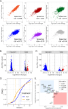
A. Scatter plots and Spearman’s correlation of Crm1 and Nup cleavage over upstream activating sequences (UASs; −700 to −125 from transcriptional start site) vs Rpb1-MNase cleavage for all yeast genes. B & C. SLAMseq analysis of nascent and total mRNA from either a crm1-T539C strain treated for 30 minutes with leptomycin B (B) or a Nup2-AID strain treated with auxin for 16h (C). D. Growth analysis of Nup2-AID strain ± auxin. The density of cultures of Nup2-AID strains was compared with Nup2-AID strains lacking non-essential Mediator subunits. The maximal growth rate (slope at the indicated timepoints) and the lag time (i.e., the time required to achieve the maximal growth rate) are highlighted. The effect of the loss of Nup2 was determined from the effects of adding auxin to the Nup2-AID strain. The fitness effect of loss of the Mediator mutations alone was determined from the ratio of these values to those of the Nup2-AID strain in the absence of auxin. The ratio of the maximal growth rate and lag time for each double mutant was compared with the expected value from multiplying the two growth defects (E). The blue square highlights buffering interactions and the red square highlights negative genetic interactions.
Figure 5.
Figure 5.. Crm1 and Nup2 interactions at the NPC overlap with the parts of the NPC that can contact chromatin.
Crm1-x-GFP (panel A) or Nup2 (panel C) were purified from yeast using LaG94–10 anti-GFP nanobody magnetic beads. A. Lanes 2 and 4 were from the Crm1-x-GFP strain; lanes 1 and 3 are a mock purification from a lysate lacking a GFP tagged protein. For lanes 1 and 2, the proteins were eluted by heating in LDS sample buffer. For lanes 3 and 4, the proteins were eluted by PreScission protease (PPX) cleavage. Samples were subjected to mass spectrometry and label free quantification. B & D. The abundance of Nups from Crm1 (B) and Nup2 (D) was normalized to their stoichiometry within the pore to highlight over-represented hits. C. Lane 1 is a purification from a strain expressing Genetically Encoded Multimeric particles tagged with GFP ; lane 2 is a purification from a strain expressing Nup2-x-GFP. Samples were eluted by heating in SDS and subjected to mass spectrometry and label free quantification. D. The 31 known nuclear pore proteins were identified in the top 224 hits. E. Heatmap of peripheral localization of URA3 induced by fusing LexA to each of 27 Nups in a strain having a LacO array and LexA binding site at URA3 (Figure S5).
Figure 6.
Figure 6.. Biochemical reconstitution of TF-NPC bridging complex.
A. GCN4 strains transformed with the indicated constructs were grown in medium ± histidine and the localization of either HIS4 or HIS5 was determined. B-E. Recombinant His-tagged Crm1 (5μM) and His-tagged Gsp1/Ran (± GDP or GTP; 10μM) and untagged Gcn4 PD-DBD were incubated with the indicated GST fusion proteins on magnetic beads. Bead-bound proteins were washed twice, eluted with 40mM glutathione and separated by SDS-PAGE. The GST fusion proteins, Crm1 Gcn4 and Ran are indicated. D. Wild type or pd mutant GST-PD-DBD (amino acids 181–281) bound to magnetic beads were incubated with the following concentrations of recombinant Crm1: 338nM, 675nM, 1.35μM, 2.7μM and 6.75μM. F. 200nM Gcn4 DBD or PD-DBD were incubated with fluorescent Gcn4 binding site in the presence of the following concentrations of 1μM Crm1 ± 2.5μMRan-GTP.
Figure 7.
Figure 7.. Model for Crm1 function as an adaptor for TF-NPC docking.
Crm1 (likely bound to Ran-GTP) associates with TFs bound to DNA in the nucleoplasm. This TF-Crm1 complex undergoes random subdiffusion in the nucleus . When it encounters the NPC, it interacts with Nup2 (and potentially other Nups) and forms a docking complex.

Similar articles

References

    1. Lin D.H., and Hoelz A. (2019). The Structure of the Nuclear Pore Complex (An Update). Annu. Rev. Biochem. 88, 1–59. 10.1146/annurev-biochem-062917-011901. - DOI - PMC - PubMed
    1. Akey C.W., Singh D., Ouch C., Echeverria I., Nudelman I., Varberg J.M., Yu Z., Fang F., Shi Y., Wang J., et al. (2022). Comprehensive structure and functional adaptations of the yeast nuclear pore complex. Cell 185, 361–378.e25. 10.1016/j.cell.2021.12.015. - DOI - PMC - PubMed
    1. Dultz E., Wojtynek M., Medalia O., and Onischenko E. (2022). The Nuclear Pore Complex: Birth, Life, and Death of a Cellular Behemoth. Cells 11, 1456. 10.3390/cells11091456. - DOI - PMC - PubMed
    1. Gozalo A., Duke A., Lan Y., Pascual-Garcia P., Talamas J.A., Nguyen S.C., Shah P.P., Jain R., Joyce E.F., and Capelson M. (2020). Core Components of the Nuclear Pore Bind Distinct States of Chromatin and Contribute to Polycomb Repression. Mol Cell 77, 67–81.e7. 10.1016/j.molcel.2019.10.017. - DOI - PMC - PubMed
    1. Vosse D.W.V. de, Wan Y., Wozniak R.W., and Aitchison J.D. (2011). Role of the nuclear envelope in genome organization and gene expression. Wiley Interdiscip Rev Syst Biology Medicine 3, 147–166. 10.1002/wsbm.101. - DOI - PMC - PubMed

Publication types