[Preprint]. 2024 May 14:2024.05.12.592631.
doi: 10.1101/2024.05.12.592631.

Structural Basis of Aquaporin-4 Autoantibody Binding in Neuromyelitis Optica

Affiliations

Structural Basis of Aquaporin-4 Autoantibody Binding in Neuromyelitis Optica

Meghna Gupta et al. bioRxiv. .

Update in

Abstract

Neuromyelitis Optica (NMO) is an autoimmune disease of the central nervous system where pathogenic autoantibodies target the human astrocyte water channel aquaporin-4 causing neurological impairment. Autoantibody binding leads to complement dependent and complement independent cytotoxicity, ultimately resulting in astrocyte death, demyelination, and neuronal loss. Aquaporin-4 assembles in astrocyte plasma membranes as symmetric tetramers or as arrays of tetramers. We report molecular structures of aquaporin-4 alone and bound to Fab fragments from patient-derived NMO autoantibodies using cryogenic electron microscopy. Each antibody binds to epitopes comprised of three extracellular loops of aquaporin-4 with contributions from multiple molecules in the assembly. The structures distinguish between antibodies that bind to the tetrameric form of aquaporin-4, and those targeting higher order orthogonal arrays of tetramers that provide more diverse bridging epitopes.


Conflict of interest statement

Competing interests: Authors declare that they have no competing interests.

Figures
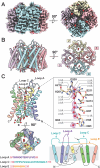
Figure 1.
Figure 1.. Structure of human AQP4 tetramer in nanodiscs.
(A) AQP4 density map determined by cryoEM at 2.1 Å resolution, side view and top view showing the extracellular surface of the same. Each monomer is colored differently, blue, pink, lemon, thistle anticlockwise. The four individual water channels contain water molecules (shown in red). (B) Side view and top view cartoon of the AQP4 tetramer model built using the density map in (A). The monomers are colored as (A) and numbered anticlockwise (C) AQP4 monomer shown in rainbow (from N to C terminus) in the side and top views representing location of water molecules. The three extracellular loops A, C, E are shown in the side view. Key amino acid residues interacting with the waters are highlighted and experimentally determined waters in the density maps are shown with mesh surface. A cartoon of AQP4 monomer depicting the relative organization of transmembrane helices in rainbow. The sequence of each extracellular loop shown.
Figure 2.
Figure 2.. Fab58 binding to human AQP4 M1 tetramer.
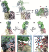
(A) Side view of the cryoEM density map at 2.5 Å resolution showing AQP4 tetramer and Fab58 interacting with the extracellular loops. Fab58 HC is shown in light steel blue and LC in pale green. A cartoon of the side view and top view of the model built using the density map with the same color scheme and numbered anticlockwise. One molecule of Fab58 makes interactions with two neighboring AQP4 monomers in the tetramer. (B) Molecular interactions between the LC of Fab58 and loop A of monomer 1 of AQP4 tetramer. Bond length in Å is mentioned in blue and the bonds are shown with a black dotted line. (C) Molecular interactions between the HC of Fab58 and loop C of monomer 1 of AQP4 tetramer mediated by a water molecule. (D) Molecular interactions between the HC of Fab58 and AQP4 loop C of monomer 2 of AQP4 tetramer. Two water molecules in red play an important role in this interaction.
Figure 3.
Figure 3.. Fab186 binding to human AQP4 M1 tetramer.
(A) Side view of the cryoEM density map surrounded by a detergent micelle at 2.9 Å resolution showing AQP4 tetramer and three Fab186 molecules interacting with the extracellular loops. Fab186 HC is shown in light sea green and LC in medium purple. A cartoon of the side view and top view of the model built using the density map with the same color scheme and numbered anticlockwise. One Fab186 makes interactions with two neighboring AQP4 monomers in the tetramer. (B) Molecular interactions between the HC of Fab186 and loop C of monomer 2 of AQP4 tetramer. Bond length in Å is in blue and hydrogen bonds are shown with black dotted line. (C) Hydrogen bonded interactions between the HC of Fab186 and loop C of monomer 1 of AQP4 tetramer. (D) Hydrogen bonded interactions between the LC of Fab186 and loop A of monomer 2 of AQP4 tetramer.
Figure 4.
Figure 4.. Comparison between Fab58 and Fab186 binding on AQP4 tetramer.
(A) Fab58 binds on AQP4 surface with an angle of 95.5° between selected labelled Cα atoms while (B) Fab186 binds at 109.7° measured using the orthologous amino acid residues to compare Fab58 and Fab186 binding. (C) Occupancy of Fab58 on AQP4 tetramer shown in the top view exhibiting steric limitation to accommodate another Fab58 molecule. (D) Fab186 binding at a steeper angle enables efficient binding of four Fabs. We see full density for three molecules of Fab while fourth molecule has a weak density as shown in figure S9. (E) Overlay of Fab58 and Fab186 structures shows their different loops relative to a constant template. Fab186 has extended regions and one of these take part in key interaction with H151 of loop C differentiating it from Fab58.
Figure 5.
Figure 5.. Understanding potential rAB186 association with OAPs.
(A) Human AQP4 structure superposed on the two neighboring tetramers of rat AQP4 M23 isoform tetramers from 2D crystal lattice structure based on electron diffraction analysis (PDB ID 2D57). Two of the monomers in one of the tetramers were replaced with a minimal Fab186 bound structure consisting of two monomers of AQP4 and one molecule of Fab186. These space filling representations emphasize the proximity of the Fab186 to the neighboring tetramer, favored by AQP4 M23 OAPs. The side view and top view exhibiting the possibility of Fab186 interaction spanning over two AQP4 tetramers. (B) Closeup of the Fab186 HC potential interactions (as in A) with loop A and loop C key of the neighboring tetramer.

References

    1. Jarius S. et al., Neuromyelitis optica. Nat Rev Dis Primers 6, 85 (2020). - PubMed
    1. Bennett J. L., Owens G. P., Neuromyelitis Optica: Deciphering a Complex Immune-Mediated Astrocytopathy. J Neuroophthalmol 37, 291–299 (2017). - PMC - PubMed
    1. Wrzos C. et al., Early loss of oligodendrocytes in human and experimental neuromyelitis optica lesions. Acta Neuropathol 127, 523–538 (2014). - PMC - PubMed
    1. Herwerth M. et al., In vivo imaging reveals rapid astrocyte depletion and axon damage in a model of neuromyelitis optica-related pathology. Ann Neurol 79, 794–805 (2016). - PMC - PubMed
    1. Bennett J. L. et al., Intrathecal pathogenic anti-aquaporin-4 antibodies in early neuromyelitis optica. Ann Neurol 66, 617–629 (2009). - PMC - PubMed

Publication types