[Preprint]. 2024 May 17:rs.3.rs-4402792.
doi: 10.21203/rs.3.rs-4402792/v1.

De novo designed proteins neutralize lethal snake venom toxins

Affiliations

De novo designed proteins neutralize lethal snake venom toxins

Susana Vázquez Torres et al. Res Sq. .

Update in

  • De novo designed proteins neutralize lethal snake venom toxins.
    Vázquez Torres S, Benard Valle M, Mackessy SP, Menzies SK, Casewell NR, Ahmadi S, Burlet NJ, Muratspahić E, Sappington I, Overath MD, Rivera-de-Torre E, Ledergerber J, Laustsen AH, Boddum K, Bera AK, Kang A, Brackenbrough E, Cardoso IA, Crittenden EP, Edge RJ, Decarreau J, Ragotte RJ, Pillai AS, Abedi M, Han HL, Gerben SR, Murray A, Skotheim R, Stuart L, Stewart L, Fryer TJA, Jenkins TP, Baker D. Vázquez Torres S, et al. Nature. 2025 Mar;639(8053):225-231. doi: 10.1038/s41586-024-08393-x. Epub 2025 Jan 15. Nature. 2025. PMID: 39814879 Free PMC article.

Abstract

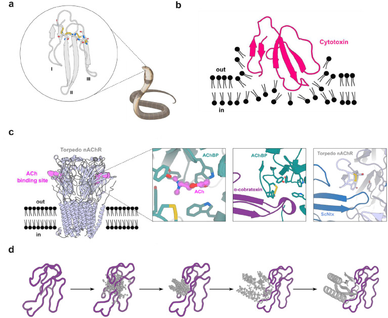
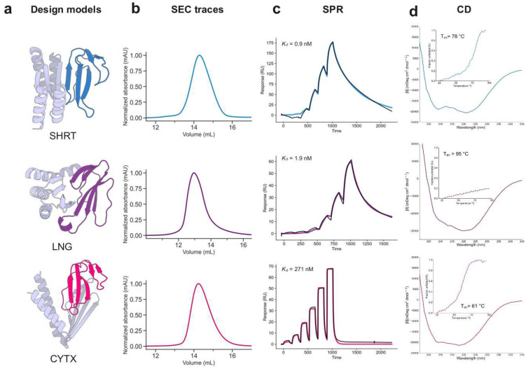
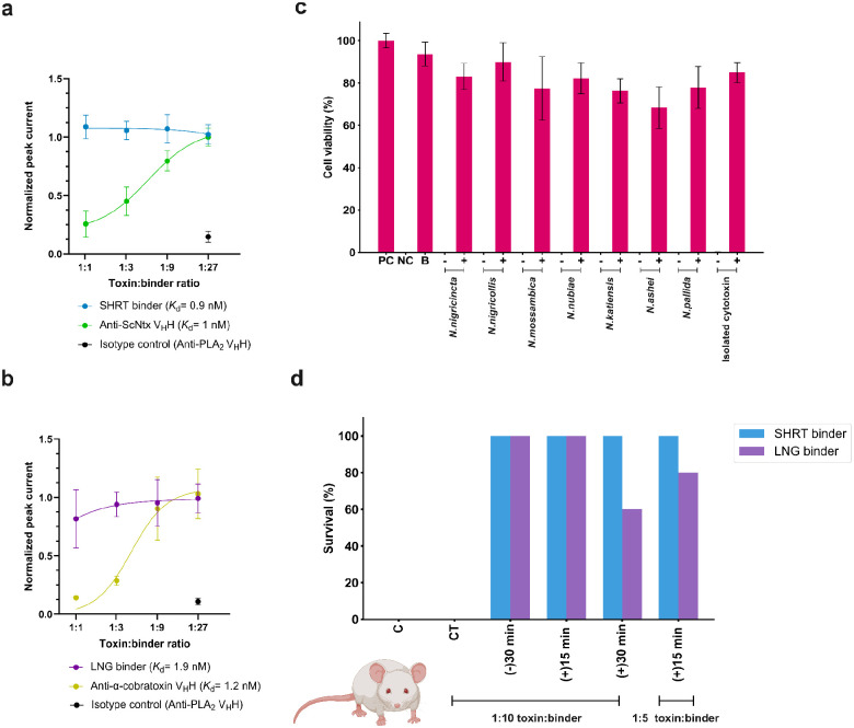
Snakebite envenoming remains a devastating and neglected tropical disease, claiming over 100,000 lives annually and causing severe complications and long-lasting disabilities for many more1,2. Three-finger toxins (3FTx) are highly toxic components of elapid snake venoms that can cause diverse pathologies, including severe tissue damage3 and inhibition of nicotinic acetylcholine receptors (nAChRs) resulting in life-threatening neurotoxicity4. Currently, the only available treatments for snakebite consist of polyclonal antibodies derived from the plasma of immunized animals, which have high cost and limited efficacy against 3FTxs5,6,7. Here, we use deep learning methods to de novo design proteins to bind short- and long-chain α-neurotoxins and cytotoxins from the 3FTx family. With limited experimental screening, we obtain protein designs with remarkable thermal stability, high binding affinity, and near-atomic level agreement with the computational models. The designed proteins effectively neutralize all three 3FTx sub-families in vitro and protect mice from a lethal neurotoxin challenge. Such potent, stable, and readily manufacturable toxin-neutralizing proteins could provide the basis for safer, cost-effective, and widely accessible next-generation antivenom therapeutics. Beyond snakebite, our computational design methodology should help democratize therapeutic discovery, particularly in resource-limited settings, by substantially reducing costs and resource requirements for development of therapies to neglected tropical diseases.


Figures

Figure 1.
Figure 1.. Targets of 3 finger snake toxins (3FTxs).
(a) Structure of 3FTxs (PDB ID: 1QKD). Highly conserved cysteine residues are highlighted in sticks and each of the three fingers indicated (I-III). (b) Representation of a type IA cytotoxin (dark pink) (PDB ID: 5NQ4) interacting with a lipid bilayer. (c) Muscle acetylcholine (Torpedo) receptor (light blue) (PDB ID: 7Z14). Acetylcholine (ACh) binding site is depicted in violet. Left inset: Close-up of the acetylcholine binding protein (AChBP) (teal) (PDB ID: 3WIP) bound to Ach (violet). A set of aromatic residues form a cage around the neurotransmitter. Middle: Close-up of α-cobratoxin (dark purple) blocking access to the ACh binding site in AChBP (teal) (PDB ID: 1YI5). Right: Close-up of ScNtx (dark blue) blocking access to the ACh binding site in the Torpedo receptor (light blue) (PDB ID: 7Z14). (d) Schematic showing α-cobratoxin binder design using RFdiffusion. Starting from a random distribution of residues around the specified β-strands in the target toxin (dark purple), successive RFdiffusion denoising steps progressively remove the noise leading at the end of the trajectory to a folded structure interacting with α-cobratoxin β-strands. Panels (a), (b), and (c) were created with BioRender.com
Figure 2.
Figure 2.. Experimental characterization of 3FTx binding proteins.
(a) Design models of protein binders (gray) bound to their 3FTx targets (dark blue: ScNtx, dark purple: α-cobratoxin, dark pink: consensus cytotoxin). (b) SEC traces of purified proteins. (c) SPR binding affinity measurements. Colored solid lines represent fits using the heterogeneous ligand model, with dissociation constant (Kd) values derived from these fits. (d) CD data confirms the presence of αβ-secondary structure in the 3FTx binding proteins and their thermal stability (inset).
Figure 3.
Figure 3.. Crystal structures of 3FTx binding proteins closely match design models.
(a) Apo-state crystal structure of SHRT design. Left: Hydrogen bonding between the carbonyl oxygen of Cys41 in ScNtx (dark blue) and the side chain of Tyr45 in the SHRT design model (gray). Middle: Overlay of SHRT design model (gray) with crystal structure (wheat). Right: Backbone hydrogen bonding between the SHRT design model (gray) and ScNtx (dark blue) β strands. (b) Crystal structure of LNG design in complex with α-cobratoxin. Left: Cross-interface hydrogen-bond network involving Arg33 at loop II in α-cobratoxin (light purple) and Glu69, Tyr40, and Tyr49 in LNG crystal structure (wheat). Middle: Overlay of LNG design model (gray) bound to α-cobratoxin (dark purple) with crystal structure of binder (wheat) bound to toxin (light purple). Right: Backbone hydrogen bonding between crystal structure of designed binder (wheat) and α-cobratoxin (light purple) β strands. (c) Crystal structure of CYTX_B10 design in complex with Naja pallida cytotoxin. Left: Cross-interface electrostatic interaction network between loops III and II of Naja pallida cytotoxin (light pink) and binder crystal structure (wheat). Middle: Overlay of CYTX_B10 design model (gray) bound to toxin (dark pink) with crystal structure of binder (wheat) bound to Naja pallida cytotoxin (light pink). Right: Salt bridge between positively charged Lys18 in cytotoxin (light pink) and Asp57 in the binder crystal structure (wheat).
Figure 4.
Figure 4.. In vitro and in vivo efficacy of designed proteins against snake venom toxins.
(a) Concentration-response curves comparing SHRT binder and anti-ScNtx VHH efficacy in preventing nAChR blocking by 1 IC80 of ScNtx. Data represent the toxin’s inhibition of ACh response, normalized to full ACh response, averaged within each group (n=16). (b) Concentration-response curves comparing the efficacy of LNG binder and anti-α-cobratoxin VHH in preventing nAChR blocking by 1 IC80 of α-cobratoxin. (c) Neutralization of the cytolytic effects of whole venoms from seven different Naja (N.) species and isolated cytotoxin by the CYTX binder. 2 IC50 of the whole venoms or toxin were pre-incubated with CYTX at a 1:5 molar ratio (toxin:binder). Keratinocyte media was used as a positive control (PC). Triton X-100 was used as a negative control (NC). CYTX binder (B) was used as a positive control. (−) denotes 2 IC50 of the whole venoms without binder, and (+) denotes venoms incubated with binder. Experiments were performed in triplicates, and results are expressed as mean ± SD. (d) Mice survival following lethal neurotoxin challenge (n=5). 3 LD50s of ScNtx or α-cobratoxin were preincubated for 30 minutes (−30 min) with the corresponding protein binders at 1:10 ratios and then administered IP into groups of five mice. Toxins administered IP following IP administration of binders at 1:10 or 1:5 molar ratios (toxin:binder) either after 15 (+15 min) or 30 minutes (+30 min) post-toxin injection. Controls included mice receiving toxins alone (C). Specificity was assessed via cross-treatment (CT) experiments, where non-target binders were preincubated with 3 LD50s of ScNtx or α-cobratoxin and administered IP. Signs of toxicity were observed, and deaths were recorded for a period of 24 hours. (d) was created with BioRender.com.

References

    1. GBD 2019 Snakebite Envenomation Collaborators et al. Global mortality of snakebite envenoming between 1990 and 2019. Nat. Commun. 13, 6160 (2022). - PMC - PubMed
    1. Gutiérrez J. M. et al. Snakebite envenoming. Nat. Rev. Dis. Primer 3, 17063 (2017). - PubMed
    1. Royte L. & Sawarkar A. Snake bite - cytotoxic effects of snake venom: a rare clinical image. Pan Afr. Med. J. 44, (2023). - PMC - PubMed
    1. Barber C. M., Isbister G. K. & Hodgson W. C. Alpha neurotoxins. Toxicon 66, 47–58 (2013). - PubMed
    1. Deka A., Gogoi A., Das D., Purkayastha J. & Doley R. Proteomics of Naja kaouthia venom from North East India and assessment of Indian polyvalent antivenom by third generation antivenomics. J. Proteomics 207, 103463 (2019). - PubMed

Publication types

LinkOut - more resources