Review
. 2024 Apr 24;6(1):tzae010.
doi: 10.1093/bjro/tzae010. eCollection 2024 Jan.

Imaging findings after a total reconstructed breast with autologous fat transfer: what the radiologist needs to know

Affiliations
Review

Imaging findings after a total reconstructed breast with autologous fat transfer: what the radiologist needs to know

Maud E P Rijkx et al. BJR Open. .

Abstract

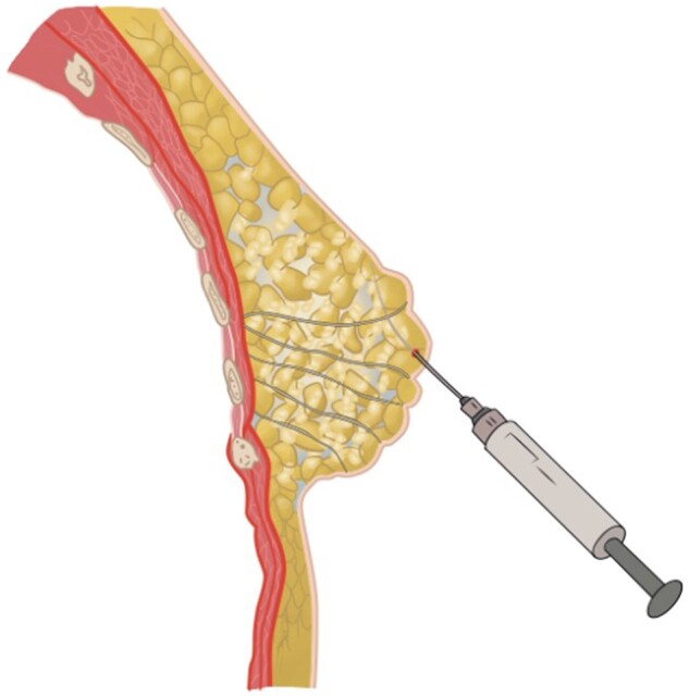
Autologous fat transfer (AFT) is an upcoming technique for total breast reconstruction. Consequently, radiological imaging of women with an AFT reconstructed breast will increase in the coming years, yet radiological experience and evidence after AFT is limited.

The surgical procedure of AFT and follow-up with imaging modalities including mammography (MG), ultrasound (US), and MRI in patients with a total breast reconstruction with AFT are summarized to illustrate the radiological normal and suspicious findings for malignancy.

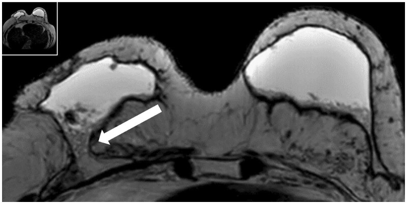
Imaging after a total breast reconstruction with AFT appears to be based mostly on benign imaging findings with an overall low biopsy rate. As higher volumes are injected in this technique, the risk for the onset of fat necrosis increases. Imaging findings most often are related to fat necrosis after AFT. On MG, fat necrosis can mostly be seen as oil cysts. The occurrence of a breast seroma after total breast reconstruction with AFT is an unfavourable outcome and may require special treatment. Fat deposition in the pectoral muscle is a previously unknown, but benign entity. Although fat necrosis is a benign entity, it can mimic breast cancer (recurrence).

In symptomatic women after total breast reconstruction with AFT, MG and US can be considered as first diagnostic modalities. Breast MRI can be used as a problem-solving tool during later stage. Future studies should investigate the most optimal follow-up strategy, including different imaging modalities, in patients treated with AFT for total breast reconstruction.

Keywords: autologous fat transfer (AFT); breast; breast reconstruction; imaging of the reconstructed breast.


Conflict of interest statement

None declared.

Figures

Figure 1.
Figure 1.
Autologous fat transfer (AFT) as a total breast reconstruction method.
Figure 2.
Figure 2.
Autologous fat transfer procedure (copyright is held by JAMA Surgery, and Ava Krueger, medical and scientific illustrator of this figure).
Figure 3.
Figure 3.
The different stages of fat necrosis. These images are of a 46-year-old female who completed her left total breast reconstruction with AFT 1 year ago. She received a target US. (A) A hyperechogenic area, where a hyperacute phase within the subcutaneous tissue is seen. (B) The inflammatory phase where an oil cyst is starting to form. It also includes a small part of fibrosis in the anterior part of the cyst. This is the third pathophysiological phase. (C) An almost complete oil cyst that only has a small part of fibrosis in the anterior part of the cyst left. Abbreviation: AFT = autologous fat transfer.
Figure 4.
Figure 4.
Benign calcifications related to fat necrosis. (A) MLO view of a 46-year-old female patient who completed her left total breast reconstruction with AFT 2 years and 10 months ago. An oval mass with hyperdense membrane can be observed in the former retro areolar region (white arrow), surrounded by multiple calcifications. The mass is corresponding with an oil cyst. (B) Corresponding CC view of the same patient. A total of 2 oil cysts can be appreciated in the former retro areolar region (white arrows). Abbreviations: AFT = autologous fat transfer; MLO = mediolateral oblique.
Figure 5.
Figure 5.
Typical aspects of fat necrosis on MRI. The images are from a 46-year-old female patient that underwent breast MRI 2 years and 10 months after her last AFT. Detailed images of the right breast demonstrate a T2 hypointense lesion (or mass) in the upper-outer quadrant of the right breast (A) without any enhancement on the T1W-sequence after contrast administration (B). Abbreviation: AFT = autologous fat transfer.
Figure 6.
Figure 6.
Breast seroma after total reconstruction with AFT on ultrasound. This is an US image of a 53-year-old female patient, 1 month after her second bilateral AFT procedure. Postoperative she suffered from bilateral seroma. (A) An example of an anechogenic fluid collection in the breast. (B) A hypoechogenic fluid collection. The increased echogenity compared to (A) suggests the presence of free-floating fat particles, which may complicate drainage. Abbreviations: AFT = autologous fat transfer; US = ultrasound.
Figure 7.
Figure 7.
Breast seroma after total breast reconstruction with AFT on MRI. This is a breast MRI of the same 53-year-old patient that had a persistent seroma despite multiple drainage treatments and sclerotherapy. The axial T2-image shows bilateral hyperintense collections. The white arrow demonstrates that the right fluid collection is continuous with the pectoral muscle and a gravity dependent gradient/layering of fluid-debris is seen. Abbreviations: AFT = autologous fat transfer; MRI = magnetic resonance imaging.
Figure 8.
Figure 8.
Radiographic change of the pectoral muscle after finishing a total breast reconstruction with AFT. This is a beast MRI of a 68-year-old patient that was 2 years after her last bilateral AFT procedure. (A) The axial T2W image of the right reconstructed breast with fat depositions within the pectoral muscle. (B) The corresponding sagittal view. Abbreviations: AFT = autologous fat transfer; MRI = magnetic resonance imaging; T2W = T2 weighted.
Figure 9.
Figure 9.
Suspicious findings for malignancy after a total breast reconstruction with AFT on mammography. This is a mammography of a 54-year-old female patient. Her prior medical history includes a DCIS stage 3, treated with a mastectomy and direct breast reconstruction. This mammography was taken 12 months after her final AFT session and shows multiple grouped and clustered pleiomorphic calcifications with a maximum diameter of up to 9 mm in the upper quadrant of the breast (BI-RADS 4). Note in addition the lucent areas within the pectoral muscle, representing pectoral muscle fat depositions. Abbreviation: AFT = autologous fat transfer.
Figure 10.
Figure 10.
Breast MRI of a 64-year-old woman with a unilateral left-sided total breast reconstruction with AFT, because of a multifocal left-sided invasive carcinoma NST (ER+PR+Her2−) treated with mastectomy. Breast MRI demonstrates an enhancing mass prepectoral on the inner side of the reconstructed breast extending through the pectoral muscle (white arrow). After core-needle biopsy, recurrent breast cancer was confirmed (invasive carcinoma NST, ER+PR+Her2−). Abbreviation: AFT = autologous fat transfer.

References

    1. Howard-McNatt MM. Patients opting for breast reconstruction following mastectomy: an analysis of uptake rates and benefit. Breast Cancer (Dove Med Press). 2013;5:9-15. - PMC - PubMed
    1. Miseré RML, van Kuijk SMJ, Claassens EL, et al.Breast-related and body-related quality of life following autologous breast reconstruction is superior to implant-based breast reconstruction—a long-term follow-up study. Breast. 2021;59:176-182. - PMC - PubMed
    1. Zehra S, Doyle F, Barry M, et al.Health-related quality of life following breast reconstruction compared to total mastectomy and breast-conserving surgery among breast cancer survivors: a systematic review and meta-analysis. Breast Cancer. 2020;27(4):534-566. - PubMed
    1. Cordeiro PG. Breast reconstruction after surgery for breast cancer. N Engl J Med. 2008;359(15):1590-1601. - PubMed
    1. Krastev TK, Schop SJ, Hommes J, et al.Meta-analysis of the oncological safety of autologous fat transfer after breast cancer. Br J Surg. 2018;105(9):1082-1097. - PMC - PubMed