. 2024 May 24:7:0371.
doi: 10.34133/research.0371. eCollection 2024.

Progesterone Enhances Niraparib Efficacy in Ovarian Cancer by Promoting Palmitoleic-Acid-Mediated Ferroptosis

Affiliations

Progesterone Enhances Niraparib Efficacy in Ovarian Cancer by Promoting Palmitoleic-Acid-Mediated Ferroptosis

Nayiyuan Wu et al. Research (Wash D C). .

Abstract

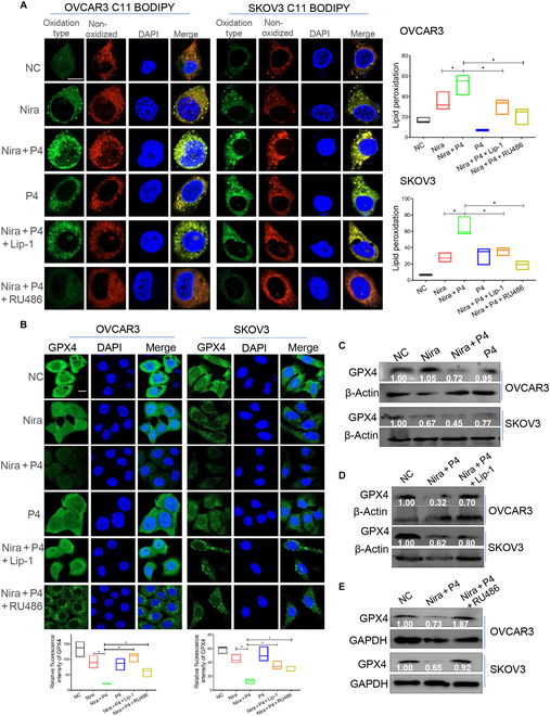
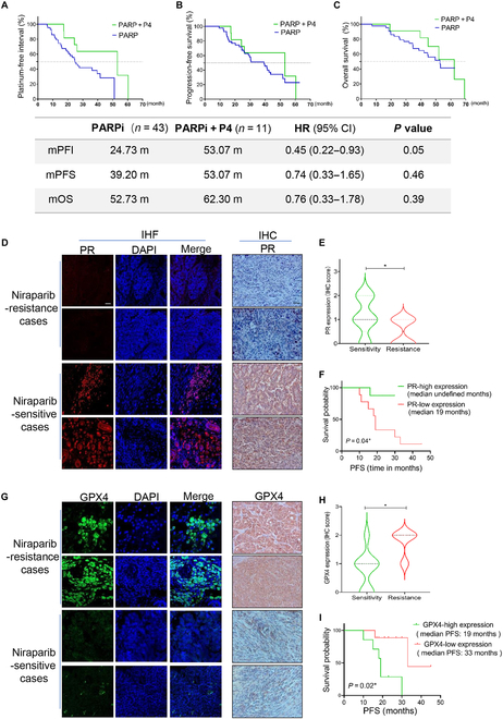
Poly (adenosine 5'-diphosphate-ribose) polymerase inhibitors (PARPi) are increasingly important in the treatment of ovarian cancer. However, more than 40% of BRCA1/2-deficient patients do not respond to PARPi, and BRCA wild-type cases do not show obvious benefit. In this study, we demonstrated that progesterone acted synergistically with niraparib in ovarian cancer cells by enhancing niraparib-mediated DNA damage and death regardless of BRCA status. This synergy was validated in an ovarian cancer organoid model and in vivo experiments. Furthermore, we found that progesterone enhances the activity of niraparib in ovarian cancer through inducing ferroptosis by up-regulating palmitoleic acid and causing mitochondrial damage. In clinical cohort, it was observed that progesterone prolonged the survival of patients with ovarian cancer receiving PARPi as second-line maintenance therapy, and high progesterone receptor expression combined with low glutathione peroxidase 4 (GPX4) expression predicted better efficacy of PARPi in patients with ovarian cancer. These findings not only offer new therapeutic strategies for PARPi poor response ovarian cancer but also provide potential molecular markers for predicting the PARPi efficacy.


Conflict of interest statement

Competing interests: The authors declare that they have no competing interests.

Figures

Fig. 1.
Fig. 1.
P4 enhances the inhibitory effect of niraparib in ovarian cancer. (A) P4 plus niraparib combination treatment at a ratio of 1:1 in human BRCA2-mutated ovarian cancer cell line PEO1 and wild-type BRCA ovarian cancer cell lines OVCAR3, SKOV3, and A2780. (B) Cell counting kit-8 (CCK8) test cell viability in PEO1, OVCAR3, SKOV3, and A2780 cells treated with different concentration gradients of vehicle (NC), P4 (blue line), niraparib (green line), and P4 plus niraparib (black line) for 48 h. (C and D) half inhibitory concentration (IC50) of PEO1, OVCAR3, SKOV3, and A2780 cells. (E) Representative images of ovarian cancer organoid tissues after the above treatment with vehicle, niraparib, P4 plus niraparib, or P4 for 5 d, acquired by light microscopy. (F) Cell viabilities in 10 ovarian cancer organoid tissue cases were tested after treatment with vehicle, niraparib, P4 plus niraparib or P4 for 5 d. *P < 0.05; **P < 0.01.
Fig. 2.
Fig. 2.
P4 enhances the sensitivity of ovarian cancer cells to niraparib by inducing DNA damage and apoptosis. Ovarian cancer cells were treated with vehicle (NC), niraparib, P4 (10 μM) plus niraparib (10 μM), or P4 (10 μM) for 24 h, and IF (A) and WB (B) were performed to assess γ-H2AX expression. (C) Flow cytometry test cell death rate. The data in the graph represent annexin-V-positive cells. *P < 0.05. Scale bar, 10 μM. FITC, fluorescein isothiocyanate.
Fig. 3.
Fig. 3.
P4 enhances the efficacy of niraparib in ovarian cancer in vivo. Representative 3-dimensional CT images (A) and CT scans (B) of ovarian in situ tumorigenesis mice before treatment (0 d) and 15 d after drug administration. (C and E) Ovarian in situ tumorigenesis mice and (D and F) intraperitoneal tumorigenesis mice treated with vehicle, niraparib (50 mg/kg), P4 plus niraparib (P4 at 5 mg/kg and niraparib at 50 mg/kg), or P4 (5 mg/kg); mice were sacrificed after 28 d of drug administration, and tumor sizes and weights were obtained. (G) Immunofluorescence (IF) test γ-H2AX expression in mouse tumor tissue samples; (H) IHC of the cell-proliferation-associated antigen Ki67 and the terminal-deoxynucleotidyl-transferase-mediated deoxyuridine triphosphate nick end labeling (TUNEL) test for apoptosis-related markers in the mouse tumor tissue. (I) Kaplan–Meier survival analysis of the survival of C57BL/6 mice in 4 different treatment groups (n = 8). *P < 0.05; **P < 0.01, ***P < 0.001. Scale bars,10 μm.
Fig. 4.
Fig. 4.
P4 enhances the activity of niraparib by up-regulating fatty acid metabolism and POA production in ovarian cancer cells. (A) Metabolomics were performed in OVCAR3 cells after treated with vehicle (NC), niraparib, P4 plus niraparib, or P4 for 24 h; the Z score heatmap of the overall profile of metabolites. (B) Enrichment analysis results of metabolomics. (C) Detection of POA and MA in OVCAR3 cells after 24 h after the above treatment. (D) CCK8 assay tested the viability of OVCAR3 cells after 24 h of treatment with vehicle, niraparib, POA or MA + niraparib, POA, or MA. (E) IF and (F) WB were performed to assess the expression of γ-H2AX in OVCAR3 and SKOV3 cells after treatment with vehicle, niraparib (10 μM), POA (200 μM) + niraparib (10 μM), or POA (200 μM) for 24 h. (G) BODIPY 581/591 C11 staining was performed to quantify lipid peroxidation in OVCAR3 administered with the above treatment. (H) IF and WB were performed to assess GPX4 expression in OVCAR3 and SKOV3 cells administered with the above treatment. *P < 0.05; **P < 0.01; ***P < 0.001. Scale bar, 10 μm.
Fig. 5.
Fig. 5.
P4 combined with niraparib promote lipid oxidation and GPX4 suppression. (A) BODIPY 581/591 C11 staining was performed to quantify lipid peroxidation in OVCAR3 and SKOV3 cells treated with vehicle (NC), P4 + niraparib, P4, P4 + niraparib + liproxstatin-1 (Lip-1) (1 μM), or P4 + niraparib + RU486 (5 μM) for 24 h. (B to E). IF and WB tested GPX4 expression in OVCAR3 and SKOV3 cells administered with the above treatments. *P < 0.05. Scale bars, 10 μm.
Fig. 6.
Fig. 6.
P4 combined with niraparib promote ferroptosis in ovarian cancer cells. (A) IF determined Fe2+ levels in OVCAR3, A2780, SKOV3, and PEO1 cells treated with vehicle (NC), niraparib, P4 + niraparib, or P4 for 24 h. (B and C) Transmission electron microscope and IF observed mitochondrial damage of the OVCAR3 cells administered with the above treatments. (D) Flow cytometry test of ROS of OVCAR3 cells administered with the above treatments. (E) CCK8-tested ovarian OVCAR3 cell viability treated with vehicle, P4 + niraparib, P4 + niraparib + NAC, or liproxstatin-1. Scale bars, 10 μm (IF) and 1 μm (transmission electron microscope). *P < 0.05.
Fig. 7.
Fig. 7.
P4 enhances PARPi efficacy in ovarian cancer second-line maintenance therapy, and PR-high/GPX4-low expression was linked to better prognosis and higher PARPi sensitivity. (A) PFI, (B) PFS, and (C) OS of patients with ovarian cancer received second-line maintenance therapy with or without concomitant P4 therapy. IF and IHC test the PR (D and E) and GPX4 (G and H) levels in PARPi-sensitive and -resistant ovarian cancer tissues. (F and I) PFS of patients with ovarian cancer administered with PARPi as maintenance therapy with different levels of PR/GPX4 expression. *P < 0.05. Scale bar, 10 μm. CI, confidence interval; HR, hazard ratio.

References

    1. Siegel RL, Giaquinto AN, Jemal A. Cancer statistics, 2024. CA Cancer J Clin. 2024;74(1):12–49. - PubMed
    1. Torre LA, Trabert B, DeSantis CE, Miller KD, Samimi G, Runowicz CD, Gaudet MM, Jemal A, Siegel RL. Ovarian cancer statistics, 2018. CA Cancer J Clin. 2018;68(4):284–296. - PMC - PubMed
    1. Groelly FJ, Fawkes M, Dagg RA, Blackford AN, Tarsounas M. Targeting DNA damage response pathways in cancer. Nat Rev Cancer. 2023;23(2):78–94. - PubMed
    1. Costa A, Chowdhury D, Shapiro GI, D’Andrea AD, Konstantinopoulos PA. Targeting replication stress in cancer therapy. Nat Rev Drug Discov. 2023;22(1):38–58. - PMC - PubMed
    1. Huang D, Kraus WL. The expanding universe of PARP1-mediated molecular and therapeutic mechanisms. Mol Cell. 2022;82(12):2315–2334. - PMC - PubMed