Case Reports
. 2024 May 20:17:507-519.
doi: 10.2147/IMCRJ.S458142. eCollection 2024.

Insights into Ocular Emergencies: case Series on Non-Arteritic Anterior Ischemic Optic Neuropathy (NAION) Secondary to Acute Angle Closure Glaucoma

Affiliations
Case Reports

Insights into Ocular Emergencies: case Series on Non-Arteritic Anterior Ischemic Optic Neuropathy (NAION) Secondary to Acute Angle Closure Glaucoma

Alia Arianti et al. Int Med Case Rep J. .

Abstract

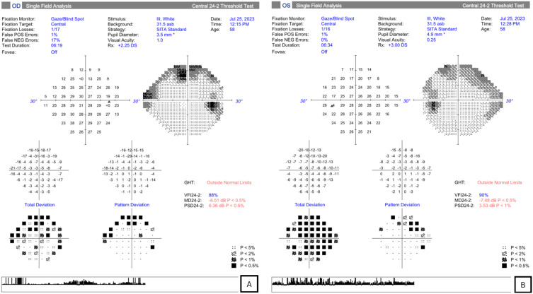
This case series aims to report the manifestation of acute secondary optic neuropathy attributed to optic nerve injury associated with a singular episode of markedly elevated intraocular pressure (IOP) during an acute glaucoma attack. The correlation between acute primary angle-closure (APAC) and non-arteritic anterior ischemic optic neuropathy (NAION) remains uncertain within the context of current knowledge. Definitive conclusions regarding the causal relationship between APAC and NAION or their mutual influence cannot be established based on the current evidence. The association between these conditions is recognized as a potential link, and comprehensive research is imperative to elucidate their interrelationship thoroughly. This case series emphasizes the importance of promptly addressing acute optic nerve injury and neuropathy associated with elevated intraocular pressure (IOP) in patients with crowded disc anatomical risk factors. It underscores the need for proactive interventions to prevent irreversible damage, highlighting the infrequent yet vision-compromising occurrence of non-arteritic anterior ischemic optic neuropathy (NAION) in acute primary angle-closure (APAC).

Keywords: AACG; Acute Glaucoma Attack; NAION; Neuropathy.


Conflict of interest statement

The authors report no conflicts of interest in this work.

Figures

Figure 1
Figure 1
(A)Dilated pupil in the right eye, glaucomflecken, and (B) patent peripheral iridectomy in the right eye.
Figure 2
Figure 2
Peripheral anterior synechiae visualized by gonioscopy.
Figure 3
Figure 3
Fundus photography of (A) right eye; swollen optic nerve head and (B) left eye; normal optic nerve head.
Figure 4
Figure 4
OCT of the ONH in the RE and LE was conducted. The optical coherence tomography (OCT) of the optic nerve head revealed that the analysis of the retinal nerve fiber layer showed 360° optic nerve edema in the right eye, while no optic nerve edema was detected in the left eye, with a small crowded cup-to-disc ratio (CDR).
Figure 5
Figure 5
AS-OCT on the RE and LE. The anterior chamber angles in both eyes exhibited similarity, with both being shallow. Pink arrows in the ACT imaging showed the plane where the calculations were taken.
Figure 6
Figure 6
HFA 30–2 test in the right (A) and left (B) eye. Marked scotoma in the right eye with low test reliability of the LE.
Figure 7
Figure 7
Case 1 - OCT ONH exhibiting diffuse optic disc edema, with a swollen optic nerve head diffusely, displaying increased thickness markers in the superior and inferior quadrants. This condition led to a significant scotoma in the Humphrey visual field analysis (HFA) of the right eye.
Figure 8
Figure 8
(A) OCT of the optic nerve head shows optic nerve edema in the left eye, with crowded disc (0.029) in the fellow eye. (B) Ganglion OCT shows no abnormality in both eyes.
Figure 9
Figure 9
HFA visual field testing demonstrates superior arcuate field defect in the left eye (A). HVF testing in the right eye was not reliable (B).
Figure 10
Figure 10
(A, B) HVF testing a week after initial diagnosis with better reliability with defect in both eyes.
Figure 11
Figure 11
(A) ONH OCT with marked disc edema 360 in all quadrants of the right eye compared to the left eye (B) Ganglion OCT showed no significant defect in both eyes.
Figure 12
Figure 12
(A, B) Visual field test results obtained using the Humphrey visual field analyzer (HFA), showing no significant defects in the right eye.

References

    1. Kong JH, Park SP, Na KI. Differences in optic nerve head structure between acute angle-closure glaucoma and open-angle glaucoma. Sci Rep. 2023;13(1):7935. doi:10.1038/s41598-023-35020-y - DOI - PMC - PubMed
    1. Wang Y, Chen D, Yang W, et al. Primary acute angle-closure glaucoma: three-dimensional reconstruction imaging of optic nerve heard structure in based on optical coherence tomography (OCT). Med Sci Monit. 2019;25:3647. doi:10.12659/MSM.913541 - DOI - PMC - PubMed
    1. Liu B, Yu Y, Liu W, Deng T, Xiang D Risk factors for non-arteritic anterior ischemic optic neuropathy: a large scale meta-analysis. Front Med. 2021; Availablr from: https://www.frontiersin.org/articles/10.3389/fmed.2021.618353. Accessed Oct 6 2023. - DOI - PMC - PubMed
    1. Miller NR, Arnold AC. Current concepts in the diagnosis, pathogenesis and management of nonarteritic anterior ischaemic optic neuropathy. Eye. 2015;29(1):65–79. doi:10.1038/eye.2014.144 - DOI - PMC - PubMed
    1. Fraser JA, Rueløkke LL, Malmqvist L, Hamann S. Prevalence of optic disc drusen in young patients with nonarteritic anterior ischemic optic neuropathy: a 10-year retrospective study. J Neuroophthalmol. 2021;41(2):200. doi:10.1097/WNO.0000000000000974 - DOI - PubMed

Publication types