. 2024 May 10:15:1395945.
doi: 10.3389/fimmu.2024.1395945. eCollection 2024.

Depletion of donor dendritic cells ameliorates immunogenicity of both skin and hind limb transplants

Affiliations

Depletion of donor dendritic cells ameliorates immunogenicity of both skin and hind limb transplants

Muhammad Imtiaz Ashraf et al. Front Immunol. .

Abstract

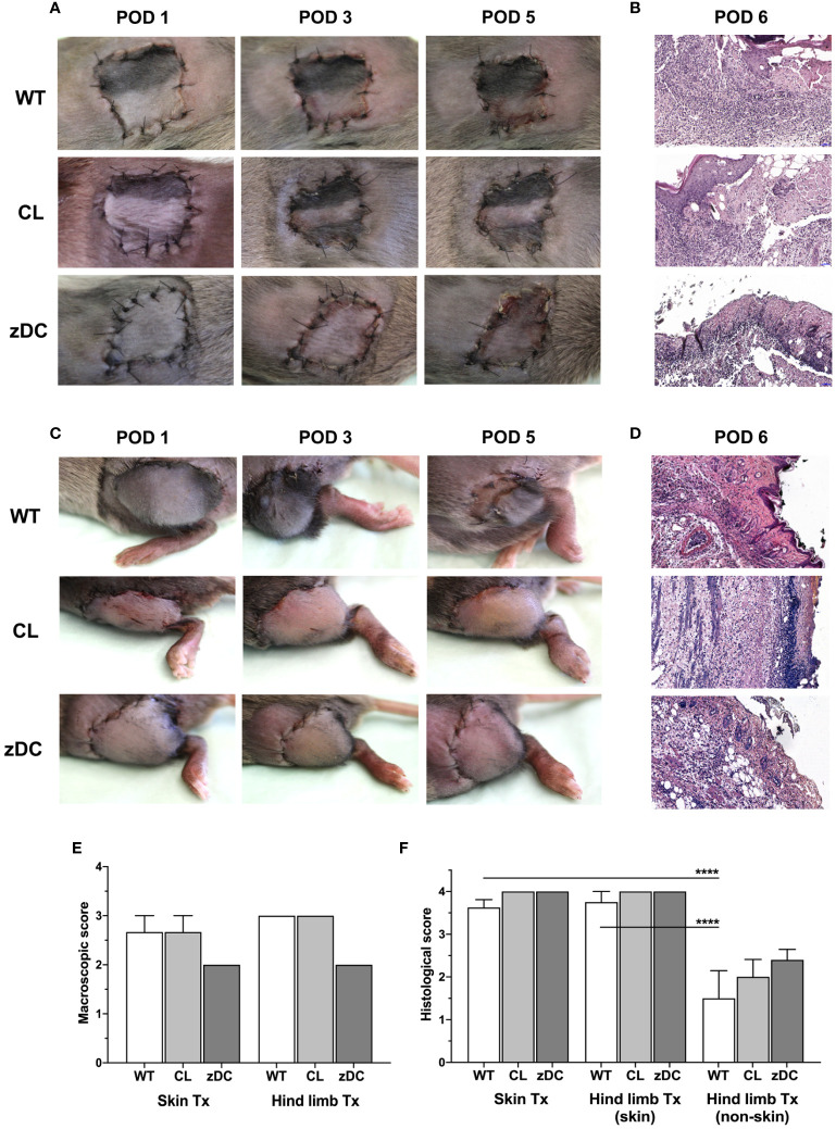
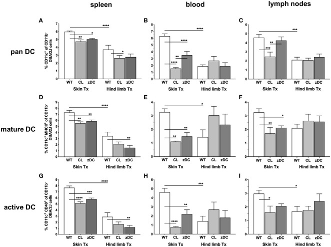
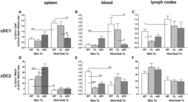
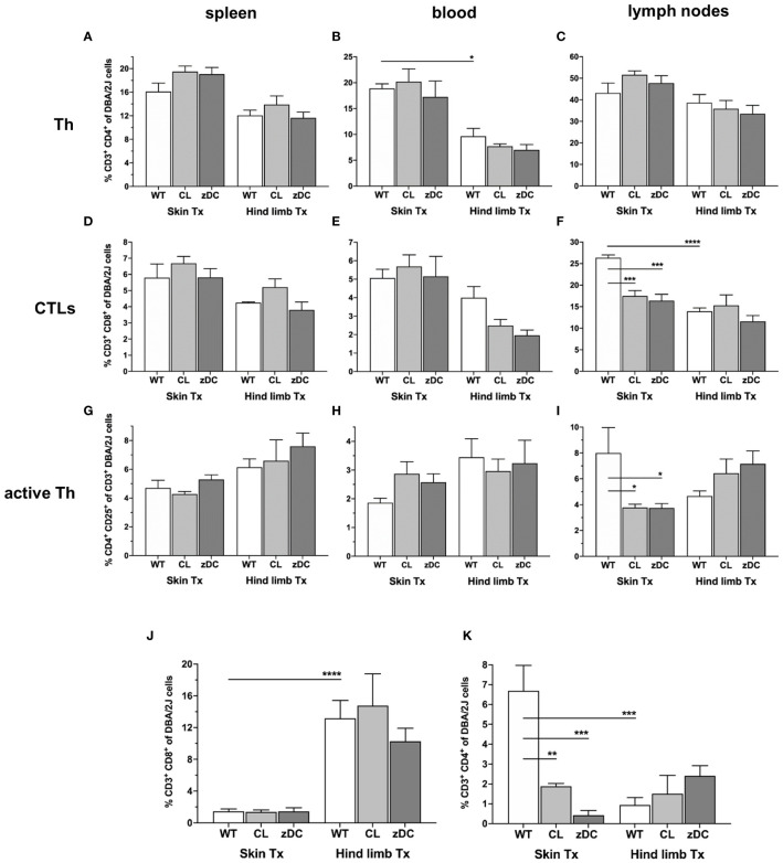
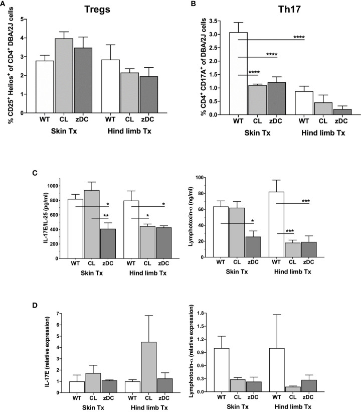
Acute cellular rejection remains a significant obstacle affecting successful outcomes of organ transplantation including vascularized composite tissue allografts (VCA). Donor antigen presenting cells (APCs), particularly dendritic cells (DCs), orchestrate early alloimmune responses by activating recipient effector T cells. Employing a targeted approach, we investigated the impact of donor-derived conventional DCs (cDCs) and APCs on the immunogenicity of skin and skin-containing VCA grafts, using mouse models of skin and hind limb transplantation. By post-transplantation day 6, skin grafts demonstrated severe rejections, characterized by predominance of recipient CD4 T cells. In contrast, hind limb grafts showed moderate rejection, primarily infiltrated by CD8 T cells. Notably, the skin component exhibited heightened immunogenicity when compared to the entire VCA, evidenced by increased frequencies of pan (CD11b-CD11c+), mature (CD11b-CD11c+MHCII+) and active (CD11b-CD11c+CD40+) DCs and cDC2 subset (CD11b+CD11c+ MHCII+) in the lymphoid tissues and the blood of skin transplant recipients. While donor depletion of cDC and APC reduced frequencies, maturation and activation of DCs in all analyzed tissues of skin transplant recipients, reduction in DC activities was only observed in the spleen of hind limb recipients. Donor cDC and APC depletion did not impact all lymphocyte compartments but significantly affected CD8 T cells and activated CD4 T in lymph nodes of skin recipients. Moreover, both donor APC and cDC depletion attenuated the Th17 immune response, evident by significantly reduced Th17 (CD4+IL-17+) cells in the spleen of skin recipients and reduced levels of IL-17E and lymphotoxin-α in the serum samples of both skin and hind limb recipients. In conclusion, our findings underscore the highly immunogenic nature of skin component in VCA. The depletion of donor APCs and cDCs mitigates the immunogenicity of skin grafts while exerting minimal impact on VCA.

Keywords: Th17 immune response; acute cellular rejection; allograft immunogenicity; antigen presenting cells (APCs); conventional dendritic cells (cDCs); mouse models of skin and hind limb transplantation; vascularized composite-tissue allografts (VCA).


Conflict of interest statement

The authors declare that the research was conducted in the absence of any commercial or financial relationships that could be construed as a potential conflict of interest.

Figures

Figure 1
Figure 1
Effect of donor APC and cDC depletion on mouse hind limb and skin allografts. Hind limb and skin grafts of untreated control (WT) and APC depleted (CL; clodronate treated) C57Bl/6 mice and cDC depleted (zDC; DT treated zDC-DTR) mice were transplanted in DBA/2 mice. The grafts were macroscopically evaluated for acute rejection as previously defined (20). Representative images of the hind limb (A) and skin (C) transplants on post-operative day (POD) 1, 3 and 5 are shown. Representative images of H&E-stained sections of the hind limb (B) and skin (D) allografts of untreated (WT), APC depleted (CL) and cDC depleted (zDC) donor mice, harvested on POD 6 are shown (20x magnification) (n=5). The rejection scores of the macroscopic evaluation (E) (n=3) and Banff classification (F) (n=8 skin Tx, n=5 hind limb Tx) of the hind limb and skin allografts are presented by the bar graphs. ****p<0.0001.
Figure 2
Figure 2
Donor APC and cDC depletion variably impacts abundance, maturation, and activation of recipient DCs, following murine skin and hind limb transplantation. Immune cells were isolated from blood and lymphoid tissues (spleen and lymph nodes) of the recipients (DBA/2 mice) of skin and hind limb grafts-derived from untreated (WT), APC depleted (CL; clodronate treated) and cDC depleted (zDC; DT treated zDC-DTR) donor mice at POD 6 and analyzed by flow cytometry. The bar graphs indicate frequencies of pan (CD11b-CD11c+) (A–C), mature (CD11b-CD11c+MHCII+) (D–F) and active (CD11b-CD11c+CD40+) (G–I) DCs gated on recipient H2Kd+ CD45+ cells. The gating strategies are described in Supplementary Figure 2 . All values are given in % of the described population on the ordinate/”y-axis”. (n=8 skin Tx, n=5 hind limb Tx). *p<0.05, **p<0.01, ***p<0.001, ****p<0.0001.
Figure 3
Figure 3
Donor APC and cDC depletion demonstrate diverse impacts on two major subsets of DCs, following murine skin and hind limb transplantation. Immune cells were isolated from blood and lymphoid tissues (spleen and lymph nodes) of the recipients (DBA/2 mice) of skin and hind limb grafts-derived from untreated (WT), APC depleted (CL; clodronate treated) and cDC depleted (zDC; DT treated zDC-DTR) donor mice at POD 6 and analyzed by flow cytometry. The bar graphs indicate frequencies of two major subsets of cDCs, cDC1 (CD11b-B220-CD11c+CD8+) (A–C) and cDC2 (CD11b+CD11c+MHCII+) (D–F) in the specified organs. The cDCs were gated on recipient-specific H2Kd+ CD45+ cells. The gating strategies are described in Supplementary Figure 3 . All values are given in % of the described population on the ordinate/”y-axis”. (n=8 skin Tx, n=5 hind limb Tx). *p<0.05, **p<0.01, ***p<0.001, ****p<0.0001.
Figure 4
Figure 4
Effect of donor APC and cDC depletion on recipient T lymphocytes, following murine skin and hind limb transplantation. Immune cells were isolated from blood, lymphoid tissues (spleen and lymph nodes) and the allografts of the recipients (DBA/2 mice) of skin and hind limb grafts-derived from untreated (WT), APC depleted (CL; clodronate treated) and cDC depleted (zDC; DT treated zDC-DTR) donor mice at POD 6 and analyzed by flow cytometry. The bar graphs indicate frequencies of two major subsets of T lymphocytes, Th (CD3+CD4+) (A–C) and CTLs (CD3+CD8+) (D–F) and active Th cells (CD3+CD4+CD25+) (G–I) isolated from spleen, blood and lymph nodes of the recipients. (J, K) The bar graphs indicate frequencies of recipient CD8+ (J) and CD4+ (K) T cells infiltrated in the skin and hind limb grafts of the recipients at POD 6 in untreated (WT), APC depleted (CL) and cDC depleted (zDC) groups. The lymphocytes were gated on recipient-specific H2Kd+ CD45+ cells. The gating strategies are described in Supplementary Figure 2 . All values are given in % of the described population on the ordinate/”y-axis”. (n=8 skin Tx, n=5 hind limb Tx). *p<0.05, **p<0.01, ***p<0.001, ****p<0.0001.
Figure 5
Figure 5
Effect of donor APC and DC depletion on Th17 mediated alloimmune response. Splenocytes were isolated from the recipients (DBA/2 mice) of skin and hind limb grafts-derived from untreated (WT), APC depleted (CL; clodronate treated) and cDC depleted (zDC; DT treated zDC-DTR) donor mice at POD 6 and analyzed for the expression of IL-17 and Hellios in CD4+ T cells using flow cytometry. The bar graphs indicate frequencies of Tregs (CD4+Hellios+) (A) and Th17 (CD4+IL17+) (B) cells, respectively. The CD3+ T cells were gated on CD3+CD45+ cells. The gating strategies are described in Supplementary Figure 4 . All values are given in % of the described population on the ordinate/”y-axis”. Serum was isolated from the blood samples of the skin and hind limb recipients at POD 6 and subjected to multiplex analysis for the levels of Th17-related cytokines IL17E/IL-25 and lymphotoxin-α. The representative bar graphs are shown (C). (D) Quantitative RT-PCR analysis of IL17/IL-25 and lymphotoxin-α expression in the skin and hind limb grafts at POD 6. (n=8 skin Tx, n=5 hind limb Tx). *p<0.05, **p<0.01, ***p<0.001, ****p<0.0001.

References

    1. Shores JT, Brandacher G, Lee WPA. Hand and upper extremity transplantation: an update of outcomes in the worldwide experience. Plast Reconstr Surg. (2015) 135:351e–60e. doi: 10.1097/PRS.0000000000000892 - DOI - PubMed
    1. Siemionow M. The past the present and the future of face transplantation. Curr Opin Organ Transplant. (2020) 25:568–75. doi: 10.1097/MOT.0000000000000812 - DOI - PubMed
    1. Lantieri L, Cholley B, Lemogne C, Guillemain R, Ortonne N, Grimbert P, et al. . First human facial retransplantation: 30-month follow-up. Lancet. (2020) 396:1758–65. doi: 10.1016/S0140-6736(20)32438-7 - DOI - PubMed
    1. Brannstrom M. Womb transplants with live births: an update and the future. Expert Opin Biol Ther. (2017) 17:1105–12. doi: 10.1080/14712598.2017.1347633 - DOI - PubMed
    1. Kaufman CL, Marvin MR, Chilton PM, Hoying JB, Williams SK, Tien H, et al. . Immunobiology in VCA. Transpl Int. (2016) 29:644–54. doi: 10.1111/tri.12764 - DOI - PubMed