. 2024 Mar 11;10(5):956-968.
doi: 10.1021/acscentsci.3c01461. eCollection 2024 May 22.

Flipping the GPCR Switch: Structure-Based Development of Selective Cannabinoid Receptor 2 Inverse Agonists

Affiliations

Flipping the GPCR Switch: Structure-Based Development of Selective Cannabinoid Receptor 2 Inverse Agonists

Miroslav Kosar et al. ACS Cent Sci. .

Abstract

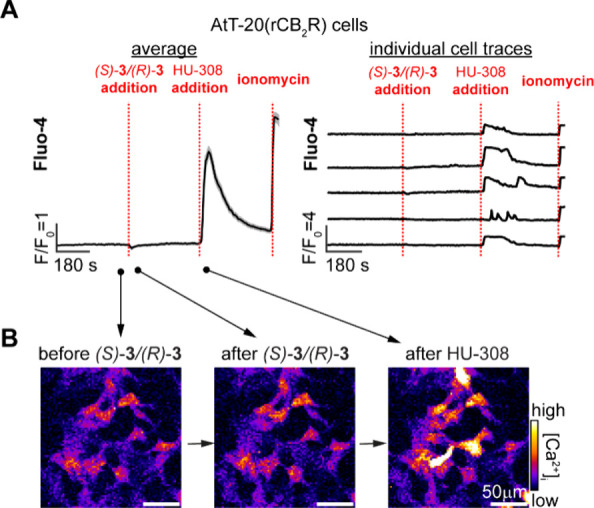
We report a blueprint for the rational design of G protein coupled receptor (GPCR) ligands with a tailored functional response. The present study discloses the structure-based design of cannabinoid receptor type 2 (CB2R) selective inverse agonists (S)-1 and (R)-1, which were derived from privileged agonist HU-308 by introduction of a phenyl group at the gem-dimethylheptyl side chain. Epimer (R)-1 exhibits high affinity for CB2R with Kd = 39.1 nM and serves as a platform for the synthesis of a wide variety of probes. Notably, for the first time these fluorescent probes retain their inverse agonist functionality, high affinity, and selectivity for CB2R independent of linker and fluorophore substitution. Ligands (S)-1, (R)-1, and their derivatives act as inverse agonists in CB2R-mediated cAMP as well as G protein recruitment assays and do not trigger β-arrestin-receptor association. Furthermore, no receptor activation was detected in live cell ERK1/2 phosphorylation and Ca2+-release assays. Confocal fluorescence imaging experiments with (R)-7 (Alexa488) and (R)-9 (Alexa647) probes employing BV-2 microglial cells visualized CB2R expressed at endogenous levels. Finally, molecular dynamics simulations corroborate the initial docking data in which inverse agonists restrict movement of toggle switch Trp2586.48 and thereby stabilize CB2R in its inactive state.


Conflict of interest statement

The authors declare the following competing financial interest(s): M.K., R.C.S., B.K., W.G., U.G., and E.M.C. have filed a patent on CB2R selective modulators and fluorescent probes.

Figures

Scheme 1
Scheme 1. Novel Structure-Based Design of HU-308-Derived CB2R-Selective Inverse Agonists That Actuate Trp2586.48 Toggle Switch
Figure 1
Figure 1
Comparison of active (A, PDB 8GUS, ligand HU-308) and inactive (B, PDB 5ZTY, ligand AM10257) CB2R conformations. (C) Docking study of HU-308-derived putative inverse agonist (R)-1 in the inactive CB2R conformation (PDB 5ZTY). (R)-1 reaches into the secondary pocket occupied by the toggle switch responsible for CB2R activation, Trp2586.48 (orange), and shares binding interactions virtually identical with those of AM10257.
Figure 2
Figure 2
Design of inverse agonists (S)-1 and (R)-1.
Scheme 2
Scheme 2. Synthesis of Novel CB2R-Selective HU-308-Derived Inverse Agonists
Reagents and conditions: (a) 1-chloro-5-iodopentane, t-BuLi, n-pentane, Et2O, −78 °C to rt, 83%; (b) KHMDS, CS2, THF, −78 °C to rt and then MeI, 40 °C, 95%; (c) BBr3, CH2Cl2, 0 °C, 97%; (d) Pd/C, H2, EtOAc, rt, 97%; (e) semipreparative SFC, (S)-14, 25%, >99% ee, (R)-14, 20%, 96% ee; (f) 15, pTsOH·H2O, CH2Cl2, rt, 64–71%; (g) (MeO)2SO2, K2CO3, acetone, rt, 78–82%; (h) NaN3, DMF, 50 °C, 88–96%; (i) N2H4·H2O, (E)/(Z)-crotyl alcohol, EtOH, 75 °C, 76–94%; (j) for conjugation conditions and details, see Supporting Information; (k) H2O, microwave irradiation, 150 °C, 99%; (l) 4-nitrobenzoyl chloride, NEt3, DMAP, CH2Cl2, rt, 85%.
Figure 3
Figure 3
TR-FRET-based saturation binding profile of (R)-7 (Alexa488) at CB2R determined at 37 °C. Nonspecific binding was determined in the presence of SR-144,528 (10 μM). Data shown as a mean ± SEM, N = 3.
Figure 4
Figure 4
BRET-based Gi-CASE membrane assay to characterize G protein recruitment at CB2R. Efficacy, Emax, of the compounds is shown as a mean ± SEM, N = 3–4.
Figure 5
Figure 5
BRET-based assay to characterize β-arrestin recruitment at CB2R. L = ligand. Data are a representative of N = 3.
Figure 6
Figure 6
Live cell AlphaScreen SureFire phospho-ERK assay with CB2R inducible breast cancer HCC1954 cell line. Cells were optionally induced with doxycycline (DOX) for 24 h to stimulate expression of CB2R followed by incubation with a vehicle (0.1% DMSO), agonist JWH133 (1 μM), or (R)-2 (1 μM) for 30 min. Statistical significance was examined by one-way ANOVA followed by Tukey’s post hoc test. ns = nonsignificant, ∗∗, p < 0.01; ∗∗∗, p < 0.001. Data are an average of three independent biological replicates.
Figure 7
Figure 7
Live cell fluorescent Ca2+ imaging in rat CB2R overexpressing AtT-20 cells [AtT-20(rCB2R)] loaded with Fluo-4AM (2 μM). After initial equilibration, (S)-3/(R)-3 (20 μM) was added, followed by HU-308 (20 μM) and ionomycin (10 μM). Shown are the average responses of 200 cells (A, left), individual traces of five representative cells (A, right), and representative fluorescence images from different time points (B). Averaged data plotted as mean ± SEM, T = 4.
Figure 8
Figure 8
Confocal imaging of (R)-7 in live cell lines. (A) AtT-20(SNAP-hCB2R) cells were labeled for 15 min with (R)-7 (Alexa488, 625 nM, green), SNAP-JF549i (JF549i, 500 nM, red), and Hoechst33342 (Hoechst, 1 μM, blue) to visualize CB2R, SNAP-tags, and nuclei, respectively. Fluorescence intensity profiles across the white line for Alexa488, JF549i, and Hoechst33342 are shown on the right. (B) AtT-20(rCB2R) cells (left) and AtT-20(WT) cells (right) were treated with (R)-7 (625 nM, green) and Hoechst33342 (1 μM, blue) for 15 min and imaged by confocal microscopy. (C) Live BV-2 microglial cells that endogenously express CB2R were incubated with (R)-7 (2.5 μM, green) and Hoechst33342 (1 μM, blue) for 15 min and imaged by confocal microscopy.
Figure 9
Figure 9
Representative frames from molecular dynamics (MD) simulations of CB2R (PDB 5ZTY) in complex with (S)-3 or (R)-3. Superimposition at level of protein backbone of (A) CB2R X-ray structure with AM10257 (light gray) and (R)-3 MD complex (ligand in gold and protein in light yellow) and of (B) the two inverse agonist complexes (S)-3 (ligand in blue and protein light blue) and (R)-3 (ligand in gold and protein in light yellow).

References

    1. Maccarrone M.; Di Marzo V.; Gertsch J.; Grether U.; Howlett A. C.; Hua T.; Makriyannis A.; Piomelli D.; Ueda N.; van der Stelt M. Goods and Bads of the Endocannabinoid System as a Therapeutic Target: Lessons Learned after 30 Years. Pharmacol. Rev. 2023, 75, 885–958. 10.1124/pharmrev.122.000600. - DOI - PMC - PubMed
    1. Munro S.; Thomas K. L.; Abu-Shaar M. Molecular characterization of a peripheral receptor for cannabinoids. Nature 1993, 365, 61–65. 10.1038/365061a0. - DOI - PubMed
    1. Goncalves E. D.; Dutra R. C. Cannabinoid receptors as therapeutic targets for autoimmune diseases: where do we stand?. Drug Discovery Today 2019, 24, 1845–1853. 10.1016/j.drudis.2019.05.023. - DOI - PubMed
    1. Rossi F.; Punzo F.; Umano G. R.; Argenziano M.; Miraglia Del Giudice E. Role of Cannabinoids in Obesity. Int. J. Mol. Sci. 2018, 19, 2690–2700. 10.3390/ijms19092690. - DOI - PMC - PubMed
    1. Kumawat V. S.; Kaur G. Therapeutic potential of cannabinoid receptor 2 in the treatment of diabetes mellitus and its complications. Eur. J. Pharmacol. 2019, 862, 172628–172633. 10.1016/j.ejphar.2019.172628. - DOI - PubMed

LinkOut - more resources