Review
. 2023;2(1):3.
doi: 10.1186/s44201-023-00018-9. Epub 2023 Mar 6.

Lung cancer in the emergency department

Affiliations
Review

Lung cancer in the emergency department

Jeremy R Walder et al. Emerg Cancer Care. 2023.

Abstract

Background: Though decreasing in incidence and mortality in the USA, lung cancer remains the deadliest of all cancers. For a significant number of patients, the emergency department (ED) provides the first pivotal step in lung cancer prevention, diagnosis, and management. As screening recommendations and treatments advance, ED providers must stay up-to-date with the latest lung cancer recommendations. The purpose of this review is to identify the many ways that emergency providers may intersect with the disease spectrum of lung cancer and provide an updated array of knowledge regarding detection, management, complications, and interdisciplinary care.

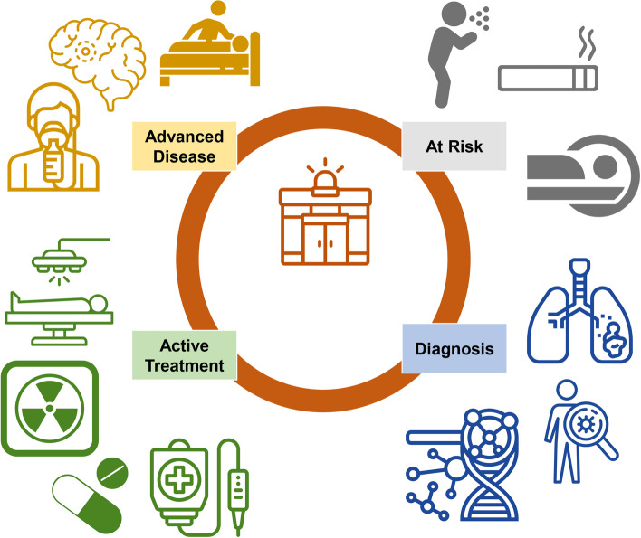
Findings: Lung cancer, encompassing 10-12% of cancer-related emergency department visits and a 66% admission rate, is the most fatal malignancy in both men and women. Most patients presenting to the ED have not seen a primary care provider or undergone screening. Ultimately, half of those with a new lung cancer diagnosis in the ED die within 1 year. Incidental findings on computed tomography are mostly benign, but emergency staff must be aware of the factors that make them high risk. Radiologic presentations range from asymptomatic nodules to diffuse metastatic lesions with predominately pulmonary symptoms, and some may present with extra-thoracic manifestations including neurologic. The short-term prognosis for ED lung cancer patients is worse than that of other malignancies. Screening offers new hope through earlier diagnosis but is underutilized which may be due to racial and socioeconomic disparities. New treatments provide optimism but lead to new complications, some long-term. Multidisciplinary care is essential, and emergency medicine is responsible for the disposition of patients to the appropriate specialists at inpatient and outpatient centers.

Conclusion: ED providers are intimately involved in all aspects of lung cancer care. Risk factor modification and referral for lung cancer screening are opportunities to further enhance patient care. In addition, with the advent of newer cancer therapies, ED providers must stay vigilant and up-to-date with all aspects of lung cancer including disparities, staging, symptoms of disease, prognosis, treatment, and therapy-related complications.

Keywords: Emergency department; Lung cancer; Lung cancer screening.


Conflict of interest statement

Competing interestsThe authors declare that they have no competing interests.

Figures

Fig. 1
Fig. 1
Lung cancer and emergency department. ED providers may encounter lung cancer along the cancer continuum. The ED may play a role in identifying those at risk of developing lung cancer or referring for further screening those with concerning findings. Incidental lung cancer may present with or without symptoms, and appropriate staging and tissue diagnosis with molecular markers are crucial. Sequelae from active treatment may include pain, radiation-induced lung injury, or drug-related pneumonitis. In those with advanced or metastatic disease, stabilization, symptom management, and supportive care are often required
Fig. 2
Fig. 2
Lung cancer staging. T refers to the primary tumor size and ranges from no primary tumor (T0) to a tumor more than 7 cm in the greatest dimension (T4). N describes regional lymph node involvement and broadly includes no regional lymph node involvement (N0) to contralateral mediastinal, hilar, scalene, or supraclavicular or ipsilateral scalene or supraclavicular lymph node metastasis (N3). The presence of distant metastasis is categorized as M0 (no distant metastasis) and M1 (distant metastasis). M1 is further delineated into subclasses where M1a consists of secondary nodules in the contralateral lung, pericardium, or pleura, with or without the presence of a malignant pleural or pericardial effusion. M1b involves one extra-thoracic metastasis, and M1c accounts for multiple extra-thoracic metastases. Information derived from [96]
Fig. 3
Fig. 3
ED presentations of lung cancer. A Elderly woman who is a never-smoker presents with cough and fatigue. Imaging revealed a 1.8-cm right upper lobe lung nodule (arrow) without hilar or mediastinal lymphadenopathy. She underwent lobectomy (stage IA, pT1c pN0 cM0). B Middle-aged man with metastatic adenocarcinoma of the lung was sent for anemia and fatigue. Imaging reveals innumerable pulmonary nodules (arrows) with mediastinal, bilateral hilar, and subcarinal lymphadenopathy (asterisk). C Middle-aged man presents with hemoptysis and dyspnea. Imaging reveals endobronchial disease in the right main bronchus at the level of the carina (arrowhead), moderate pleural effusion (P), and lung mass (arrow). D Middle-aged man with chronic myeloid leukemia on imatinib transferred for airway exacerbation. Imaging incidentally revealed a left upper lobe mass (arrow) contiguous with mediastinal and hilar lymphadenopathy consistent with primary lung malignancy
Fig. 4
Fig. 4
Manifestations of disease. A Ultrasound image of suspected malignant pleural effusion (P) with nodule (arrowhead) on the diaphragm (arrow). B Lung cancer on immune checkpoint inhibitor with multiple nodules and ground glass infiltrates (arrows, left > right) concerning immune-mediated pneumonitis. C Extensive small cell lung cancer with metastatic disease to spine (T4, T5) and extension of epidural tumor (arrowhead). D Left lung mass (arrow) with extrinsic compression (arrowhead) of the left mainstem bronchus. E Elderly aged man with a right upper lobe lung mass (arrow) in the apex consistent with a Pancoast tumor (mass that originates in the superior sulcus of the lung apex which may present atypically with shoulder pain, Horner syndrome, and superior vena cava syndrome) presenting with right shoulder pain and cough. F Middle-aged man with metastatic poorly differentiated lung cancer presents to the emergency center after fall. Imaging reveals a 1.8-cm right posterior frontal mass (arrow) with local edema

References

    1. Sung H, Ferlay J, Siegel RL, Laversanne M, Soerjomataram I, Jemal A, et al. Global Cancer Statistics 2020: GLOBOCAN estimates of incidence and mortality worldwide for 36 cancers in 185 countries. CA Cancer J Clin. 2021;71(3):209–249. doi: 10.3322/caac.21660. - DOI - PubMed
    1. Kino A, Boiselle PM, Raptopoulos V, Hatabu H. Lung cancer detected in patients presenting to the emergency department studies for suspected pulmonary embolism on computed tomography pulmonary angiography. Eur J Radiol. 2006;58(1):119–123. doi: 10.1016/j.ejrad.2005.11.008. - DOI - PubMed
    1. Hall WB, Truitt SG, Scheunemann LP, Shah SA, Rivera MP, Parker LA, et al. The prevalence of clinically relevant incidental findings on chest computed tomographic angiograms ordered to diagnose pulmonary embolism. Arch Intern Med. 2009;169(21):1961–1965. doi: 10.1001/archinternmed.2009.360. - DOI - PubMed
    1. Lehman SJ, Abbara S, Cury RC, Nagurney JT, Hsu J, Goela A, et al. Significance of cardiac computed tomography incidental findings in acute chest pain. Am J Med. 2009;122(6):543–549. doi: 10.1016/j.amjmed.2008.10.039. - DOI - PubMed
    1. Scholtz JE, Lu MT, Hedgire S, Meyersohn NM, Oliveira GR, Prabhakar AM, et al. Incidental pulmonary nodules in emergent coronary CT angiography for suspected acute coronary syndrome: impact of revised 2017 Fleischner Society Guidelines. J Cardiovasc Comput Tomogr. 2018;12(1):28–33. doi: 10.1016/j.jcct.2017.11.005. - DOI - PMC - PubMed

LinkOut - more resources