

A case report of a childhood scurvy musculoskeletal manifestation: Radiologic findings and diagnostic implications
- PMID: 38800077
- PMCID: PMC11126872
- DOI: 10.1016/j.radcr.2024.04.027
A case report of a childhood scurvy musculoskeletal manifestation: Radiologic findings and diagnostic implications
Abstract
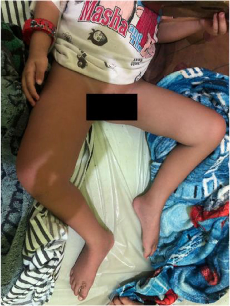
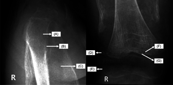
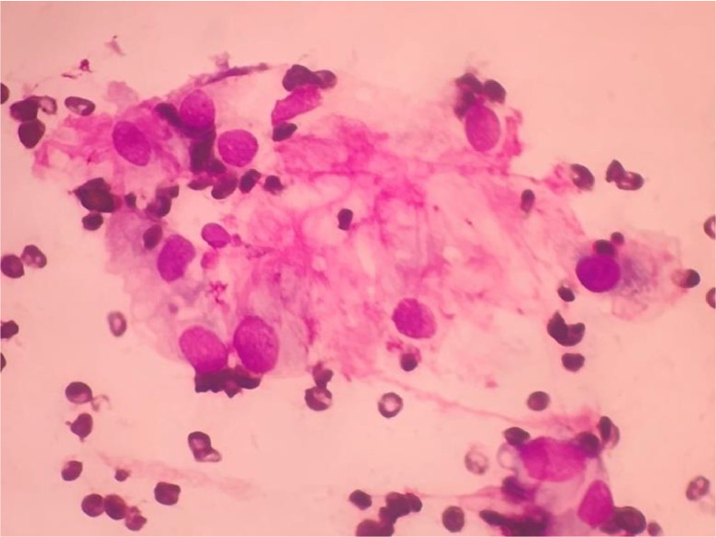
Scurvy is an infrequent pathological condition resulting from a sustained dietary vitamin C deficiency. Radiology becomes pivotal because the diagnostic process for scurvy can be intricate, given its resemblance to bone neoplasms. A 6-year-old boy, reported persistent pain and swelling in the right thigh for 2 months prior to hospitalization. Clinical examination revealed a mass localized in the right thigh and anemia. A radiograph of the right femur demonstrated extensive osteopenic changes, "Trümmerfeld zone", "Frankel line", "Pelkin fracture", "Wimberger ring sign", and para-epiphyseal subperiosteal hematoma. The absence of any such cases in our institution over the preceding decade emphasizes the uniqueness of this presentation. Histopathological evaluation yielded atypical results, prompting further radiographic assessment of the left femur and thorax. The subsequent findings corroborated the classic "scorbutic rosary" presentation, indicative of scurvy. The patient's symptoms gradually resolved with high-dose supplementation of vitamin C. Scurvy predominantly presents with musculoskeletal manifestations. Plasma vitamin C level assessment is the gold standard for the diagnosis, but it is currently inaccessible in our nation. Consequently, radiographic evaluation reveals pathognomonic features of the disorder. In thoracic radiographs, the "scorbutic rosary" presentation is evident. In contrast, long bones exhibit hallmarks of scurvy: diffuse osteopenia, "Frankel line", "Trümmerfeld zone", "Pelkin fracture", "Wimberger ring sign", and para-epiphyseal subperiosteal hematoma. Prompt intervention with vitamin C thwarts the progression to severe complications. Radiology is an indispensable tool in diagnosing pediatric scurvy, especially in developmental countries where the assessment of vitamin C serum levels is inaccessible.
Keywords: Musculoskeletal; Pediatric; Radiology; Scurvy; Vitamin C.
© 2024 The Authors. Published by Elsevier Inc. on behalf of University of Washington.
Figures


Similar articles
-
Modern Day Scurvy in Pediatric Orthopaedics: A Forgotten Illness.J Pediatr Orthop. 2021 Mar 1;41(3):e279-e284. doi: 10.1097/BPO.0000000000001731. J Pediatr Orthop. 2021. PMID: 33528119
-
Scurvy: Forgotten diagnosis, but still exist.Int J Surg Case Rep. 2020;68:263-266. doi: 10.1016/j.ijscr.2020.03.002. Epub 2020 Mar 7. Int J Surg Case Rep. 2020. PMID: 32199252 Free PMC article.
-
Scurvy in a 10-month-old boy.Int J Dermatol. 2007 Feb;46(2):194-8. doi: 10.1111/j.1365-4632.2007.02856.x. Int J Dermatol. 2007. PMID: 17269976
-
Scurvy in pediatric age group - A disease often forgotten?J Clin Orthop Trauma. 2015 Jun;6(2):101-7. doi: 10.1016/j.jcot.2014.12.003. Epub 2015 Jan 5. J Clin Orthop Trauma. 2015. PMID: 25983516 Free PMC article. Review.
-
Musculoskeletal manifestations of scurvy.Joint Bone Spine. 2005 Mar;72(2):124-8. doi: 10.1016/j.jbspin.2004.01.007. Joint Bone Spine. 2005. PMID: 15797491 Review.
References
Publication types
LinkOut - more resources
Full Text Sources
