

Spontaneous Cerebrospinal Fluid Rhinorrhea With Meningoencephalocele Recurrence After Placement of a Lumboperitoneal Shunt: A Case Report
- PMID: 38800265
- PMCID: PMC11116915
- DOI: 10.7759/cureus.58896
Spontaneous Cerebrospinal Fluid Rhinorrhea With Meningoencephalocele Recurrence After Placement of a Lumboperitoneal Shunt: A Case Report
Abstract
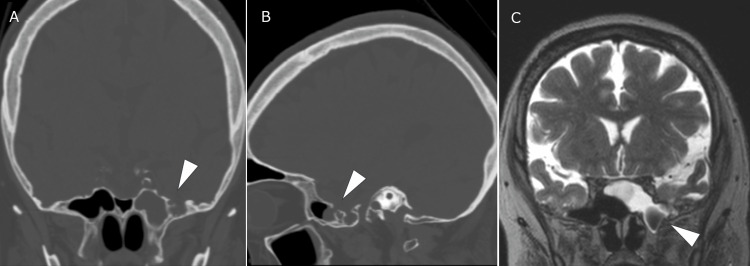
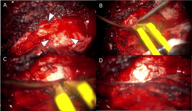
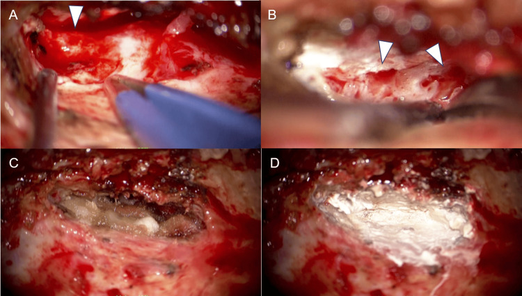
Cerebrospinal fluid rhinorrhea associated with meningoencephalocele is usually treated surgically. During the perioperative period, cerebrospinal fluid diversion may be employed to control intracranial pressure, but there are few indications for this method. A 51-year-old female presented with cerebrospinal fluid rhinorrhea associated with meningoencephalocele and underwent surgical repair followed by the placement of a lumboperitoneal shunt. However, cerebrospinal fluid leakage recurred, requiring a second surgery. Lumbar drainage effectively controls intracranial pressure, but it does not cure bone defects. The use of these devices should be carefully considered based on the patient's condition.
Keywords: cerebrospinal fluid diversion; cerebrospinal fluid rhinorrhea; lumbar drainage; lumboperitoneal shunt; meningoencephalocele.
Copyright © 2024, Natsuhara et al.
Conflict of interest statement
The authors have declared that no competing interests exist.
Figures
Similar articles
-
Spontaneous cerebrospinal fluid rhinorrhea as a primary presentation of idiopathic intracranial hypertension, management strategies, and clinical outcome.Surg Neurol Int. 2024 Dec 11;15:458. doi: 10.25259/SNI_560_2024. eCollection 2024. Surg Neurol Int. 2024. PMID: 39777167 Free PMC article.
-
Successful Treatment of Spontaneous Cerebrospinal Fluid Rhinorrhea With Endoscopic Third Ventriculostomy and Lumboperitoneal Shunt: A Case Report.Front Neurosci. 2020 Jan 31;14:57. doi: 10.3389/fnins.2020.00057. eCollection 2020. Front Neurosci. 2020. PMID: 32082119 Free PMC article.
-
Effect of lumbar drain placement on recurrence of cerebrospinal rhinorrhea after endoscopic repair.Int Forum Allergy Rhinol. 2012 May-Jun;2(3):222-6. doi: 10.1002/alr.21023. Epub 2012 Feb 16. Int Forum Allergy Rhinol. 2012. PMID: 22344940
-
Analysis of the Causes and Experience in the Diagnosis and Treatment of Meningocele Caused by Sternberg's Canal of the Sphenoid Sinus: Two Case Reports and a Review of the Literature.Curr Med Imaging. 2023;19(9):1063-1070. doi: 10.2174/1573405619666230206103036. Curr Med Imaging. 2023. PMID: 36748216 Review.
-
Ventriculoperitoneal shunt strategy for cerebrospinal fluid rhinorrhea repair: a case report and review of the literature.Pediatr Neurol. 2012 Nov;47(5):369-72. doi: 10.1016/j.pediatrneurol.2012.07.010. Pediatr Neurol. 2012. PMID: 23044021 Review.
References
-
- Cerebrospinal fluid rhinorrhoea: diagnosis and management. Abuabara A. http://www.medicinaoral.com/pubmed/medoralv12_i5_pE397.pdf Med Oral Patol Oral Cir Bucal. 2007;12:397–400. - PubMed
-
- Spontaneous encephaloceles of the temporal lobe. Wind JJ, Caputy AJ, Roberti F. Neurosurg Focus. 2008;25:0. - PubMed
-
- Spontaneous sphenoid lateral recess cerebrospinal fluid leaks arise from intracranial hypertension, not Sternberg's canal. Illing E, Schlosser RJ, Palmer JN, Curé J, Fox N, Woodworth BA. Int Forum Allergy Rhinol. 2014;4:246–250. - PubMed
-
- Management of cerebrospinal fluid rhinorrhea: an evidence-based review with recommendations. Oakley GM, Orlandi RR, Woodworth BA, Batra PS, Alt JA. Int Forum Allergy Rhinol. 2016;6:17–24. - PubMed
Publication types
LinkOut - more resources
Full Text Sources