. 2024 May 15:39:25-40.
doi: 10.1016/j.bioactmat.2024.04.014. eCollection 2024 Sep.

A tantalum-containing zirconium-based metallic glass with superior endosseous implant relevant properties

Affiliations

A tantalum-containing zirconium-based metallic glass with superior endosseous implant relevant properties

Yunshu Wu et al. Bioact Mater. .

Abstract

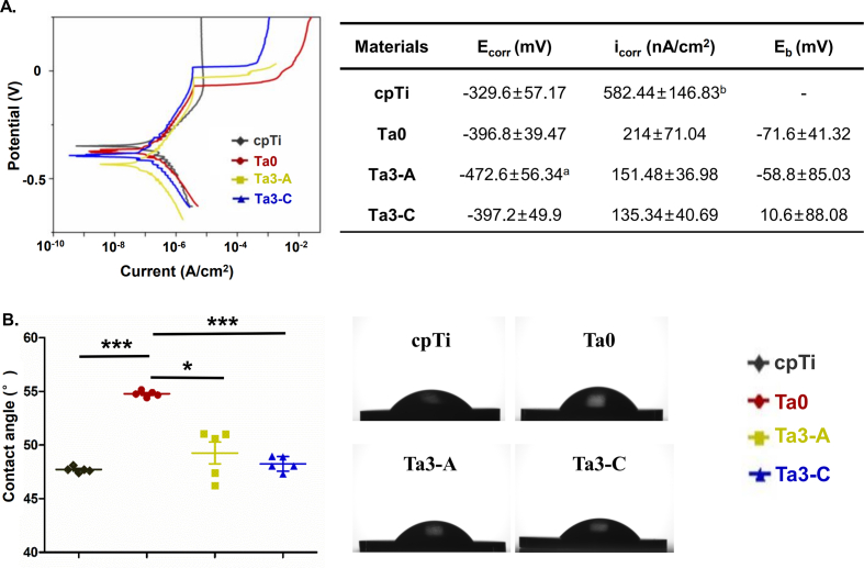
Zirconium-based metallic glasses (Zr-MGs) are demonstrated to exhibit high mechanical strength, low elastic modulus and excellent biocompatibility, making them promising materials for endosseous implants. Meanwhile, tantalum (Ta) is also well known for its ideal corrosion resistance and biological effects. However, the metal has an elastic modulus as high as 186 GPa which is not comparable to the natural bone (10-30 GPa), and it also has a relative high cost. Here, to fully exploit the advantages of Ta as endosseous implants, a small amount of Ta (as low as 3 at. %) was successfully added into a Zr-MG to generate an advanced functional endosseous implant, Zr58Cu25Al14Ta3 MG, with superior comprehensive properties. Upon carefully dissecting the atomic structure and surface chemistry, the results show that amorphization of Ta enables the uniform distribution in material surface, leading to a significantly improved chemical stability and extensive material-cell contact regulation. Systematical analyses on the immunological, angiogenesis and osteogenesis capability of the material are carried out utilizing the next-generation sequencing, revealing that Zr58Cu25Al14Ta3 MG can regulate angiogenesis through VEGF signaling pathway and osteogenesis via BMP signaling pathway. Animal experiment further confirms a sound osseointegration of Zr58Cu25Al14Ta3 MG in achieving better bone-implant-contact and inducing faster peri-implant bone formation.

Keywords: Atomic structure; Endosseous implant; Surface chemistry; Tantalum; Zirconium-based metallic glass.


Conflict of interest statement

The authors declare no conflict of interest.

Figures

Image 1
Graphical abstract
Fig. 1
Fig. 1
Structural properties, thermal parameters and surface chemical compositions of Ta0, Ta3-A and Ta3–C. A) Representative XRD pattern of Ta0 and Ta3-A. B) Representative DSC data of Ta0 and Ta3-A, showing their transition temperature (Tg) and crystallization temperature (Tx). C) Representative XRD pattern of Ta3–C, showing major crystal phases of CuZr2, Zr0.18Ta0.82, Zr4Al3 and Cu3Al. D) Microstructure and SAED pattern of Ta3-A, showing microstructural homogeneity and the amorphous ring. E) Microstructure and SAED pattern of Ta3–C, showing the presence of nanocrystalline and crystalline spots. F) XPS survey spectrum and narrow scan spectra of Ta for Ta3-A. Ta2O5 was detected in the oxide film formed on the surface of Ta3-A. G) XPS survey spectrum and narrow scan spectra of Ta for Ta3–C. Ta2O5 was less potentially presented in the oxide film formed on the surface of Ta3–C.
Fig. 2
Fig. 2
Electrochemical results and surface hydrophility. A) Potentiodynamic-polarization curves and corrosion parameters of all samples in simulated body fluid (SBF), indicating a slower corrosion rate of Ta0, Ta3-A and Ta3–C, compared to cpTi. Data are presented as mean ± SD. a: Ta3-A vs cpTi p < 0.01; b: cpTi vs the other three p < 0.001. B) Contact angles of all samples. Ta0 showed significantly higher contact angle than the other three. Data are presented as mean ± SE. *p < 0.05 and ***p < 0.001.
Fig. 3
Fig. 3
Cell adhesion, morphology and viability features. A) (Left) Representative cell adhesion figures at 1 h, 4 h and 24 h. Ta0 and Ta3–C exhibited inferior initial cell adhesion of BMSCs at 1 h and 4 h. (Right) Phalloidin staining of BMSCs F-actin at 24 h. B) SEM images of BMSCs cell morphology on material surface at 24 h. C) Relative BMSCs cell viability fold change of all samples, indicating that Ta3–C significantly inhibited cell viability at day 5 and day 7. Data are presented as mean ± SE. **p < 0.01. D) Flow cytometry result of FV520 staining of all samples, indicating a population shift from living to necrotic BMSCs that co-cultured with Ta3–C for 5 days.
Fig. 4
Fig. 4
Ion release features related to material surface chemistry and atomic structure. A) The release of Cu ions in α-MEM at 37 °C measured by ICP-MS. A dramatic increase of ion releases was observed after 5 days in Ta3–C. Data are presented as mean ± SD. B) The intracellular Cu ion levels detected by ICP-MS. After incubating with Ta3–C for 5 days, BMSCs absorbed significantly more Cu ions, compromising cell viability. Data are presented as mean ± SE. *p < 0.05. C) Chemical analysis of Ta3-A and Ta3–C before and after being immersed in α-MEM at 37 °C for 5 days. High angle angular dark field detector (HADDF) images showed Cu depletion at the near surface of Ta3–C.
Fig. 5
Fig. 5
Macrophage responses to different surface chemistry. A) ELISA results of IL-1β, TNF-α and IL-10. Ta0 and Ta3-A significantly suppressed the secretion of pro-inflammatory cytokines. B) qRT-PCR results of M1 and M2 activity related markers. Ta0 and Ta3-A significantly reduced the expression of pro-inflammatory markers. C) Heatmap of representative overlapped genes between Ta3-A vs cpTi and Ta0 vs cpTi comparisons, showing an overall decrease in pro-inflammatory genes. D) Western blot result of iNos, Cd80, Cd206 and β-actin. Data are presented as mean ± SE. *p < 0.05, **p < 0.01 and ***p < 0.001.
Fig. 6
Fig. 6
The angiogenesis reaction to different surface chemistry. A) (Top) Scratch assay and quantitative measurement of HUVECs cultured in material's extract, indicating better wound healing in Ta0 and Ta3-A. (Bottom) Representative images and quantitative measurement of tube formation assay at 4 h, 8 h and 12 h, indicating that Ta3-A could efficiently promote vasculature development. B) qRT-PCR of angiogenesis marker, VEGF and CD31. C) GSEA of GO found expression enrichment of Ta3-A in VEFG production (GO: 0010573) when compared to that of cpTi. D) Immunofluorescent staining of VEGF, confirming that Ta3-A up-regulated the protein level of VEGF in comparison with cpTi. Data are presented as mean ± SE. *p < 0.05, **p < 0.01 and ***p < 0.001.
Fig. 7
Fig. 7
The osteogenic potential of different surface chemistry. A) (Top) ALP staining on cpTi, Ta0 and Ta3-A surface, and quantitative measurement of ALP activity. Ta3-A showed enhanced ALP activity. (Bottom) ARS staining on cpTi, Ta0 and Ta3-A surface, and dissolved ARS OD value measured at A562 nm. Ta3-A showed exhibited better calcium nodules formation. B) qRT-PCR of osteogenic markers. Ta3-A significantly increased the expression of SP7, ALPL and BGLAP. C) Heatmap showing the expression of representative osteogenic genes in investigated materials. D) GSEA of KEGG found expression enrichment of Ta3-A in regulating TGF-beta signaling pathway (HSA04350) in comparison with cpTi and Ta0. Note that in KEGG pathway, TGF-beta signaling pathway encompassed “TGF-β signaling pathway” and “BMP signaling pathway” (https://www.kegg.jp/pathway/map=map04350&keyword=TGF-Beta). E) qRT-PCR results confirmed the elevated expression of BMP4 and SMAD9 in Ta3-A. F) Western blot of BMP4, total SMAD9, phosphorylated SMAD1/5/9, ALPL and β-actin. Results indicated an up-regulated and activated BMP signaling pathway in Ta3-A. Data are presented as mean ± SE. *p < 0.05 and **p < 0.01.
Fig. 8
Fig. 8
Osseointegration on different surface chemistry. A) 3D reconstruction of peri-implant bone at 4- and 8-weeks post-implantation. B) Micro-CT analysis of bone volume to total volume fraction, indicating an inferior quantity of peri-implant bone around cpTi implants than that of Ta3-A. C) Representative images of Stevenel's blue and Van Gieson's picrofuchsin staining showing new bone formation around implants. Ta3-A implants were able to induce more bone formation that was in direct close contact to implant surfaces. D) Quantitative measurement of bone area percentage around implants and relative bone-implant-contact length. Ta3-A implants achieved better osseointegration than Ta0. E) Quantitative analysis of dynamic mineral apposition rate (MAR) between 4 and 8 weeks. Ta3-A implants induced faster bone formation rate. F) Immunofluorescent images merged from ARS at 4 weeks and calcein green at 8 weeks. The distance between the two immunofluorescent lines represented the new bone formed during 4–8 weeks. Data are presented as mean ± SE. *p < 0.05, **p < 0.01 and ***p < 0.001.

References

    1. Li H.F., Zheng Y.F. Recent advances in bulk metallic glasses for biomedical applications. Acta Biomater. 2016;36:1–20. https://10.1016/j.actbio.2016.03.047 - DOI - PubMed
    1. Sun K., Fu R., Liu X.W., Xu L.M., Wang G., Chen S.Y., Zhai Q.J., Pauly S. Osteogenesis and angiogenesis of a bulk metallic glass for biomedical implants. Bioact. Mater. 2022;8:253–266. https://10.1016/j.bioactmat.2021.06.018 - DOI - PMC - PubMed
    1. Liu X., Ding C., Chu P.K. Surface modification of titanium, titanium alloys, and related materials for biomedical applications. Mater. Sci. Eng. R. 2004;47:49–121. https://10.1016/j.mser.2004.11.001 - DOI
    1. Gautam S., Bhatnagar D., Bansal D., Batra H., Goyal N. Recent advancements in nanomaterials for biomedical implants. Biomed. Eng. Adv. 2022;3 https://10.1016/j.bea.2022.100029 - DOI
    1. Nagarajan S., Mohana M., Sudhagar P., Raman V., Nishimura T., Kim S., Kang Y.S., Rajendran N. Nanocomposite coatings on biomedical grade stainless steel for improved corrosion resistance and biocompatibility. ACS Appl. Mater. Interfaces. 2012;4:5134–5141. https://10.1021/am301559r - DOI - PubMed