. 2024 Sep;138(5):2021-2036.
doi: 10.1007/s00414-024-03249-5. Epub 2024 May 27.

Oxidative stress and NF-KB/iNOS inflammatory pathway as innovative biomarkers for diagnosis of drowning and differentiating it from postmortem submersion in both fresh and saltwater in rats

Affiliations

Oxidative stress and NF-KB/iNOS inflammatory pathway as innovative biomarkers for diagnosis of drowning and differentiating it from postmortem submersion in both fresh and saltwater in rats

Rana Adel et al. Int J Legal Med. 2024 Sep.

Abstract

Background: Finding a dead body in water raises an issue concerning determining the cause of death as drowning because of the complex pathophysiology of drowning. In addition, the corpse may be submersed postmortem.

Objective: Evaluate the role of oxidative stress markers and NF-KB/iNOS inflammatory pathway as diagnostic biomarkers in drowning and whether they could differentiate freshwater from saltwater drowning.

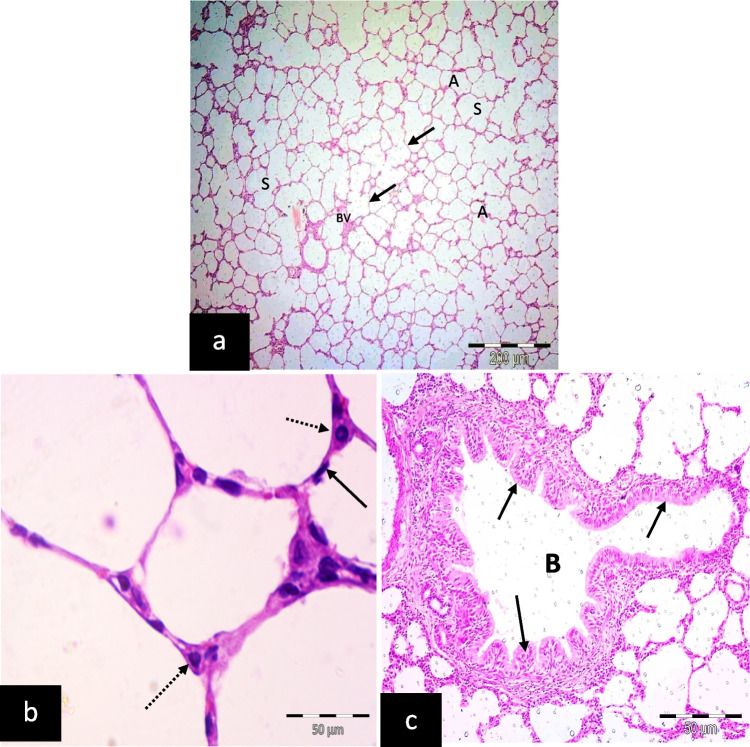
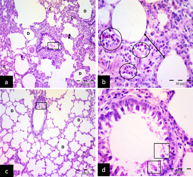
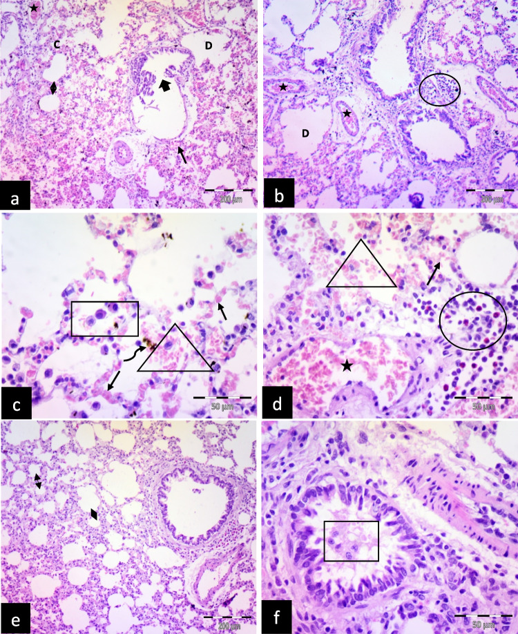
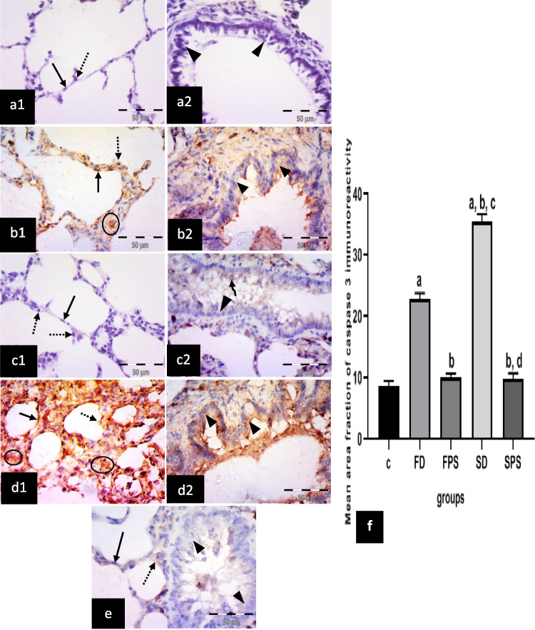
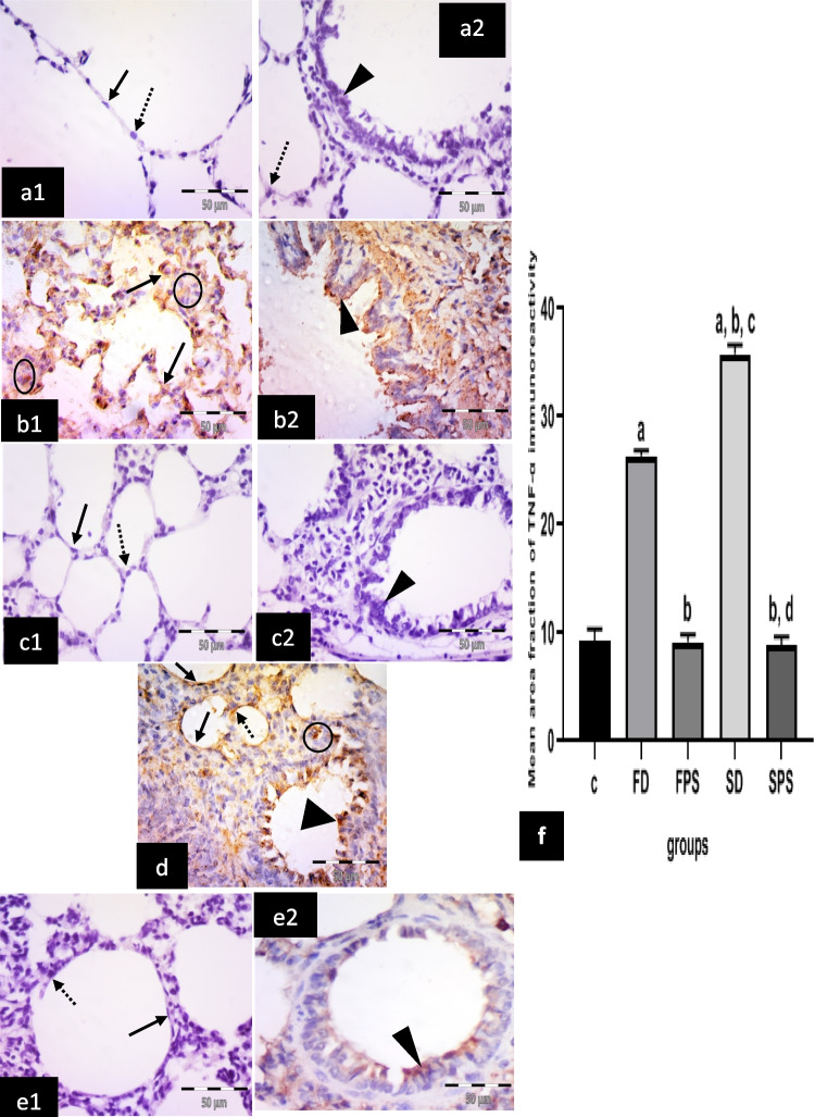
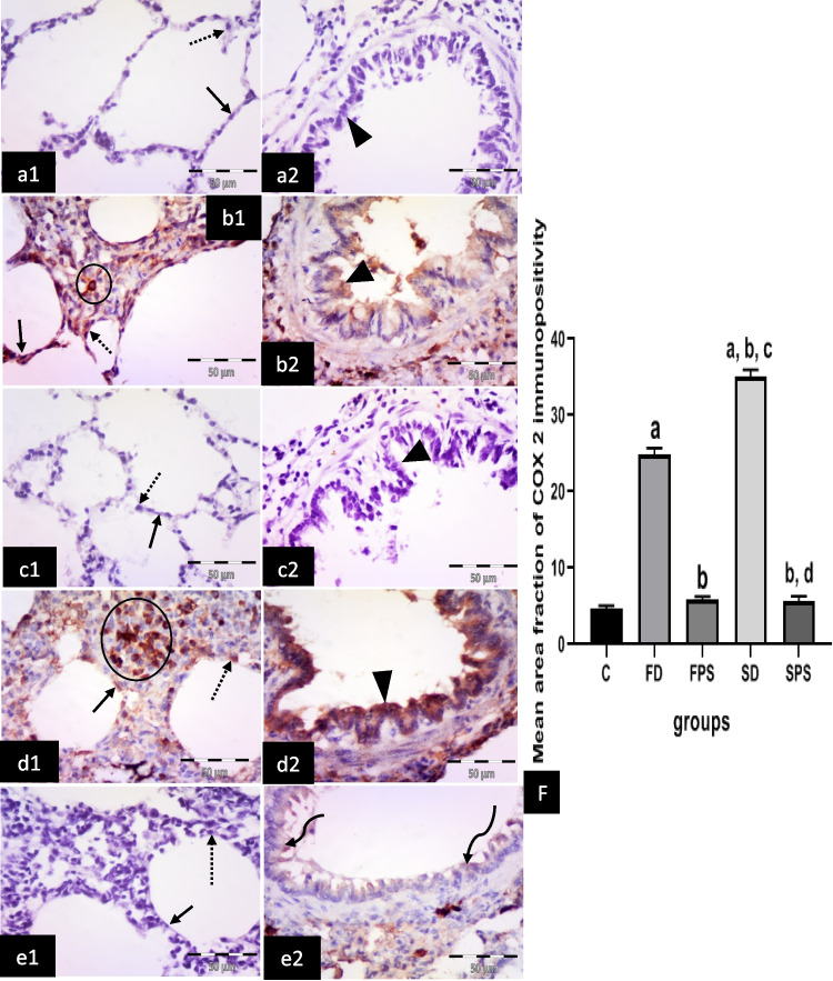
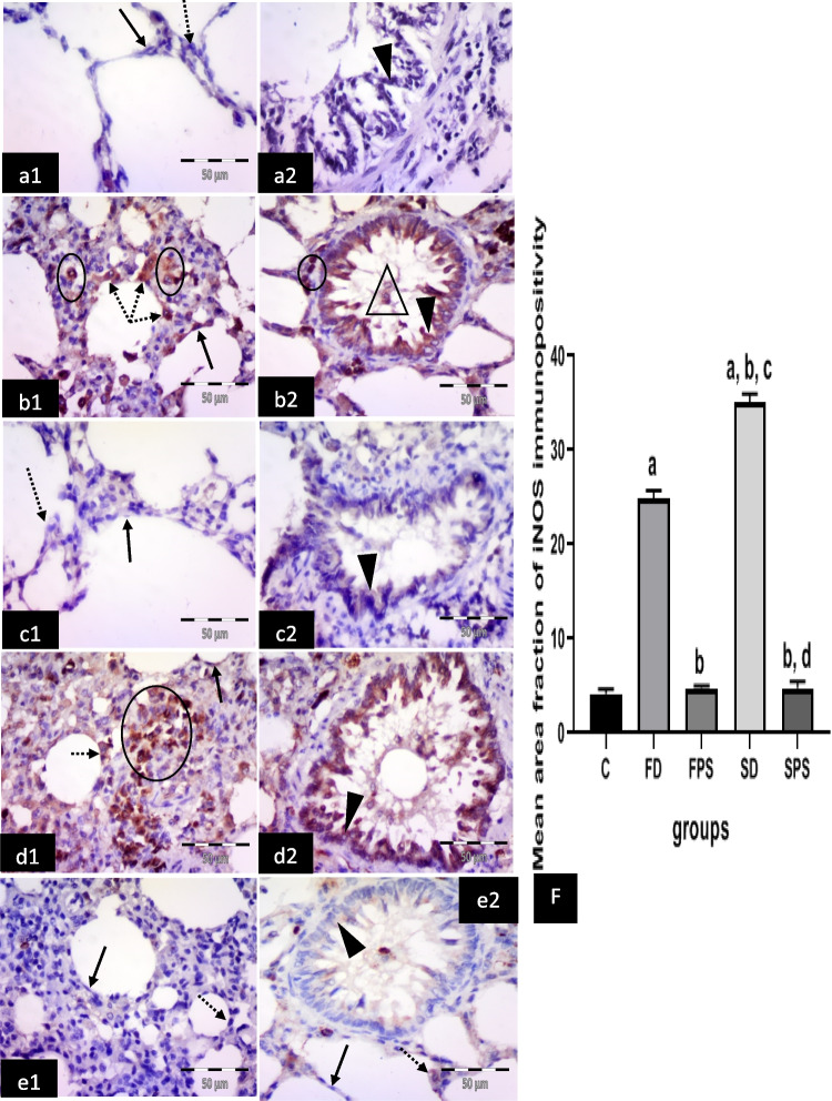
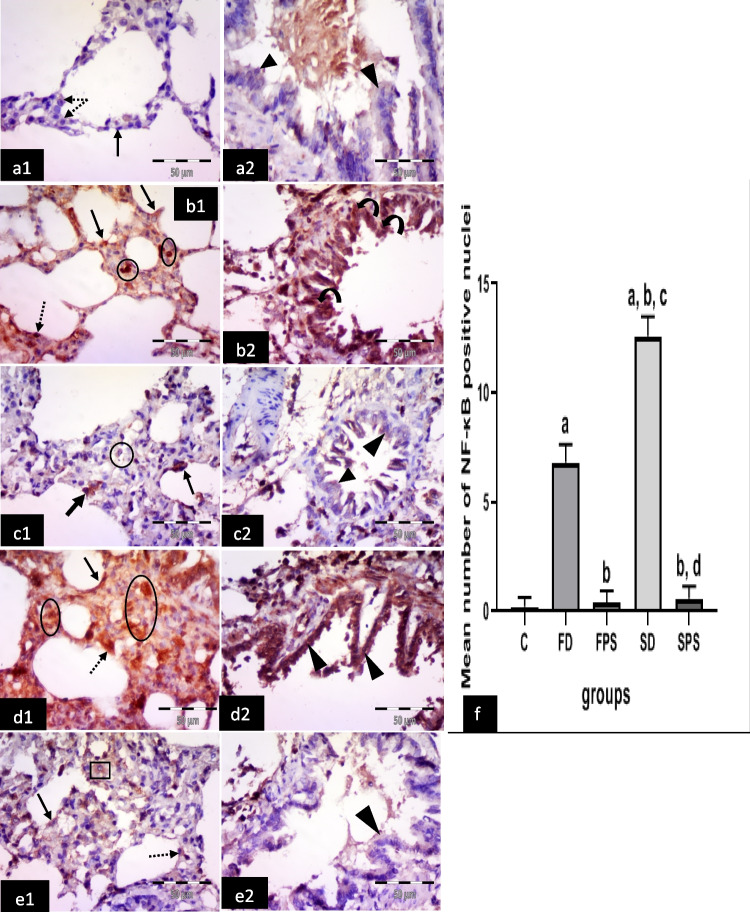
Methods: This study included forty-five adult male albino rats classified into five groups: control group (C), Freshwater-drowned group (FD), Freshwater postmortem submersion group (FPS), saltwater-drowned group (SD), and saltwater postmortem submersion group (SPS). After the autopsy, the rats' lungs in each group were prepared for histological, immunohistochemical (caspase 3, TNF-α, NF-kB, COX-2 & iNOS), biochemical studies; MDA, NOx, SOD, GSH, VCAM-1, COX-2; and RT-PCR for the relative quantification of NF-kB and iNOS genes expression.

Results: Lung oxidative markers were significantly affected in drowned groups than in postmortem submersion groups. Inflammatory pathway markers were also significantly increased in the drowned groups, with concern that all markers were significantly affected more in saltwater than in freshwater drowned group.

Conclusions: It is concluded that the tested markers can be used accurately in diagnosing drowning and differentiating it from postmortem submersion with a better understanding of the mechanism of death in drowning as both mechanisms, inflammatory and oxidative stress, were revealed and involved.

Keywords: Acute lung injury; Drowning; Freshwater; Postmortem submersion; Saltwater.


Conflict of interest statement

The authors declare that there is no competing interest.

Figures

Fig. 1
Fig. 1
Illustrative photomicrographs of the lung of adult male albino rats of the control group; a showing alveoli (A), alveolar sacs (S), and blood vessels(B.V). Notice thin inter-alveolar septa (arrows). b showing interalveolar septum formed of a single layer of cells: pneumocyte type I (arrow) and pneumocyte type II (dotted arrows). c showing bronchiole (B) lined with simple columnar epithelium (arrows). (H&E; a X 100, scale bar = 200 μm; b, c X 400, scale bar = 50 μm)
Fig. 2
Fig. 2
Illustrative photomicrographs of the lung of adult male albino rats from; a, b freshwater-drowned group (FD), showing thickening of the inter-alveolar septa (double head arrows) with inflammatory cellular infiltration (circles). Some alveoli are collapsed (c), and others are dilated (D). Notice: exfoliated epithelial cells (square) inside the bronchioles. c, d freshwater postmortem submersion group (FPS), showing dilated alveoli (D) and exfoliated epithelial cells (squares) inside the bronchioles. (H&E; a, c, X 100, scale bar = 200 μm; b, d X 400, scale bar = 50 μm)
Fig. 3
Fig. 3
Illustrative photomicrographs of the rats' lungs from different experimental groups in (a, b, c, d) saltwater-drowned group (SD); showing thickening of the inter-alveolar septa (double head arrow) with congested blood vessels (stars), extravasated RBCs in the interalveolar septa (thin arrows) and inside the alveoli (triangles) with inflammatory cellular infiltration (circles). Some alveoli are collapsed (c), and others are dilated (D). Notice: destructed bronchioles (thick arrow), hemosiderin-laden macrophages (curved arrow), and intra-alveolar lymphocytes (square). e, f Saltwater postmortem submersion group (SPS): Showed thickening of the inter-alveolar septa (double head arrows) and exfoliated epithelial cells (square) inside the bronchioles. (H&E; a, b, e X 100, scale bar = 200 μm; c, d, f X 400, scale bar = 50 μm)
Fig. 4
Fig. 4
Illustrative photomicrographs of the lung from all experimental groups in adult male albino rats immunolabelled for caspase 3 in; a1, 2 control group(C); showing negative immuno-expression in type I pneumocytes (arrow), type II pneumocytes (dotted arrow) and epithelial lining of the bronchiole (arrow heads). b1, 2 freshwater-drowned group (FD), revealing many cells with strong positive cytoplasmic (circle) expression, most properly macrophages in the interalveolar space. Strong positive cytoplasmic expression in pneumocytes type I (arrow) and type II (dotted arrow). Moderate positive cytoplasmic expression in the bronchiolar epithelium (arrowhead) is also noticed. c1, 2 freshwater postmortem submersion group (FPS), showing negative cytoplasmic expression in type I pneumocytes (arrow) or type II pneumocytes (dotted arrows). Notice negative (head arrow) or faint (curved arrow) cytoplasmic expression in the bronchiolar epithelium. d1, 2 saltwater-drowned group(SD), showing numerous cells with strong positive cytoplasmic expression in the interalveolar septum (circles). Notice strong cytoplasmic expression in type I (arrow) and type II (dotted arrow) pneumocytes and strong cytoplasmic expression in the bronchiolar epithelium (arrowheads). e saltwater postmortem submersion group (SPS), revealing faint cytoplasmic expression in type I pneumocytes (arrow) and type II pneumocytes (dotted arrow). Notice negative or faint cytoplasmic expression in the bronchiolar epithelium (arrowheads) (n = 9) (immunohistochemistry for caspase 3 × 400, scale bar = 50 μm). f Mean area fraction of caspase 3 immunoreactivity, a significant difference from the control group, b significant difference from (FD) group, c significant difference from (FPS) group, d significant difference from (SD) group. P < 0.05. Values are expressed as mean ± SEM (n=9)
Fig. 5
Fig. 5
Illustrative photomicrographs of the lung from all experimental groups in adult male albino rats immunolabelled for TNF-α in (a1, 2) control group (C); showing negative immuno-expression in type I pneumocytes (arrow), type II pneumocytes (dotted arrow) and epithelial lining of the bronchiole (arrowhead). b1, 2 freshwater-drowned group (FD), revealing many cells with positive cytoplasmic expression (arrows) in the alveolar lining, most properly pneumocytes type II (arrows) and positive cytoplasmic expression in the bronchiolar epithelium (arrowhead) and cells in the interalveolar septum (circles). c1, 2 freshwater postmortem submersion group (FPS), showing negative cytoplasmic expression in type I (arrow) and type II (dotted arrow) pneumocytes. Notice negative (head arrow) cytoplasmic expression in bronchiolar epithelium. d saltwater-drowned group (SD), showing numerous cells with positive cytoplasmic expression in cells lining the alveoli, either type I pneumocytes (arrows) or type II pneumocytes (dotted arrow). Notice cells with positive cytoplasmic expression in the bronchiolar epithelium (arrowhead) and the interalveolar septum (circle). e1, 2 saltwater postmortem submersion group (SPS), revealing negative cytoplasmic expression in cells lining the alveoli, either type I (arrow) or type II (dotted arrow) pneumocytes. Notice some cytoplasmic expression in the bronchiolar epithelium (arrowhead). (n = 9) (immunohistochemistry for TNF-α × 400, scale bar = 50 μm). f Mean area fraction of TNF-α immunoreactivity, a significant difference from the control group, b significant difference from (FD) group, c significant difference from (FPS) group, d significant difference from (SD) group. P < 0.05. Values are expressed as mean ± SEM (n = 9)
Fig. 6
Fig. 6
Illustrative photomicrographs of the lung from all experimental groups in adult male albino rats immunolabelled for COX 2 in (a1, 2) control group (C): showing negative immuno-expression in type I pneumocytes (arrow), type II pneumocytes (dotted arrow) and epithelial lining of the bronchiole (arrowhead). b1, 2 freshwater-drowned group (FD) revealing many cells with positive cytoplasmic expression in the alveolar lining, either type I (arrow) or type II (dotted arrow) pneumocytes, in the bronchiolar epithelium (arrowhead) and connective tissue cells in the interalveolar septum (circle). c1, 2 freshwater postmortem submersion group (FPS) and (e1, 2) saltwater postmortem submersion group (SPS), showing negative cytoplasmic expression in type I (arrows) and type II (dotted arrows) pneumocytes. Notice negative (head arrow) or faint (curved arrows) cytoplasmic expression in the bronchiolar epithelium. d1, 2 saltwater-drowned group (SD), showing numerous cells with positive strong cytoplasmic expression in cells lining the alveoli, either type I (arrow) or type II (dotted arrow) pneumocytes. Notice strong positive cytoplasmic expression in the bronchiolar epithelium (arrowhead) and connective tissue cells in the interalveolar septum (circle). (n = 9) (immunohistochemistry for COX 2 × 400, scale bar = 50 μm). (f) Mean area fraction of COX 2 immunoreactivity, a significant difference from the control group, b significant difference from (FD) group, c significant difference from ( FPS) group, d significant difference from (SD) group. P < 0.05. Values are expressed as mean ± SEM (n = 9)
Fig. 7
Fig. 7
Illustrative photomicrographs of the lung from all experimental groups in adult male albino rats immunolabelled for iNOS 2 in (a1, 2) control group (C): showing negative immuno-expression in type I pneumocytes (arrow), type II pneumocytes (dotted arrow) and epithelial lining of the bronchiole (arrowhead). b1, 2 freshwater-drowned group (FD), revealing many cells with strong positive cytoplasmic expression in the alveolar lining, either type I (arrow) or type II (dotted arrows) pneumocytes, in connective tissue cells in the interalveolar septum (circles), in the bronchiolar epithelium (arrowhead) and cells in the bronchiolar lumen (triangle). c1, 2 freshwater postmortem submersion group (FPS), showing negative cytoplasmic expression in type I (arrow) and type II (dotted arrow) pneumocytes. Notice very faint (head arrow) cytoplasmic expression in the bronchiolar epithelium. d1, 2 saltwater-drowned group (SD), showing numerous cells with strong positive cytoplasmic expression in the lining of the alveoli, either type I (arrow) or type II pneumocyte (dotted arrow). Notice strong positive cytoplasmic expression in the bronchiolar epithelium (arrowhead) and connective tissue cells in the interalveolar septum (circle). e1, 2 saltwater postmortem submersion group (SPS), revealing negative or faint cytoplasmic expression in cells lining the alveoli, either type I (arrows) or type II pneumocyte (dotted arrows) and in the bronchiolar epithelium (arrowhead). (n = 9) (immunohistochemistry for iNOS × 400, scale bar = 50 μm). f Mean area fraction of iNOS immunoreactivity, a significant difference from the control group, b significant difference from (FD)group, c significant difference from (FPS) group, d significant difference from (SD) group. P < 0.05. Values are expressed as mean ± SEM (n = 9)
Fig. 8
Fig. 8
Illustrative photomicrographs of the lung from all experimental groups in adult male albino rats immunolabelled for NF-κB2 in; a1, 2 control group (C); showing negative or faint cytoplasmic and negative nuclear expression in type I pneumocytes (arrow), type II pneumocytes (dotted arrows) and epithelial lining of a bronchiole (arrow heads). b1, 2 freshwater-drowned group (FD), revealing many cells with strong positive cytoplasmic (arrows) or nuclear and cytoplasmic (dotted arrow) expression in the alveolar lining, strong nuclear expression in connective tissue cells in the interalveolar septum (circles) and strong nuclear and cytoplasmic expression in the bronchiolar epithelium (curved arrows). c1, 2 freshwater postmortem submersion group (FPS), showing some cells with positive cytoplasmic expression in the alveolar lining (arrows) and moderate cytoplasmic expression in the bronchiolar epithelium (arrowheads) and cells in the interalveolar septum (circle). d1, 2 saltwater-drowned group (SD), showing numerous cells with strong positive cytoplasmic and nuclear expression in the alveoli (type I (arrow) and type II (dotted arrow) pneumocytes), in connective tissue cells in the interalveolar septum (circle) and epithelium lining of the bronchiole (arrowheads). e1, 2 saltwater postmortem submersion group (SPS), revealing positive cytoplasmic expression in cells lining the alveoli, either type I (arrow) or type II pneumocyte (dotted arrow) and cells in the interalveolar septum (square) and faint cytoplasmic expression in the bronchiolar epithelium (arrowhead). (n = 9) (immunohistochemistry for NF-κBx 400, scale bar = 50 μm). f Mean number of NF-κB positive nuclei, a significant difference from the control group, b significant difference from (FD) group, c significant difference from (FPS) group, d significant difference from (SD) group. P < 0.05. Values are expressed as mean ± SEM (n = 9)

Similar articles

Cited by

References

    1. Lee SY, Ha EJ, Cho HW, Kim HR, Lee D, Eom YB (2019) Potential forensic application of receptor for advanced glycation end products (RAGE) and aquaporin 5 (AQP5) as novel biomarkers for diagnosis of drowning. J Forensic Leg Med 62:56–62 - PubMed
    1. Girela-López E, Beltran-Aroca CM, Dye A, Gill JR (2022) Epidemiology and autopsy findings of 500 drowning deaths. Forensic Sci Int 330:111137 - PubMed
    1. Szpilman D, Morgan PJ (2021) Management for the drowning patient. Chest 159:1473–1483 - PubMed
    1. Kim KI, Lee WY, Kim HS, Jeong JH, Ko HH (2014) Extracorporeal membrane oxygenation in near-drowning patients with cardiac or pulmonary failure. Scand J Trauma Resusc Emerg Med 22:1–7 - PMC - PubMed
    1. Tojo K, Nagamine Y, Yazawa T, Mihara T, Baba Y, Ota S, Goto T, Kurahashi K (2015) Atelectasis causes alveolar hypoxia-induced inflammation during uneven mechanical ventilation in rats. Intensive Care Med Exp 3:1–17. 10.1186/s40635-015-0056-z - PMC - PubMed