Review
. 2024 May 27;81(1):239.
doi: 10.1007/s00018-024-05277-1.

Is CAA a perivascular brain clearance disease? A discussion of the evidence to date and outlook for future studies

Affiliations
Review

Is CAA a perivascular brain clearance disease? A discussion of the evidence to date and outlook for future studies

Susanne J van Veluw et al. Cell Mol Life Sci. .

Abstract

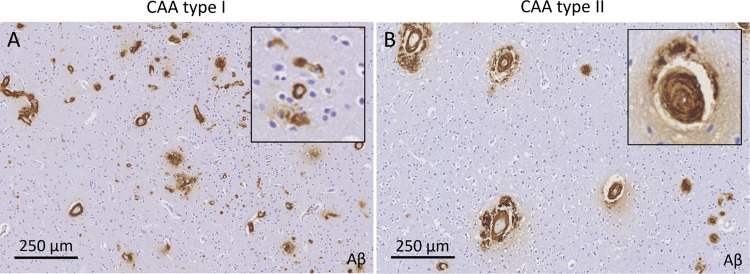
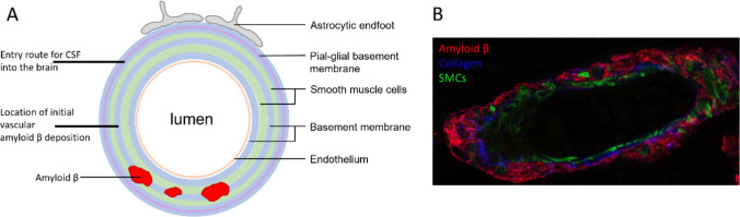
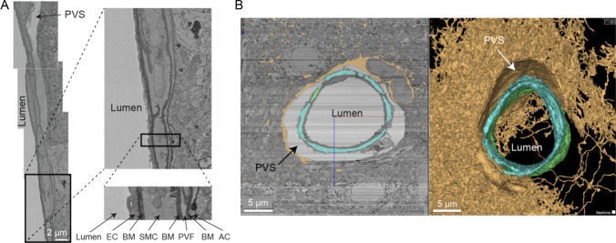
The brain's network of perivascular channels for clearance of excess fluids and waste plays a critical role in the pathogenesis of several neurodegenerative diseases including cerebral amyloid angiopathy (CAA). CAA is the main cause of hemorrhagic stroke in the elderly, the most common vascular comorbidity in Alzheimer's disease and also implicated in adverse events related to anti-amyloid immunotherapy. Remarkably, the mechanisms governing perivascular clearance of soluble amyloid β-a key culprit in CAA-from the brain to draining lymphatics and systemic circulation remains poorly understood. This knowledge gap is critically important to bridge for understanding the pathophysiology of CAA and accelerate development of targeted therapeutics. The authors of this review recently converged their diverse expertise in the field of perivascular physiology to specifically address this problem within the framework of a Leducq Foundation Transatlantic Network of Excellence on Brain Clearance. This review discusses the overarching goal of the consortium and explores the evidence supporting or refuting the role of impaired perivascular clearance in the pathophysiology of CAA with a focus on translating observations from rodents to humans. We also discuss the anatomical features of perivascular channels as well as the biophysical characteristics of fluid and solute transport.

Keywords: Brain clearance; Cerebral amyloid angiopathy; Cerebrospinal fluid; Glymphatics; IPAD; Perivascular spaces.


Conflict of interest statement

Authors E.B., H.B., R.C., S.L., G.P., and A.S., declare they have no financial interests. Author S.G. has served as a consultant for Eli Lily, as a safety monitoring committee member for Bayer and IQVIA/Washington University, and as a scientific advisory board member for MIAC, and has received industry support in the form of a sponsored research agreement from Alnylam. Author J.I. serves as the Chair for the Scientific Advisory Board of Applied Cognition, Inc. He received compensation for this work and holds an equity stake in this company. Author M.v.O has received industry support from Philips and serves as a consultant for Alnylam. Author W.V.N. has received industry support from Alnylam. Author S.v.V. has received industry support in the form of a sponsored research agreement from Therini Bio and Sanofi and has served as a consultant for Biogen and Eisai.

Figures

Fig. 1
Fig. 1
Examples from immunohistochemistry against Aβ in two autopsy cases with a clinical diagnosis of CAA, demonstrating extensive CAA with capillary involvement (A, CAA type I) and predominantly arteriolar involvement (B, CAA type II)
Fig. 2
Fig. 2
Traditional MRI markers of CAA include lobar cerebral microbleeds and cortical superficial siderosis (A). Recently, white matter features in the form of severe degree of MRI-visible perivascular spaces (B) and white matter hyperintensities in a subcortical multi-spot pattern (C, arrows) were added to the version 2.0 of the Boston criteria for the clinical diagnosis of CAA during life. SWI: susceptibility-weighted imaging, FLAIR: fluid-attenuated inversion recovery
Fig. 3
Fig. 3
Schematic of a human brain arteriole with the proposed entry route for CSF into the brain as well as the location of initial vascular Aβ depositions indicated (A). An example of an (X40 SP8) confocal microscopy image of a cross-section of a human brain arteriole with CAA, stained for Aβ (red), collagen IV (blue), and laminin (green) (B). Note that the Aβ is surrounding the smooth muscle cells in tunica media. SMCs: smooth muscle cells
Fig. 4
Fig. 4
The wall and perivascular space of a penetrating arteriole from the MICrONS Cortical MM^3 volume EM dataset (A). Magnified views of the arteriole wall showing the perivascular space diminishing in size and becoming compact layers of different vascular cell types with greater cortical depth (pial surface is toward the top of the image). Specifically, the perivascular space appears to become continuous with the perivascular fibroblast layer and the basement membrane that flanks these cells. EC: endothelial cell, BM: basement membrane, SMC: smooth muscle cell, PVF: perivascular fibroblast, AC: astrocyte. Adapted from [101]. 2D cross-section and volume rendering of penetrating arteriole from mouse cortex showing ultrastructure of perivascular compartment (B). Yellow = one astrocyte. Blue/green = two individual smooth muscle cells
Fig. 5
Fig. 5
Visualization of penetrating arteriolar perivascular space bounded by astroglial endfeet and vascular smooth muscle cells in vivo using multiphoton microscopy. Images were taken in an isoflurane anesthetized mouse. Astrocytes (green) are labeled by AAV-GFAP-Lck-GFP and smooth muscle cells (SMCs; red) are endogenous to PDGFRβ-tdTomato mice
Fig. 6
Fig. 6
Arterial Spin Labeling (ASL) is a suitable translational MRI technique that non-invasively captures blood-to-CSF water exchange in the rat and human brain. By employing long labeling durations, long post-labeling delay times and a long echo time, the arrival of labeled spins in the CSF can be captured. The long echo time ensures that signal from brain tissue is already nulled, leaving only signal in water-like regions, like ventricular and cortical CSF. Note that exchange is not limited to the ventricles but is also found around the cortex

References

    1. Iliff JJ, Wang M, Zeppenfeld DM, Venkataraman A, Plog B, a, Liao Y, Deane R, Nedergaard M, (2013) Cerebral arterial pulsation drives paravascular CSF-interstitial fluid exchange in the murine brain. J Neurosci 33:18190–18199. 10.1523/JNEUROSCI.1592-13.2013 - PMC - PubMed
    1. Wardlaw JM, Benveniste H, Nedergaard M, Zlokovic BV, Mestre H, Lee H, Doubal FN, Brown R, Ramirez J, MacIntosh BJ, Tannenbaum A, Ballerini L, Rungta RL, Boido D, Sweeney M, Montagne A, Charpak S, Joutel A, Smith KJ, Black SE (2020) Perivascular spaces in the brain: anatomy, physiology and pathology. Nat Rev Neurol 16:137–153. 10.1038/s41582-020-0312-z - PubMed
    1. Ball KK, Cruz NF, Mrak RE, Dienel GA (2010) Trafficking of glucose, lactate, and amyloid-beta from the inferior colliculus through perivascular routes. J Cereb blood flow Metab Off J Int Soc Cereb Blood Flow Metab 30:162–176. 10.1038/jcbfm.2009.206 - PMC - PubMed
    1. Greenberg SM, Bacskai BJ, Hernandez-Guillamon M, Pruzin J, Sperling R, van Veluw SJ (2020) Cerebral amyloid angiopathy and Alzheimer disease—one peptide, two pathways. Nat Rev Neurol 16:30–42. 10.1038/s41582-019-0281-2 - PMC - PubMed
    1. Koemans EA, Chhatwal JP, van Veluw SJ, van Etten ES, van Osch MJP, van Walderveen MAA, Sohrabi HR, Kozberg MG, Shirzadi Z, Terwindt GM, van Buchem MA, Smith EE, Werring DJ, Martins RN, Wermer MJH, Greenberg SM (2023) Progression of cerebral amyloid angiopathy: a pathophysiological framework. Lancet Neurol 22:632–642. 10.1016/S1474-4422(23)00114-X - PubMed

MeSH terms

Substances

LinkOut - more resources