Case Reports
. 2024 Dec;13(6):565-572.
doi: 10.1007/s13730-024-00889-9. Epub 2024 May 27.

A case of immunoglobulin G4-related kidney disease manifesting after dipeptidyl peptidase-4 inhibitor treatment

Affiliations
Case Reports

A case of immunoglobulin G4-related kidney disease manifesting after dipeptidyl peptidase-4 inhibitor treatment

Yoshifumi Ubara et al. CEN Case Rep. 2024 Dec.

Abstract

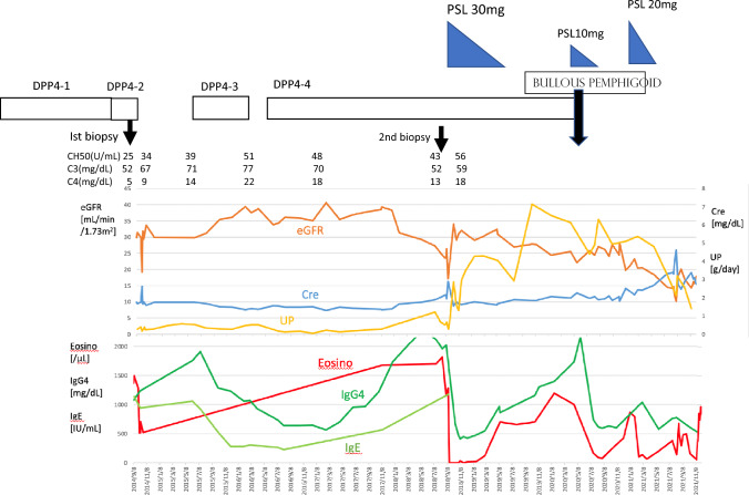
A 68-year-old man with type 2 diabetes mellitus was admitted with decreased renal function. He had high IgG4 (1070 mg/dL) and hypocomplementemia (CH50, 25 U/mL). Kidney biopsy showed tubulointerstitial nephritis with IgG4-positive plasma cell infiltration. Four years later, a second kidney biopsy revealed a new manifestation of membranous nephropathy and tubulointerstitial nephritis with exacerbated fibrosis formation. Six years later, the patient developed bullous pemphigoid, which was thought to be caused by DPP4 inhibitors, so DPP4 inhibitor treatment was discontinued. The use of DPP4 inhibitors correlated with changes in renal function, and the patient was diagnosed with IgG4-related kidney disease related to DPP4 inhibitors.

Keywords: Dipeptidyl peptidase-4 (DPP4) inhibitors; IgG4-related kidney disease; IgG4-related tubulointerstitial nephritis.


Conflict of interest statement

Declarations. Conflicts of interest: The authors declare no competing financial interests and no conflicts of interest. Ethical approval: The present report was produced in conformity with the Declaration of Helsinki, and the patient gave his written informed consent for the case report to be published.

Figures

Fig. 1
Fig. 1
First kidney biopsy. a Infiltration of inflammatory cells, predominantly plasma cells, was prominent in the tubulointerstitium, as shown by hematoxylin and eosin stain (HE). (Original magnification ×200). b Periodic acid methenamine silver staining showed little fibrosis. (Original magnification ×100). c CD138-positive cells with plasma cell organelles were prominent. (Original magnification ×50). d Immunoglobulin (Ig)G4-positive plasma cells were also prominent, and the IgG4-to-CD138 ratio was 60–70%. (Original magnification ×50)
Fig. 2
Fig. 2
Second kidney biopsy. a An inflammatory cell infiltrate predominantly composed of plasma cells, including an eosinophilic infiltrate, was shown in the tubulointerstitium by HE staining. (Original magnification ×200). b In PAM staining, fibrosis formation was more pronounced than in the first kidney biopsy. (Original magnification ×100). c In PAM staining, the bird’s eye appearance, with plasma cells surrounded by a high degree of fibrosis, was more pronounced than in the first kidney biopsy. (Original magnification ×400). d Immunoglobulin (Ig)G4-positive plasma cells were present to the same extent as in the first kidney biopsy. (Original magnification ×200). e Glomeruli showed a tendency to be swollen but no obvious spike formation. (Original magnification ×400). f Immunofluorescence IgG staining showed granular deposits along the glomerular basement membrane (GBM) (small white arrow), Bowman’s capsule (large white arrow), and proximal tubules (yellow arrow). (Original magnification ×400). g Electron microscopy showed a thickened GBM (600 nm), a small subepithelial electron-dense deposit (EDD) (small black arrow), foot process effacement (large black arrow), and subendothelial edema (yellow arrow). h An EDD (arrow) was also seen in the basement membrane of the proximal tubules. i An EDD (arrow) was also seen in the basement membrane of the Bowman’s capsule
Fig. 2
Fig. 2
Second kidney biopsy. a An inflammatory cell infiltrate predominantly composed of plasma cells, including an eosinophilic infiltrate, was shown in the tubulointerstitium by HE staining. (Original magnification ×200). b In PAM staining, fibrosis formation was more pronounced than in the first kidney biopsy. (Original magnification ×100). c In PAM staining, the bird’s eye appearance, with plasma cells surrounded by a high degree of fibrosis, was more pronounced than in the first kidney biopsy. (Original magnification ×400). d Immunoglobulin (Ig)G4-positive plasma cells were present to the same extent as in the first kidney biopsy. (Original magnification ×200). e Glomeruli showed a tendency to be swollen but no obvious spike formation. (Original magnification ×400). f Immunofluorescence IgG staining showed granular deposits along the glomerular basement membrane (GBM) (small white arrow), Bowman’s capsule (large white arrow), and proximal tubules (yellow arrow). (Original magnification ×400). g Electron microscopy showed a thickened GBM (600 nm), a small subepithelial electron-dense deposit (EDD) (small black arrow), foot process effacement (large black arrow), and subendothelial edema (yellow arrow). h An EDD (arrow) was also seen in the basement membrane of the proximal tubules. i An EDD (arrow) was also seen in the basement membrane of the Bowman’s capsule
Fig. 3
Fig. 3
Clinical Course. DPP4-1 sitagliptin, DPP4-2 linagliptin, DPP4-3 alogliptin, DPP4-4 teneligliptin, PSL prednisolone

Similar articles

Cited by

References

    1. Kawano M, Saeki T, Ubara Y, Matsui S. Recent advances in IgG4-related kidney disease. Mod Rheumatol. 2023;33(2):242–51. - PubMed
    1. Inenaga J, Ueno T, Kawada M, Imafuku A, Mise K, Sumida K, Hiramatsu R, Hasegawa E, Hayami N, Suwabe T, Hoshino J, Sawa N, Takaichi K, Fujii T, Ohashi K, Okaneya T, Ubara Y. IgG4-related disease: a mass lesion in the intrarenal sinus near the renal pelvis. Intern Med. 2015;54(15):1897–900. - PubMed
    1. Culver EL, Sadler R, Bateman AC, Makuch M, Cargill T, Ferry B, et al. Increases in IgE, eosinophils, and mast cells, can be used in diagnosis and to predict relapse of IgG4-related disease. Clin Gastroenterol Hepatol. 2017;15(9):1444-1452.e6. - PMC - PubMed
    1. Michailidou D, Schwartz DM, Mustelin T, Hughes GC. Allergic aspects of IgG4-related disease: implications for pathogenesis and therapy. Front Immunol. 2021;7(12):693192. - PMC - PubMed
    1. Inoue D, Yoshida K, Yoneda N, et al. IgG4-related disease: dataset of 235 consecutive patients. Medicine (Baltimore). 2015;94(15):e680. - PMC - PubMed

Publication types

MeSH terms

Substances

LinkOut - more resources