. 2024 Aug;115(8):2673-2685.
doi: 10.1111/cas.16204. Epub 2024 May 27.

Hexokinase 2 nonmetabolic function-mediated phosphorylation of IκBα enhances pancreatic ductal adenocarcinoma progression

Affiliations

Hexokinase 2 nonmetabolic function-mediated phosphorylation of IκBα enhances pancreatic ductal adenocarcinoma progression

Yingying Tong et al. Cancer Sci. 2024 Aug.

Abstract

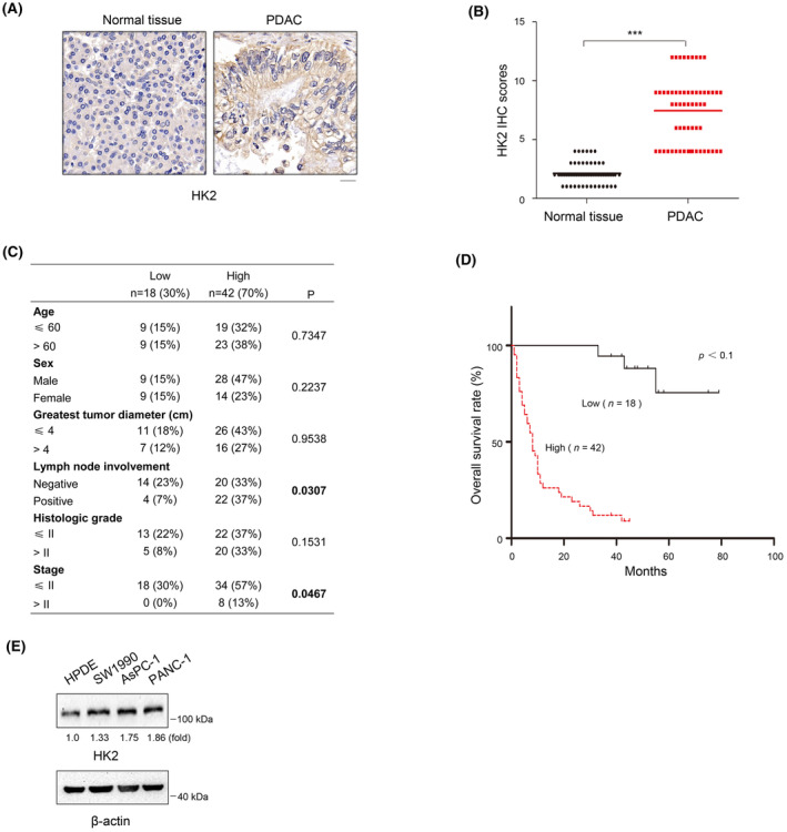
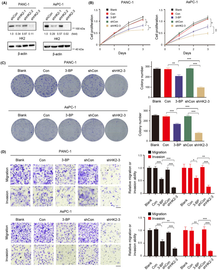
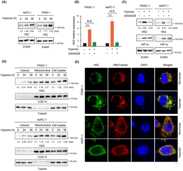
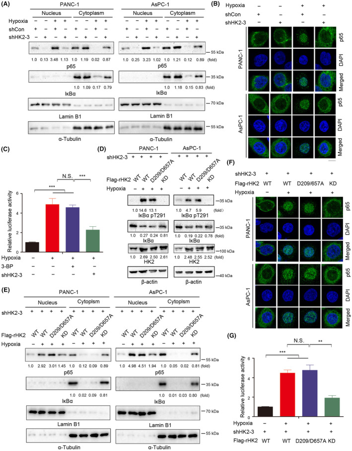
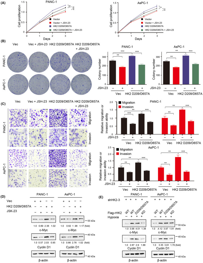
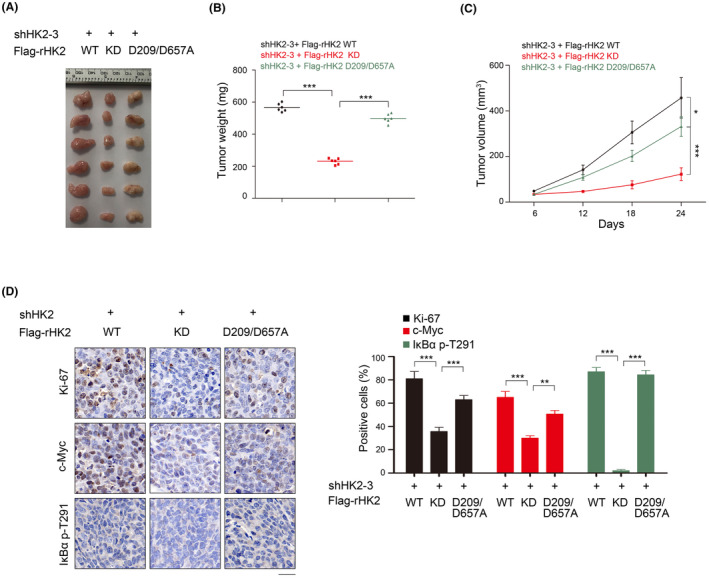
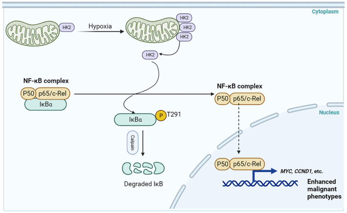
Aberrant signaling in tumor cells induces nonmetabolic functions of some metabolic enzymes in many cellular activities. As a key glycolytic enzyme, the nonmetabolic function of hexokinase 2 (HK2) plays a role in tumor immune evasion. However, whether HK2, dependent of its nonmetabolic activity, plays a role in human pancreatic ductal adenocarcinoma (PDAC) tumorigenesis remains unclear. Here, we demonstrated that HK2 acts as a protein kinase and phosphorylates IκBα at T291 in PDAC cells, activating NF-κB, which enters the nucleus and promotes the expression of downstream targets under hypoxia. HK2 nonmetabolic activity-promoted activation of NF-κB promotes the proliferation, migration, and invasion of PDAC cells. These findings provide new insights into the multifaceted roles of HK2 in tumor development and underscore the potential of targeting HK2 protein kinase activity for PDAC treatment.

Keywords: HK2; IκBα; nonmetabolic activity; pancreatic ductal adenocarcinoma; tumor progression.


Conflict of interest statement

The authors declare no conflict of interest.

Figures

FIGURE 1
FIGURE 1
HK2 is highly expressed in PDAC and correlated with poor prognosis of patients with PDAC. (A) Expression of HK2 in 60 samples of human PDAC tissues and matched adjacent normal tissues by immunohistochemical (IHC) staining with an anti‐HK2 antibody. Representative images are shown. Scale bars, 20 μm. (B) Comparative analysis of HK2 expression between PDAC tissues and matched adjacent normal tissues. ***p < 0.001. (C) Correlations between HK2 expression levels and PDAC clinicopathological parameters. (D) Kaplan–Meier plots and p‐values of the log‐rank test for comparing survivals of PDAC patients with high (staining score, 5–12) and low (staining score, 0–4) expression of HK2. (E) Lysates of the indicated cells were prepared. Immunoblot analyses were performed with the indicated antibodies.
FIGURE 2
FIGURE 2
HK2 promotes pancreatic ductal adenocarcinoma (PDAC) cell proliferation, migration, and invasion. (A) HK2 was depleted in the indicated cells by expressing HK2 shRNA. (B) Indicated cells were cultured in the presence or absence of 3‐bromopyruvate (3‐BP) (25 μM) for the indicated time. Cell proliferation was examined using a Cell Counting Kit‐8 (CCK‐8) assay. Data are presented as the means ± SD from three independent experiments (n = 3). *p < 0.05; **p < 0.01. (C) Indicated PANC‐1 and AsPC‐1 cells were plated for 2 weeks in the presence or absence of 3‐BP (25 μM) before counting colony numbers. Data are presented as the means ± SD from three independent experiments (n = 3). **p < 0.01; ***p < 0.001. (D) The migration and invasion of the indicated cells in the presence or absence of 3‐BP (25 μM) were examined by transwell assay. The membrane was photographed using a digital camera mounted onto a microscope. Scale bars, 50 μm. Data are presented as mean ± SD *p < 0.05; **p < 0.01; ***p < 0.001.
FIGURE 3
FIGURE 3
Hypoxia promotes the transcription and the mitochondrial dissociation of HK2. (A, C, D) Immunoblot analyses were performed with the indicated antibodies. (A) PANC‐1 and AsPC‐1 cells were cultured under normoxic or hypoxic condition for the indicated time. (B, C) PANC‐1 and AsPC‐1 cells were cultured under normoxic or hypoxic condition for 24 h in the absence or presence of GN44028 (20 nM). RT‐PCR (B) and immunoblot analyses (C) were performed. N.S., not significant for the indicated comparison; ***p < 0.001. (D) Mitochondrial and cytosolic fractions of PANC‐1 and AsPC‐1 cells cultured under normoxic or hypoxic condition for 24 h were prepared. (E) PANC‐1 and AsPC‐1 cells were stimulated with or without hypoxia for 24 h. Immunofluorescence analyses were performed with an anti‐HK2 antibody. Scale bars, 5 μm.
FIGURE 4
FIGURE 4
Hypoxia promotes NF‐κB activation dependent on a HK2‐mediated IκBα T291 phosphorylation. (A, D, E) Immunoblot analyses were performed with the indicated antibodies. (A, B) PANC‐1 and AsPC‐1 cells with or without HK2 depletion were cultured for 24 h under normoxia or hypoxia. Cytosolic and nuclear fractions of the indicated cells were prepared (A). PANC‐1 and AsPC‐1 cells with or without HK2 depletion were stimulated with or without hypoxia for 24 h. Immunofluorescence analyses were performed with an anti‐p65 antibody (B). Scale bars, 10 μm. (C) PANC‐1 cells with or without HK2 depletion were cultured in the absence or presence of 3‐bromopyruvate (3‐BP) (25 μM). The indicated cells were transfected with luciferase reporter plasmid (NF‐κB‐Luc) and stimulated with or without hypoxia for 12 h. The data are presented as the mean ± SD of triplicate samples. N.S., not significant for the indicated comparison; ***p < 0.001. (D) PANC‐1 and AsPC‐1 cells with depleted HK2 and reconstituted expression of WT rHK2, rHK2 D209/D657A, or KD were cultured with or without hypoxia for 24 h. Cytosolic and nuclear fractions of the indicated cells were prepared. (E, F) PANC‐1 cells with depleted HK2 and reconstituted expression of WT rHK2, rHK2 D209/D657A, or KD were cultured with or without hypoxia for 24 h. Cytosolic and nuclear fractions of the indicated cells were prepared (E). Immunofluorescence analyses were performed with an anti‐p65 antibody. Scale bars, 10 μm. (G) WT rHK2, rHK2 D209/D657A, or KD was expressed in PANC‐1 cells with the depletion of HK2. The indicated cells were transfected with luciferase reporter plasmid (NF‐κB‐Luc) and stimulated with or without hypoxia for 12 h. The data are presented as the mean ± SD of triplicate samples. N.S., not significant for the indicated comparison; **p < 0.01; ***p < 0.001.
FIGURE 5
FIGURE 5
NF‐κB inhibitor abrogates HK2 nonmetabolic function‐induced proliferation, migration, and invasion of pancreatic ductal adenocarcinoma (PDAC) cells in vitro. (D, E) Immunoblot analyses were performed with the indicated antibodies. (A) PANC‐1 and AsPC‐1 with or without HK2 D209/D657A expression were cultured in the presence or absence of JSH‐23 (10 μM) for the indicated time. Cell proliferation was examined using a Cell Counting Kit‐8 (CCK‐8) assay. Data are presented as the means ± SD from three independent experiments (n = 3). *p < 0.05; **p < 0.01; ***p < 0.001. (B) PANC‐1 and AsPC‐1 cells with or without HK2 D209/D657A expression were cultured in the presence or absence of JSH‐23 (10 μM) for 2 weeks before counting colony numbers. Data are presented as the means ± SD from three independent experiments (n = 3). **p < 0.01; ***p < 0.001. (C) The migration and invasion of the indicated cells in the presence or absence of JSH‐23 (10 μM) were examined by transwell assay. The membrane was photographed using a digital camera mounted onto a microscope. Scale bars, 50 μm. Data are presented as mean ± S.D. **p < 0.01; ***p < 0.001. (D) PANC‐1 and AsPC‐1 cells with or without HK2 D209/D657A expression were cultured in the presence or absence of JSH‐23 (10 μM) for 24 h. (E) PANC‐1 and AsPC‐1 cells with depleted HK2 and reconstituted expression of WT rHK2, rHK2 D209/D657A, or KD were cultured with or without hypoxia for 24 h.
FIGURE 6
FIGURE 6
HK2 nonmetabolic functions promote PDAC development in vivo. (A–D) PANC‐1 cells with depleted HK2 and reconstituted expression of WT rHK2, rHK2 D209/D657A, or KD were intracranially injected into athymic nude mice. Tumor growth was examined 21 days after injection. Representative tumor xenografts were shown (n = 6 mice per group) (A). Tumor weights were calculated (B). Tumor growth was measured every other day beginning on day 6, and tumor volumes were calculated (C). Immunohistochemical analyses of tumor sections with the indicated antibodies were performed. Representative images are shown (D). Scale bar, 10 mm. The percentages of Ki67‐, c‐Myc‐, and IκBα pT291‐positive cells were calculated. *p < 0.05; **p < 0.01; ***p < 0.001.
FIGURE 7
FIGURE 7
Diagram displaying the regulation of NF‐κB signaling by the nonmetabolic function of HK2. HK2‐mediated phosphorylation of IκBα at T291 in PDAC cells, leading to IκBα degradation and subsequent activation of NF‐κB for the upregulation of downstream target transcription. HK2 nonmetabolic activity‐promoted activation of NF‐κB stimulates the proliferation, migration, and invasion of PDAC cells.

Similar articles

Cited by

References

    1. Hay N. Reprogramming glucose metabolism in cancer: can it be exploited for cancer therapy? Nat Rev Cancer. 2016;16:635‐649. - PMC - PubMed
    1. Wilson JE. Isozymes of mammalian hexokinase: structure, subcellular localization and metabolic function. J Exp Biol. 2003;206:2049‐2057. - PubMed
    1. Robey RB, Hay N. Mitochondrial hexokinases, novel mediators of the antiapoptotic effects of growth factors and Akt. Oncogene. 2006;25:4683‐4696. - PubMed
    1. Xu S, Catapang A, Doh HM, et al. Hexokinase 2 is targetable for HK1 negative, HK2 positive tumors from a wide variety of tissues of origin. J Nucl Med. 2018;60:212‐217. - PMC - PubMed
    1. Mathupala SP, Ko YH, Pedersen PL. Hexokinase II: cancer's double‐edged sword acting as both facilitator and gatekeeper of malignancy when bound to mitochondria. Oncogene. 2006;25:4777‐4786. - PMC - PubMed