. 2024 Sep;20(9):2076-2091.
doi: 10.1080/15548627.2024.2356490. Epub 2024 May 27.

Development and characterization of phospho-ubiquitin antibodies to monitor PINK1-PRKN signaling in cells and tissue

Affiliations

Development and characterization of phospho-ubiquitin antibodies to monitor PINK1-PRKN signaling in cells and tissue

Jens O Watzlawik et al. Autophagy. 2024 Sep.

Abstract

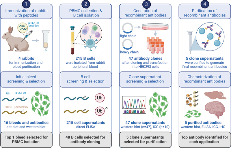
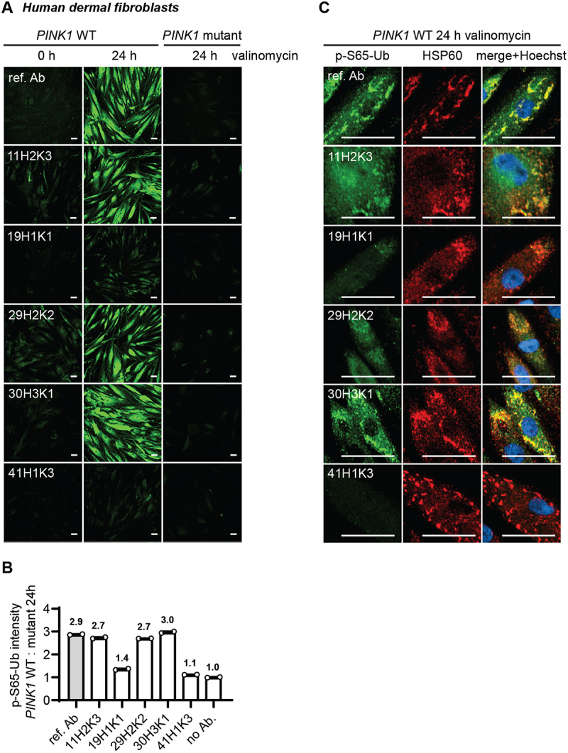
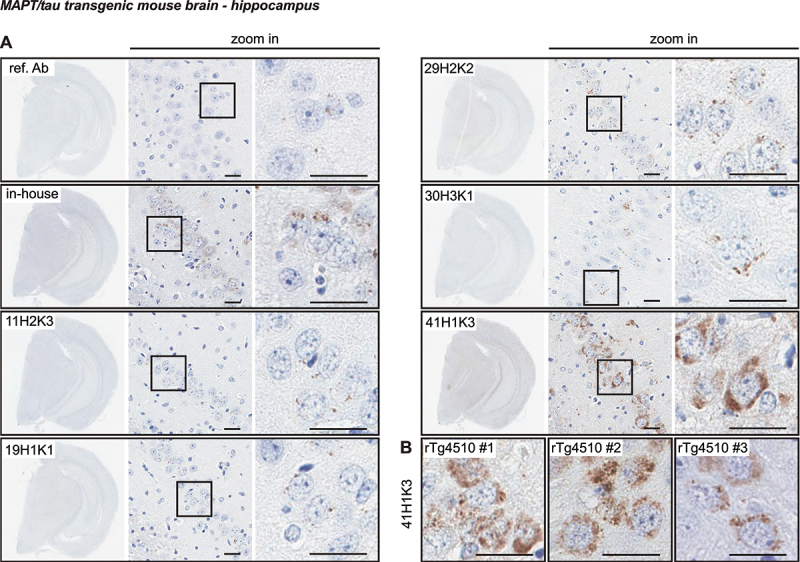
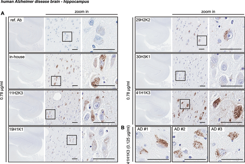
The selective removal of dysfunctional mitochondria, a process termed mitophagy, is critical for cellular health and impairments have been linked to aging, Parkinson disease, and other neurodegenerative conditions. A central mitophagy pathway is orchestrated by the ubiquitin (Ub) kinase PINK1 together with the E3 Ub ligase PRKN/Parkin. The decoration of damaged mitochondrial domains with phosphorylated Ub (p-S65-Ub) mediates their elimination though the autophagy system. As such p-S65-Ub has emerged as a highly specific and quantitative marker of mitochondrial damage with significant disease relevance. Existing p-S65-Ub antibodies have been successfully employed as research tools in a range of applications including western blot, immunocytochemistry, immunohistochemistry, and enzyme-linked immunosorbent assay. However, physiological levels of p-S65-Ub in the absence of exogenous stress are very low, therefore difficult to detect and require reliable and ultrasensitive methods. Here we generated and characterized a collection of novel recombinant, rabbit monoclonal p-S65-Ub antibodies with high specificity and affinity in certain applications that allow the field to better understand the molecular mechanisms and disease relevance of PINK1-PRKN signaling. These antibodies may also serve as novel diagnostic or prognostic tools to monitor mitochondrial damage in various clinical and pathological specimens.Abbreviations: AD: Alzheimer disease; CCCP: carbonyl cyanide 3-chlorophenylhydrazone; ELISA: enzyme-linked immunosorbent assay; HEK293E cell: human embryonic kidney E cell; ICC: immunocytochemistry; IHC: immunohistochemistry: KO: knockout; LoB: limit of blank; LoD: limit of detection; LoQ: limit of quantification; MEF: mouse embryonic fibroblast; MSD: Meso Scale Discovery; n.s.: non-significant; nonTg: non-transgenic; PBMC: peripheral blood mononuclear cell; PD: Parkinson disease; p-S65-PRKN: phosphorylated PRKN at serine 65; p-S65-Ub: phosphorylated Ub at serine 65; Ub: ubiquitin; WT: wild-type.

Keywords: Autophagy; PINK1; Parkinson disease; mitochondria; mitophagy; ubiquitin.


Conflict of interest statement

Mayo Clinic, F.C.F., and W.S. hold a patent related to PRKN activators (Small Molecule Activators of Parkin Enzyme Function, US patent, 11401255B2; August 02, 2022). Additional funding sources to disclose but not pertinent to the current study include a grant from Amazentis SA (to W.S.). Z.K.W. serves as PI or Co-PI on projects/grants from Biohaven Pharmaceuticals, Inc. and Vigil Neuroscience, Inc., and is an external advisory board member for Vigil Neuroscience, Inc., and a consultant on neurodegenerative medical research for Eli Lilly & Company. M.M.K.M. is a member of the Scientific Advisory Board of Montara Therapeutics Inc and scientific consultant to Merck Sharp and Dohme. J.B.F. was CEO at 21st Century Biochemicals Inc., K.P. is an employee of ImmunoPrecise Antibodies Ltd., and R.K. is an employee of Abcam Plc. All other authors declare they have no competing interests. This research was conducted in compliance with Mayo Clinic conflict of interest policies.

Figures

Figure 1.
Figure 1.
Schematic overview of recombinant p-S65-Ub antibody clone generation, selection, and validation. The illustration summarizes the entire process of recombinant, rabbit monoclonal antibody production starting from testing of the initial polyclonal bleeds to the isolation and screening of B cells, and the subsequent cloning of variable heavy and light chain antibody regions into an IgG vector frame. Recombinant antibodies were then sequenced, expressed in HEK293 cells and supernatants were screened by western blot and ICC. The five most promising antibody clones were affinity purified and re-validated by a series of biochemical and imaging analyses using recombinant proteins as well as cells and brain tissues from mouse and human origin. PBMC: peripheral blood mononuclear cell; ELISA: enzyme-linked immunosorbent assay; ICC: immunocytochemistry; IHC: immunohistochemistry. Created with BioRender.com.
Figure 2.
Figure 2.
Western blot assessment of the selected affinity-purified p-S65-Ub antibodies in cells. (A) HEK293E cells and (B) mouse embryonic fibroblasts with (WT) or without PINK1 expression (PINK1 KO or pink1 KO) were treated with mitochondrial depolarizer. All western blots were run and developed side-by-side. Representative western blot from three total replicates are shown for the top five p-S65-Ub antibody clones in each cell type plus reference antibody. Note that concentrations of the tested p-S65-Ub antibodies are five times higher compared to the reference antibody. For both WT HEK293 cells and WT MEFs, we used different dilutions of the WT lysates, namely 30 (1× diluted), 15 (2× diluted) and 7.5 µg (4× diluted) for WT HEK293 cells plus 30 µg of PINK1 KO HEK293 cells (1× diluted) respective 10 (1× diluted), 5 (2× diluted), 2.5 µg (4× diluted) total protein for WT MEFs and 10 µg for pink1 KO MEFs (1× diluted). GAPDH was used as loading control. A densitometric quantification of p-S65-Ub raw data is shown for all antibodies in both cell types with the entire lane being quantified (instead of a single band). KO: knockout; ref. Ab: reference antibody; WT: wild-type.
Figure 3.
Figure 3.
Assessing the top p-S65-Ub antibodies in ELISA using recombinant protein. (A) Sandwich ELISA results for the detection of recombinant K48 (solid line) and K63 (dash line) p-S65-Ub tetramers (Ub4) that were serially diluted to the range of 1–1,000,000 pg/ml. (B) Direct ELISA results for the detection of p-S65-PRKN and nonphosphorylated PRKN recombinant proteins that were serially diluted to the range of 1–1,000,000 pg/ml. N = 2. Data are shown as mean with SD. ref. Ab: reference antibody.
Figure 4.
Figure 4.
Assessing the top p-S65-Ub antibodies by sandwich ELISA using lysates from cells, mouse brain, and human brain. (A) ELISA detection of p-S65-Ub levels in cell lysates from CCCP or DMSO treated WT and PINK1 KO HEK293E cells. One-way ANOVA. N = 2. Data are shown as mean with SEM. (B) ELISA detection of p-S65-Ub levels in hemibrain lysates from unstimulated WT and pink1 KO mice. Unpaired t-test. N = 6 per group. Data are shown as mean with SEM. (C) ELISA detection of p-S65-Ub levels in human brain lysates from neurological normal controls and AD cases. Mann-Whitney U test. N = 10 per group. Data are shown as median with interquartile range. * p < 0.05, ** p < 0.005, *** p < 0.0001. Asterisks (*) indicate the comparison to the untreated WT or the comparison between two groups under the brackets. ref. Ab: reference antibody; AD: Alzheimer disease; CON: controls; KO: knockout; n.S.: non-significant; WT: wild-type.
Figure 5.
Figure 5.
Comparison of the top p-S65-Ub antibodies using ICC of human dermal fibroblasts. All five p-S65-Ub recombinant antibodies and the reference antibody were evaluated by ICC in human primary dermal fibroblasts treated with 2 μM valinomycin for 0 or 24 h. All antibodies were used at 1 μg/ml. (A) Representative images of p-S65-Ub immunoreactive signals (green) in fibroblasts carrying WT or homozygous PINK1Q456X mutation. (B) Fluorescence intensities of each antibody was quantified by high content imaging and compared signals from treated WT to the signals from the corresponding PINK1 mutant fibroblasts. Fold changes are labeled at top of each bar. Samples stained without primary antibody was used a negative control (no Ab). N = 2. Data are shown as mean with SEM. (C) Representative zoom-in images of p-S65-Ub and HSP60 immunoreactive signals in treated WT fibroblasts. 24 h of valinomycin treatment induced mitochondrial aggregation (HSP60, red) and the accumulation of p-S65-Ub (green). Nuclei were stained with Hoechst (blue). Scale bar: 50 μm. ref. Ab: reference antibody; WT: wild-type.
Figure 6.
Figure 6.
Characterization of the top p-S65-Ub antibodies by IHC staining of mouse brain. (A) All five p-S65-Ub recombinant antibodies together with our in-house p-S65-Ub antibody and the reference antibody were evaluated by IHC in serial brain sections from a 9-month-old rTg4510 mouse. Same concentration (1.56 μg/ml) was used for all antibodies. The entire region of the stained hemibrain is shown to the left at 1.5× magnification. Zoom-in images of the highlighted hippocampal regions are shown to the right. Typical punctate p-S65-Ub immunopositive structures (dark brown) were detected by all clones but with different intensity. (B) Brain sections from three different rTg4510 mice were stained with clone 41H1K3 and showed consistent immunostaining pattern. Scale bar: 50 μm. ref. Ab: reference antibody.
Figure 7.
Figure 7.
Characterization of the top p-S65-Ub antibodies by IHC staining of Alzheimer disease brain. (A) All five p-S65-Ub recombinant antibodies together with our in-house p-S65-Ub antibody and the reference antibody were evaluated by IHC of serial hippocampal sections from Alzheimer disease brain. Same concentration (0.78 μg/ml) was used for all antibodies. The entire stained hippocampal region is shown to the left at 1.5× magnification. Zoom-in images of the highlighted regions are shown to the right. Typical punctate p-S65-Ub immunopositive structures (dark brown) were detected by most of the antibodies at different sensitivity. (B) Hippocampal sections from three different Alzheimer disease cases were stained with 41H1K3 and showed consistent immunostaining pattern. The superior sensitivity of 41H1K3 allowed detection of p-S65-Ub at a much lower concentration (0.125 μg/ml) compared to other antibodies. Scale bar: 50 μm. AD: Alzheimer disease; ref. Ab: reference antibody.

Update of

References

    1. Tanaka K. The PINK1–Parkin axis: an overview. Neurosci Res. 2020. Oct;159:9–15. doi: 10.1016/j.neures.2020.01.006 - DOI - PubMed
    1. Lazarou M, Jin SM, Kane LA, et al. Role of PINK1 binding to the TOM complex and alternate intracellular membranes in recruitment and activation of the E3 ligase Parkin. Dev Cell. 2012 Feb 14;22(2):320–333. doi: 10.1016/j.devcel.2011.12.014 - DOI - PMC - PubMed
    1. Okatsu K, Uno M, Koyano F, et al. A dimeric PINK1-containing complex on depolarized mitochondria stimulates Parkin recruitment. J Biol Chem. [2013 Dec 20];288(51):36372–36384. doi: 10.1074/jbc.M113.509653 - DOI - PMC - PubMed
    1. Kane LA, Lazarou M, Fogel AI, et al. PINK1 phosphorylates ubiquitin to activate Parkin E3 ubiquitin ligase activity. J Cell Bio. [2014 Apr 28];205(2):143–153. doi: 10.1083/jcb.201402104 - DOI - PMC - PubMed
    1. Kazlauskaite A, Kondapalli C, Gourlay R, et al. Parkin is activated by PINK1-dependent phosphorylation of ubiquitin at Ser65. Biochem J. [2014 May 15];460(1):127–139. doi: 10.1042/BJ20140334 - DOI - PMC - PubMed

LinkOut - more resources