. 2024 May 27;15(1):4504.
doi: 10.1038/s41467-024-48843-8.

Hairpin protein partitioning from the ER to lipid droplets involves major structural rearrangements

Affiliations

Hairpin protein partitioning from the ER to lipid droplets involves major structural rearrangements

Ravi Dhiman et al. Nat Commun. .

Abstract

Lipid droplet (LD) function relies on proteins partitioning between the endoplasmic reticulum (ER) phospholipid bilayer and the LD monolayer membrane to control cellular adaptation to metabolic changes. It has been proposed that these hairpin proteins integrate into both membranes in a similar monotopic topology, enabling their passive lateral diffusion during LD emergence at the ER. Here, we combine biochemical solvent-accessibility assays, electron paramagnetic resonance spectroscopy and intra-molecular crosslinking experiments with molecular dynamics simulations, and determine distinct intramembrane positionings of the ER/LD protein UBXD8 in ER bilayer and LD monolayer membranes. UBXD8 is deeply inserted into the ER bilayer with a V-shaped topology and adopts an open-shallow conformation in the LD monolayer. Major structural rearrangements are required to enable ER-to-LD partitioning. Free energy calculations suggest that such structural transition is unlikely spontaneous, indicating that ER-to-LD protein partitioning relies on more complex mechanisms than anticipated and providing regulatory means for this trans-organelle protein trafficking.


Conflict of interest statement

The authors declare no competing interests.

Figures

Fig. 1
Fig. 1. Establishment of a PEGylation-based solvent-accessibility assay for opUBXD853-153mCherry single cysteine mutants in ER bilayer and LD monolayer membranes.
a Schematic depicting LD biogenesis from the ER membrane. ERTOLD hairpin proteins are considered to integrate first into the cytosolic leaflet of the ER membrane in a monotopic topology, which presumably enables them to partition from the ER bilayer to the LD monolayer membrane during LD biogenesis. NL neutral lipids. b Dual localization of opUBXD853-153mCherry to the ER and LDs. Top: Schematic outline of the opsin (op) and mCherry-tagged opUBXD853-153mCherry construct. HR hydrophobic region. Bottom: Fluorescence micrographs of oleate-treated cells transfected with opUBXD853-153mCherry representative for 3 independent experiments. LipidTox marks LDs. Scale bar: 10 µm. c Isolation of opUBXD853-153mCherry-containing LDs from cells. Left: Schematic outline for isolation of UBXD8-containing LDs. Right: immunoblot of post-nuclear supernatant (PNS), membranes (M), cytosol (C), and LD fractions derived from oleate-treated cells expressing OpUBXD853-153mCherry using anti-calnexin (ER-resident protein), anti-tubulin (cytosolic protein) and anti-mCherry antibodies. Non-transfected cells (NT) serve as specificity control for the antibody. Data are representative for 3 independent experiments. d Integration of opUBXD853-153mCherry into rough microsomes (RMs). Left: Schematic outline of co-translational protein insertion into RMs employing in vitro translation of UBXD8 mRNAs in rabbit reticulocyte lysate (RRL) with subsequent fractionation into soluble and membrane-inserted proteins by centrifugation. Right: Immunoblot of soluble (S) and membrane-inserted (M) fractions derived from in vitro translations reactions using anti-mCherry antibodies (representative for n = 3 independent experiments). mRNA encoding either opUBXD853-153mCherry or UBXD853-153mCherryOP and RMs were added to the reaction as indicated. Arrows indicate glycosylated forms of the respective proteins. eg opUBXD853-153mCherry single cysteine mutants can be PEGylated in ER bilayer and LD monolayer membranes when the cysteine is solvent-exposed. Top: Principle of solvent-accessibility probing of opUBXD853-153mCherry single cysteine mutants by PEGylation in ER bilayer and LD monolayer membranes, respectively. Only solvent-exposed cysteines are accessible to mPEG forming covalent adducts, while bilayer-embedded cysteines are not reactive with mPEG. Bottom: Proof-of-concept immunoblots probed with anti-mCherry antibodies after PEGylation reaction on RM-inserted and LD-inserted opUBXD853-153mCherry single cysteine mutants as indicated. Non-PEGylated proteins are indicated by (0 PEG) and PEGylated proteins by (1 PEG). TX-100: Triton X-100. Quantifications for multiple replicates of these experiments are shown in Fig. 2c.
Fig. 2
Fig. 2. Topological mapping of opUBXD853-153mCherry in ER bilayer and LD monolayer membranes by PEGylation suggests distinct conformations in both membranes.
a Schematic outline of opUBXD853-153mCherry. Single cysteine substitutions covering the amino acid sequence (S80-R128) including the hydrophobic region (indicated in orange) were used for probing individual solvent-accessibility by PEGylation. b Immunoblots using anti-mCherry antibodies showing the PEGylation data for the opUBXD853-153mCherry single cysteine mutants (S80C-R128C) in ER bilayer and LD monolayer membranes as indicated. First lanes: negative controls without the addition of mPEG, second lanes: samples treated with mPEG, third lanes: samples solubilized with Triton X-100 (TX-100) before subjection to mPEG serving as positive controls. c Line graphs showing the relative PEGylation of opUBXD853-153mCherry single cysteine mutants (S80C-R128C) indicated as black line (ER bilayer) and blue line (LD monolayer). Mean values derived from two independent experiments are plotted and error bars indicate standard deviation. For L91C and L92C (indicated with red asterisks) mean values ± standard deviation from n = 4 independent experiments are shown. Results obtained for P102C, I113C, and F114C in ER bilayers (marked in red) should be considered with caution as no efficient PEGylation could be detected for these residues upon solubilization with TX-100. Source data are provided as a source data file.
Fig. 3
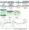
Fig. 3. Atomistic MD simulations reveal intramembrane positioning of UBXD8 in bilayer and monolayer membranes.
a Atomistic MD simulations of UBXD880-128 in a POPC bilayer membrane with a deeply inserted starting structure. Left: Starting structure Right: average structure after 2 µs. b Atomistic MD simulations of UBXD880-128 in a POPC bilayer membrane with a partially inserted starting structure. Left: Starting structure Right: average structure after 2 µs. c Atomistic MD simulations of UBXD880-128 in a POPC-triolein/cholesteryl-oleate-POPC trilayer system mimicking the LD monolayer membrane. Left: Starting structure; Middle: side view of average structure after 2 µs; Right: top view of average structure after 2 µs; d Center of mass distances of the Cα atom in P102 of UBXD880-128 and the P atom in the phospholipid headgroup (dotted line) during the simulation time of 2 µs, and upon UBXD880-128 insertion into POPC bilayers or into trilayer systems as indicated. PI partially inserted, DI deeply inserted, TRIO Triolein, CLOL (cholesteryl-oleate). e Center of mass distances of amino acid Cα atoms in UBXD880-128 and the P atom in the phospholipid headgroup (dotted line) in the average structures obtained after 2 µs simulations. UBXD880-128 was inserted into POPC bilayers or into trilayer systems as indicated. Max. penetration into bilayer is ~2 nm and into monolayer is ~1 nm. Five independent simulations with the CHARMM36m force field over 2 μs were performed. For (d) and (e): Lines and shaded areas show mean and ±SEM, respectively (n = 5 simulations). Source data are provided as a source data file.
Fig. 4
Fig. 4. Intramolecular crosslinking provides experimental evidence for angle opening of UBXD8 during ER-to-LD partitioning.
a Positioning of the amino acids L91 and L118 within the atomistic starting structure of UBXD880-128 that were mutated to a cysteine pair in opUBXD853-153L91C_L118C_mCherry. Cα atoms are highlighted as purple spheres. b Violin plots of distances between the Cα atoms of amino acids L91 and L118 in MD simulations in different membrane systems as shown in Figs. 3a, c, respectively. DI deeply inserted, TRIO Triolein, CLOL (cholesteryl-oleate). Vertical bars indicate median and maximum/minimum values of the distributions (n = 5 simulations). c Schematic representation to illustrate the principle of the combined intramolecular crosslinking - PEGylation assay. For clarity, a double-cysteine-containing peptide is only schematically depicted in a bilayer membrane and does not particularly reflect closed or open conformations of UBXD8 in different types of membranes. d, e Immunoblots using anti-mCherry antibodies showing intramolecular crosslinking/PEGylation experiments of opUBXD853-153L91C_L118C_mCherry in either RMs (d) or in isolated LDs from cells (e). Non-PEGylated protein species (0 PEG) as well as species with one mPEG (1 PEG) or two mPEG (2PEG) molecules attached, are indicated. High molecular weight adducts derived from inter-molecular crosslinking of opUBXD853-153L91C_L118C_mCherry on LDs are indicated (inter x-link). f Quantification of relative opUBXD853-153L91C_L118C_mCherry intramolecular crosslinking efficiencies in RMs versus LDs. From experiments as shown in (d, e), bands corresponding to non-PEGylated/intramolecularly crosslinked OpUBXD853-153L91C_L118CmCherry (0 PEG) were quantified. The relative increase in these bands upon addition of crosslinker (lane 2 versus negative DMSO control in lane 1) was calculated from three independent experiments and the values were normalized to the highest value, which was set to 100%. Scatter plots show the mean values with SEM from three independent experiments as well as the individual values for each replicate. Source data are provided as a source data file.
Fig. 5
Fig. 5. cwEPR spectroscopy confirms deeper membrane penetration of UBXD8 in phospholipid bilayers than in LD monolayer membranes.
a Average atomistic MD simulation structure of UBXD880-128 in a POPC bilayer (as in Fig. 3a) indicating amino acids that were substituted for single cysteines for cwEPR analyses. b Left: Schematic outline of proteo-aLDs generation from proteo-SUVs and isolation by density gradient centrifugation. Right: photograph showing density gradients after centrifugation with floating aLDs when triolein was present during the reconstitution (+). (−): negative control without triolein. c Immunoblot analysis of proteo-aLDs isolation by density gradient fractionation as indicated in (b) using anti-S-tag antibodies. Top: negative control without triolein. Arrowhead indicates MTSL-labeled sUBXD871-132His S127C in the top floating aLD fraction when triolein was present during the reconstitution. Representative for n = 3 independent experiments. d Fluorescence micrograph of the top floating aLDs fraction as shown in (b) upon reconstitution of Atto488-labeled sUBXD871-132His T130C (green). LipidTox Red marks the neutral lipid core (red). Scale bar: 10 µm. Representative for n = 3 independent experiments. e First derivative absorption cwEPR spectra of MTSL spin-labeled sUBXD871-132-His single cysteine mutants in POPC/DOPS SUVs (left) and aLDs (right). Spectra were normalized by the height of the central EPR line. Asterisks mark spectra with additional shoulders in the low-field region indicating immobile (i) and mobile (m) motional components. f Schematic illustration of how the spin-label positioning in a membrane protein affects the line shape of cwEPR spectra and the membrane depth parameter (Φ). Solvent-exposed: orange; membrane-associated: red; membrane-embedded: blue. In the bilayer midplane, the O2 concentration is the highest (green), while NiEDDA is gradually excluded from the membrane (purple). g Exemplary EPR power saturation plots of MTSL spin-labeled sUBXD871-132His Y81C and L118C single cysteine mutants reconstituted into SUVs. The peak-to-peak amplitude of the central EPR line was plotted against the square root of the applied microwave power. Power saturation curves were measured under three conditions: nitrogen gas as control (red circles), molecular O2 (black squares), and NiEDDA (blue diamonds). P1/2 values were obtained after curve fitting and are indicated with SEM. h Membrane depth parameter (Φ) analysis of MTSL spin-labeled sUBXD871-132His single cysteine mutants in SUVs using cwEPR power saturation analyses (n = 2 independent experiments). Positive Φ: membrane-embedding; negative Φ: solvent exposure. i Membrane depth parameter (Φ) analysis of MTSL spin-labeled sUBXD871-132His Y96C and I113C reconstituted into either SUVs or aLDs using cwEPR power saturation analyses (n = 2 independent experiments). Φ close to 0: proximity to solvent-membrane interface. Results for SUVs are duplicates from (h) for direct comparison with aLDs. Source data are provided as a source data file.
Fig. 6
Fig. 6. Free-energy calculations suggest that additional factors are required to assist the conformational transition of UBXD8 at the bilayer-LD interface.
a MARTINI-based coarse-grained simulation of a bilayer-embedded LD lens consisting of triolein/cholesteryl oleate. Upper panel: UBXD880-128 was integrated into the bilayer in its shallow conformation as assessed by atomistic simulations. After 5 μs, the peptides have partitioned to the LD monolayer surface where they accumulate. Lower panel: When inserted into the bilayer in the deep-V state as suggested by atomistic simulations, UBXD880-128 accumulates at the bilayer-LD rim but does not transition to the LD surface within 5 µs. b, c All-atom pulling simulations reveal high free-energy costs involved in pulling the shallow-open state of UBXD880-128 downwards (b) or the deep-V state upwards (c) within a planar POPC bilayer membrane. d All-atom simulation system of a minimal LD-bilayer system used for pulling the deep-V-inserted UBXD880-128 along the LD rim towards the open-shallow conformation. e Graphs comparing free-energy profiles during MD pulling experiments of UBXD880-128 in planar bilayers versus at the LD rim as derived from (bd). Shaded areas show SEM derived by bootstrapping from the set of 33 umbrella histograms. f Revised model for the intramembrane positioning of UBXD8 in ER bilayer versus LD monolayer membranes indicating that structural rearrangements are required for enabling the partitioning. Positive charges are indicated by “+”. Source data are provided as a source data file.

References

    1. Zechner R. FAT FLUX: enzymes, regulators, and pathophysiology of intracellular lipolysis. EMBO Mol. Med. 2015;7:359–362. doi: 10.15252/emmm.201404846. - DOI - PMC - PubMed
    1. Zadoorian A, Du X, Yang H. Lipid droplet biogenesis and functions in health and disease. Nat. Rev. Endocrinol. 2023;19:443–459. doi: 10.1038/s41574-023-00845-0. - DOI - PMC - PubMed
    1. Krahmer N, Farese RV, Jr., Walther TC. Balancing the fat: lipid droplets and human disease. EMBO Mol. Med. 2013;5:905–915. doi: 10.1002/emmm.201100671. - DOI - PMC - PubMed
    1. Olzmann JA, Carvalho P. Dynamics and functions of lipid droplets. Nat. Rev. Mol. Cell Biol. 2019;20:137–155. doi: 10.1038/s41580-018-0085-z. - DOI - PMC - PubMed
    1. Walther TC, Chung J, Farese RV., Jr. Lipid droplet biogenesis. Annu Rev. Cell Dev. Biol. 2017;33:491–510. doi: 10.1146/annurev-cellbio-100616-060608. - DOI - PMC - PubMed

MeSH terms

Substances