. 2024 May 27;7(1):644.
doi: 10.1038/s42003-024-06344-w.

Broad-spectrum ubiquitin/ubiquitin-like deconjugation activity of the rhizobial effector NopD from Bradyrhizobium (sp. XS1150)

Affiliations

Broad-spectrum ubiquitin/ubiquitin-like deconjugation activity of the rhizobial effector NopD from Bradyrhizobium (sp. XS1150)

Ying Li et al. Commun Biol. .

Abstract

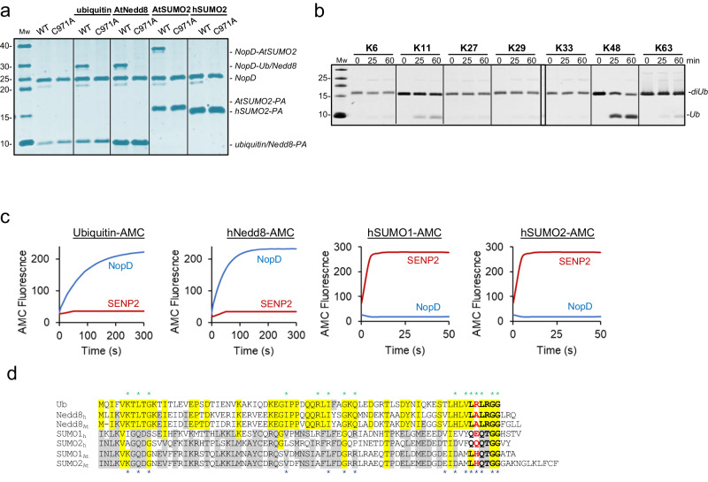
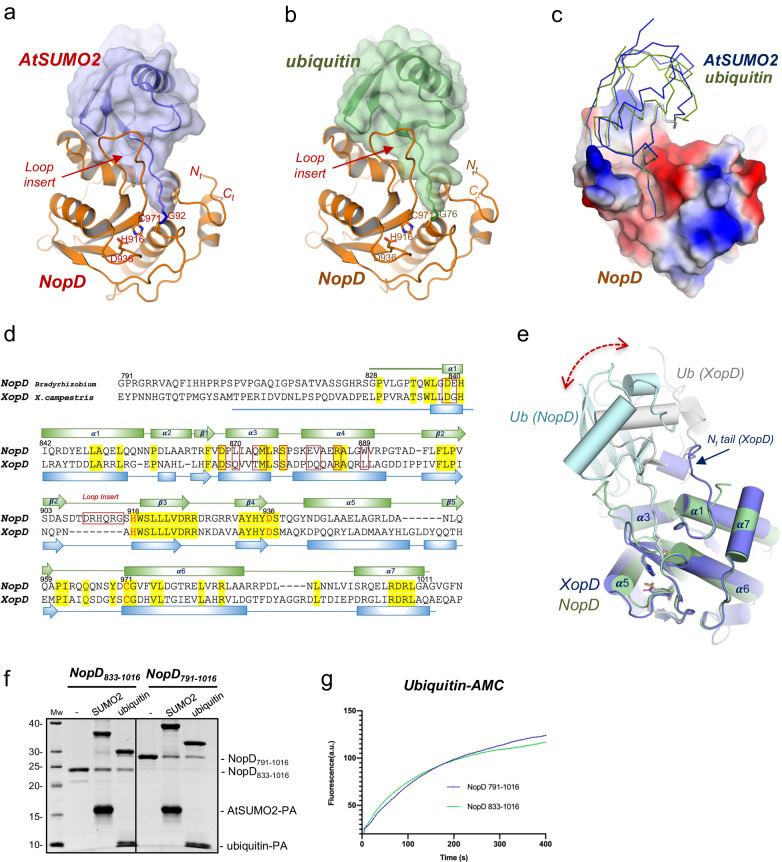
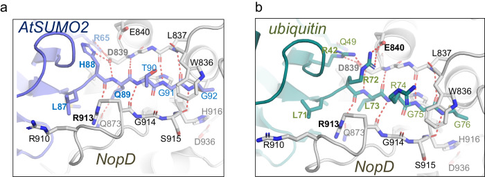
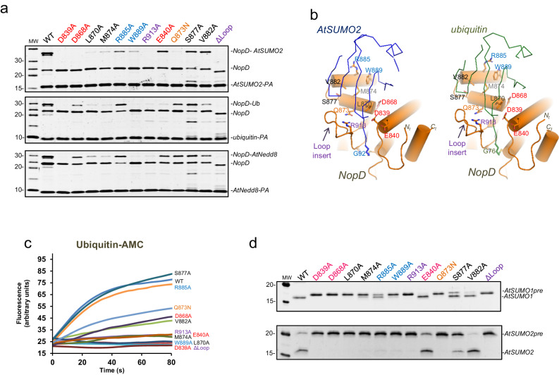
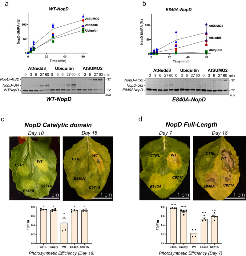
The post-translational modification of proteins by ubiquitin-like modifiers (UbLs), such as SUMO, ubiquitin, and Nedd8, regulates a vast array of cellular processes. Dedicated UbL deconjugating proteases families reverse these modifications. During bacterial infection, effector proteins, including deconjugating proteases, are released to disrupt host cell defenses and promote bacterial survival. NopD, an effector protein from rhizobia involved in legume nodule symbiosis, exhibits deSUMOylation activity and, unexpectedly, also deubiquitination and deNeddylation activities. Here, we present two crystal structures of Bradyrhizobium (sp. XS1150) NopD complexed with either Arabidopsis SUMO2 or ubiquitin at 1.50 Å and 1.94 Å resolution, respectively. Despite their low sequence similarity, SUMO and ubiquitin bind to a similar NopD interface, employing a unique loop insertion in the NopD sequence. In vitro binding and activity assays reveal specific residues that distinguish between deubiquitination and deSUMOylation. These unique multifaceted deconjugating activities against SUMO, ubiquitin, and Nedd8 exemplify an optimized bacterial protease that disrupts distinct UbL post-translational modifications during host cell infection.


Conflict of interest statement

The authors declare no competing interests.

Figures

Fig. 1
Fig. 1. Specificity of NopD for Arabidopsis SUMO, ubiquitin and Nedd8.
a SDS-PAGE of the binding between the NopD wild-type and active site mutant C971A against ubiquitin-PA, Arabidopsis Nedd8-PA, Arabidopsis SUMO2-PA and human SUMO2-PA activity-based probes for 2 h. b SDS-PAGE of the di-ubiquitin linkage specificity analysis for NopD over time. The concentration of NopD and diUb are 600 nM and 3 μM, respectively. c Time-course plots of fluorescent AMC-based substrates (100 nM) of ubiquitin, human Nedd8, human SUMO1 and SUMO2 substrates with the catalytic domains of rhizobia NopD (25 nM) and human SENP2 (25 nM) as a control. Reactions were conducted in triplicate and the average curve is displayed. d Sequence alignment of ubiquitin, human and Arabidopsis Nedd8 and, human and Arabidopsis SUMO1 and SUMO2. Yellow and grey background depict sequence conservation between ubiquitin and Nedd8, and between SUMO isoforms, respectively. The C-terminal tails before the diGly motif are marked in black bold letters. Ubiquitin Arg72 position in marked as red bold letter. Asterisks indicate binding residues of NopD with either Arabidopsis SUMO2 (blue) or ubiquitin (green), respectively.
Fig. 2
Fig. 2. Crystal structure of the complexes between NopD and AtSUMO2 and ubiquitin.
a Cartoon representation of the NopD catalytic domain with Arabidopsis AtSUMO2. b Cartoon representation of the NopD catalytic domain with ubiquitin. Catalytic triad residues are labeled and shown in stick representation. The N- and C-terminal are labeled. Loop insert in NopD is labeled and marked. c Electrostatic potential surface representation of NopD in complex with overlapped ribbon structures of Arabidopsis SUMO2 (blue) and ubiquitin (green). d Structural alignment of sequences corresponding to the catalytic domains for NopD and Xanthomonas campestris XopD. Red squares indicate interface residues to NopD. Catalytic triad residues are shown in red. Secondary structure cartoon is depicted above for NopD (green), or below for XopD (blue). e Superposition of the NopD-ubiquitin complex (green) with the Xanthomonas XopD-Ubiquitin (PDB: 5JP3) complex (blue). Double headed discontinued blue arrow indicates the displacement of ubiquitin between NopD (grey) and XopD (light blue) complexes. f SDS-PAGE of binding of NopD with a longer N-terminal extension against AtSUMO2-PA and ubiquitin-PA suicide probes for 2 h. Bands of NopD crosslink, NopD791-1016, NopD833-1016, AtSUMO2-PA, ubiquitin-PA are labeled. g Time-course of ubiquitin-AMC hydrolysis for NopD833-1016 and NopD791-1016 constructs. Ub-AMC (100 nM) was incubated with NopD (5 nM) constructs over time. Reactions were conducted in triplicate and the average curve is displayed.
Fig. 3
Fig. 3. Structural details of the NopD C-terminal interaction.
a Close-up view of the C-terminal tail of AtSUMO2 (purple) in complex with NopD (gray). b Close-up view of the C-terminal of ubiquitin (green) in complex with NopD (gray). Binding side-chain residues in the contact area of substrate and enzyme are shown in stick representation and labelled. Hydrogen bonds are represented by dashed red lines.
Fig. 4
Fig. 4. Interface between ubiquitin and AtSUMO2 globular domain with NopD.
ac Close-up view of the ribbon representation of NopD-AtSUMO2 and NopD-ubiquitin interfaces in three orthogonal views. Binding side-chain residues in the contact area are labelled and shown in stick representation. AtSUMO2 and ubiquitin residues are colored in blue and green, respectively. Hydrogen bonds and charged interactions are represented by dashed red lines.
Fig. 5
Fig. 5. In vitro catalytic analysis of the NopD SUMO/ubiquitin specificity.
a SDS-PAGE binding analysis of NopD wild-type and point mutants with AtSUMO2-PA (above), ubiquitin-PA (middle) and AtSUMO2-PA (below) activity-based propargylamine-derived probes. Reaction assays were performed with NopD at 1 μM using the PA probes at 4 μM for 2 h. b Cartoon representation of the NopD-AtSUMO2 and ubiquitin complexes. AtSUMO2 (blue) and ubiquitin (green) are shown in ribbon representation and NopD (orange) in cartoon representation. Analyzed interface residues are labeled and shown in stick representation. c Time-course plot of the hydrolysis of the fluorescent ubiquitin-AMC substrate with NopD wild type and point mutants. Reactions were conducted in triplicate and the average curve is displayed. d SDS-PAGE of end point activity assays for NopD (200 nM) using the substrates precursor of Arabidopsis SUMO1 and SUMO2 (1 μM).
Fig. 6
Fig. 6. Expression of NopD and NopD mutants in plant cells induces cell death.
Time-course plot of the SDS-PAGE binding analysis of NopD wild-type (a) and NopD E840A point mutant (b) with AtNedd8-PA, ubiquitin-PA and AtSUMO2-PA activity-based probes. Data values represent the mean ± SD, n = 3 technical replicates. Expression of the catalytic domain (c) or the full-length (d) NopD WT, NopD C971A, and NopD E840A proteins under the control of CAMV 35S promoter in different sections of a leaf was performed by infiltration of A. Tumefaciens transformed with pCAMBIA plasmids carrying the described NopD genes. Empty plasmid was used as a control for comparison. The photographs were taken during several days post infiltration (dpi). Cell death necrotic tissue, can be visualized in wtNopD (yellow-brown areas), whereas expression of NopD C971A or NopD E840A showed no visible effects. Quantification of cell death was measured by the reduction in photosynthetic efficiency of the tissue surrounding the injection point on each section of the leaf. Measurements around the injection point were taken for each section using four independent leaves. Therefore, data values represent the mean ± SD, n = 4 biological replicates. Significance was measured by a two-tailed unpaired t test relative to wild type. All data were analyzed with a 95% confidence interval. *P ≤ 0.05, **P ≤ 0.01, ***P ≤ 0.001, ****P ≤ 0.0001. Exact P values from the left to right: (a) 0.022, 0.027, 0.033, 0.030; (d) <0.0001, <0.0001, 0.0009, 0.0006.

References

    1. Deng W, et al. Assembly, structure, function and regulation of type III secretion systems. Nat. Rev. Microbiol. 2017;15:323–337. doi: 10.1038/nrmicro.2017.20. - DOI - PubMed
    1. Staehelin C, Krishnan HB. Nodulation outer proteins: double-edged swords of symbiotic rhizobia. Biochem. J. 2015;470:263–274. doi: 10.1042/BJ20150518. - DOI - PubMed
    1. Nelson MS, Sadowsky MJ. Secretion systems and signal exchange between nitrogen-fixing rhizobia and legumes. Front. Plant Sci. 2015;6:491. doi: 10.3389/fpls.2015.00491. - DOI - PMC - PubMed
    1. Bartsev AV, et al. NopL, an effector protein of Rhizobium sp. NGR234, thwarts activation of plant defense reactions. Plant Physiol. 2004;134:871–879. doi: 10.1104/pp.103.031740. - DOI - PMC - PubMed
    1. Wenzel M, Friedrich L, Göttfert M, Zehner S. The type III-secreted protein NopE1 affects symbiosis and exhibits a calcium-dependent autocleavage activity. Mol. Plant. Microbe Interact. 2010;23:124–129. doi: 10.1094/MPMI-23-1-0124. - DOI - PubMed

Publication types

MeSH terms