

Laser debonding of ultrathin occlusal veneers fabricated from different CAD/CAM ceramic materials
- PMID: 38802801
- PMCID: PMC11129369
- DOI: 10.1186/s12903-024-04314-6
Laser debonding of ultrathin occlusal veneers fabricated from different CAD/CAM ceramic materials
Abstract
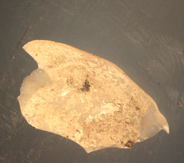
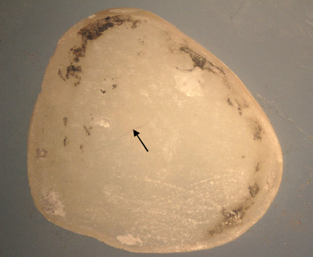
Background: Erbium lasers safely offer the possibility of reuse for debonded restorations. Since these lasers have a high affinity for water molecules, they are absorbed by resin cement causing explosive ablation of the cement and thus, the restoration debonds. The efficiency of this process depends on many factors, including the ceramic type, its chemical composition and thickness. Therefore, this study was designed to test the time taken to debond ultrathin occlusal veneers made of three types of milled ceramic materials and evaluate the integrity of these restorations after debonding.
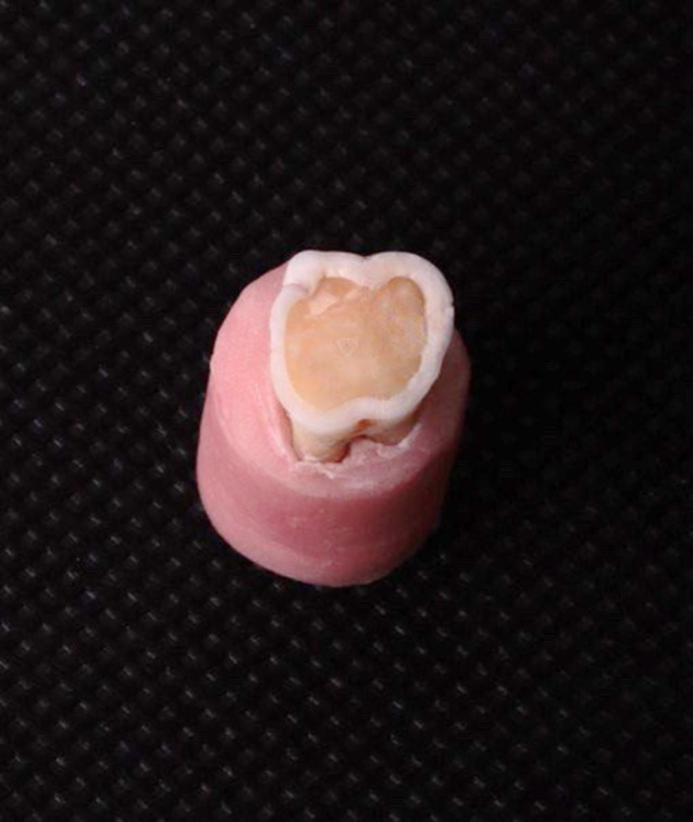
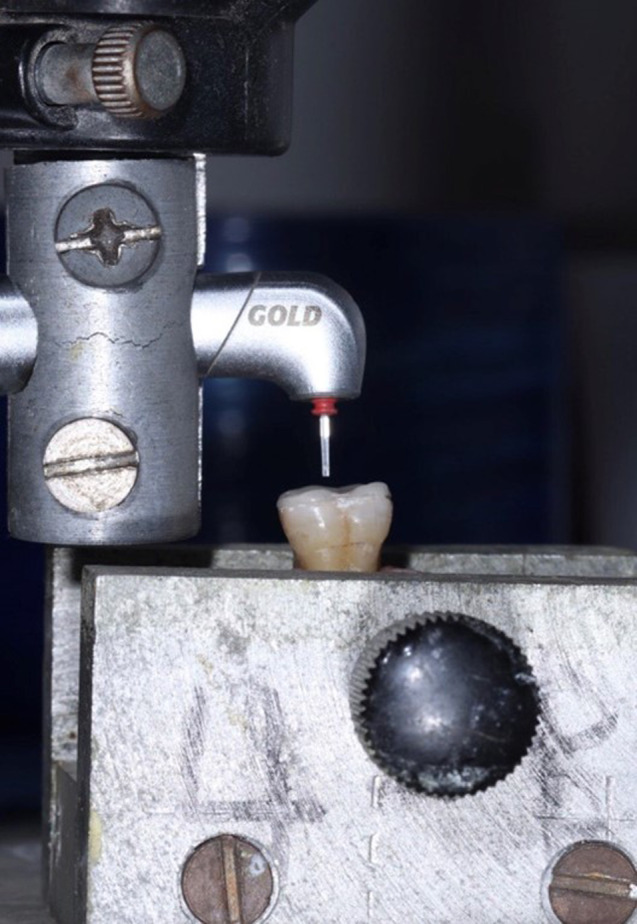
Methods: Three ceramic types were evaluated in this study: lithium disilicate (IPS Emax CAD), highly condensed lithium disilicate (GC initial®LiSi), and translucent zirconia (Katana zirconia STML). Each group consisted of 8 occlusal veneers of 0.5 mm thickness. The samples were cemented to the occlusal surfaces of the upper molar teeth. An Er; Cr: YSGG laser was applied to the occlusal veneers using the scanning method, and time until debonding was calculated. The debonded samples were then inspected under a stereomicroscope for possible damage. Numerical data are presented as the mean with 95% confidence interval (CI), standard deviation (SD), minimum (min.) and maximum (max.) values. Normality and variance homogeneity assumptions were confirmed using Shapiro-Wilk's and Levene's tests, respectively. Data were normally distributed and were analyzed using one-way ANOVA followed by Tukey's post hoc test. The significance level was set at p < 0.05 for all tests. Statistical analysis was performed with R statistical analysis software version 4.3.2 for Windows (R Core Team (2023). R: A language and environment for statistical computing. R Foundation for Statistical Computing, Vienna, Austria. URL https://www.R-project.org/).
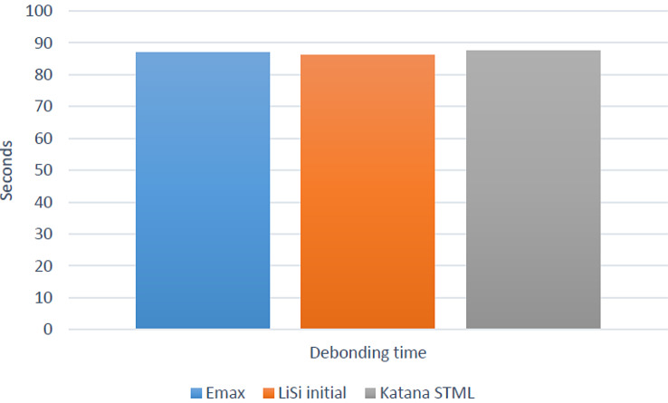
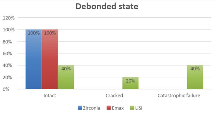
Results: There was no significant difference in debonding time between the different materials (p = 0.995). The longest debonding time was found for Katana STML (87.52 ± 20.45) (seconds), followed by Emax (86.94 ± 20.63) (seconds), while the lowest value was found for LiSi initial (86.14 ± 25.16) (seconds). In terms of damage to the debonded veneers, The Emax and zirconia samples showed no damage. However, 40% of the LiSi samples fractured during debonding, and 20% exhibited cracks. Only 40% of the LiSi samples were sound after debonding.
Conclusion: Er; Cr: YSGG laser can be used efficiently to remove ceramic occlusal veneers. However, its effect on LiSi restorations needs further research.
Keywords: Ceramics; Debonding; Er;cr:YSGG; Laser; Ultrathin occlusal veneers.
© 2024. The Author(s).
Conflict of interest statement
The authors declare no competing interests.
Figures
References
-
- Shalaby MM, Abo-Eittah MR. Influence of the preparation design and aging on the vertical marginal gap of occlusal veneers constructed of different ceramic materials. Egypt Dent J. 2020;66:1261–74. doi: 10.21608/edj.2020.28045.1108. - DOI
MeSH terms
Substances
LinkOut - more resources
Full Text Sources
Miscellaneous
