. 2024 Jul;11(28):e2403485.
doi: 10.1002/advs.202403485. Epub 2024 May 27.

USP25 Elevates SHLD2-Mediated DNA Double-Strand Break Repair and Regulates Chemoresponse in Cancer

Affiliations

USP25 Elevates SHLD2-Mediated DNA Double-Strand Break Repair and Regulates Chemoresponse in Cancer

Yunhui Li et al. Adv Sci (Weinh). 2024 Jul.

Abstract

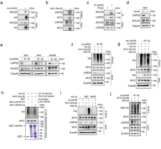
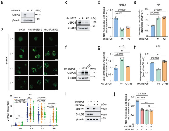
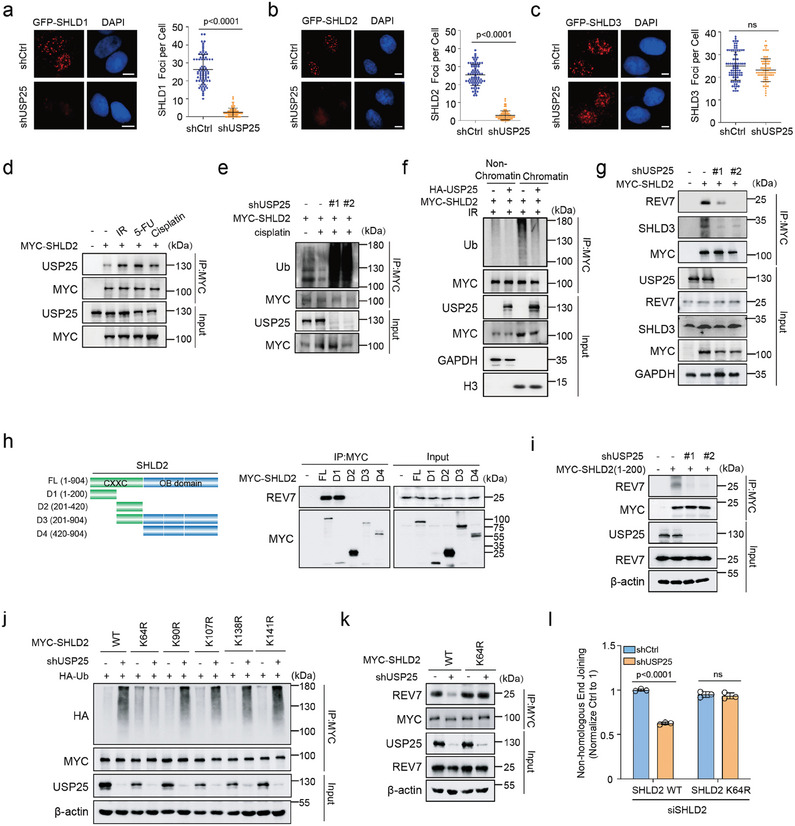
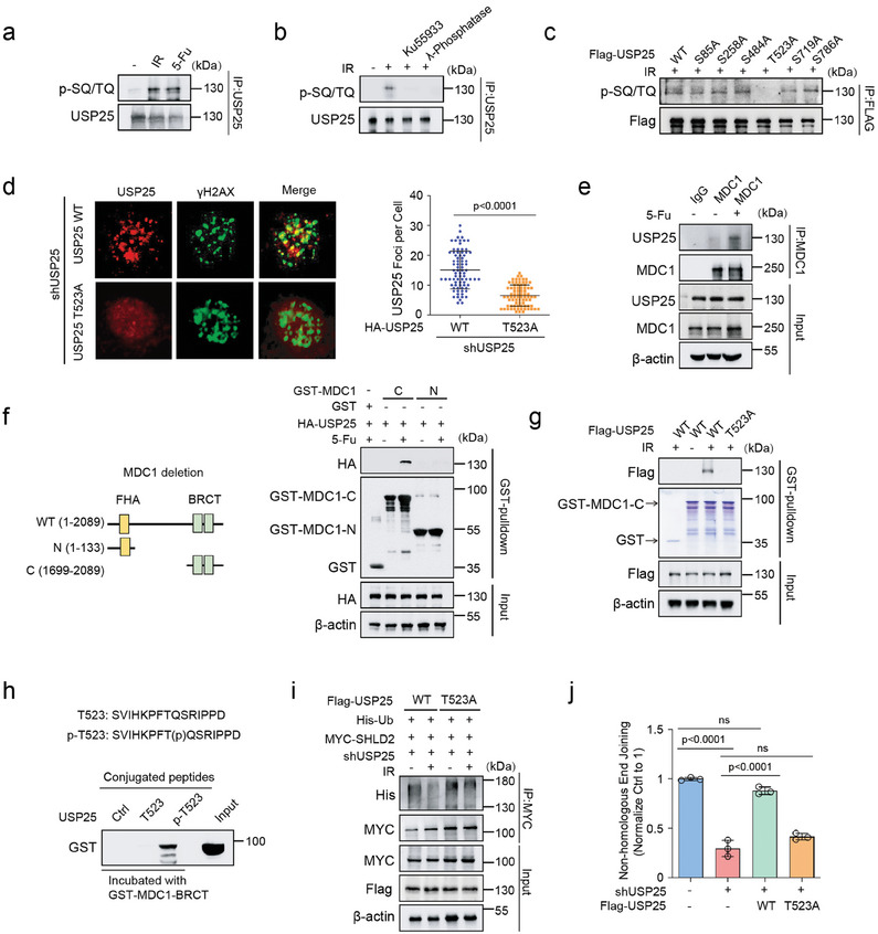
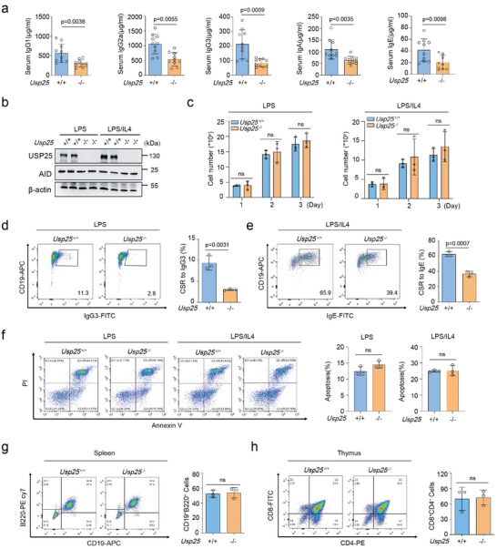
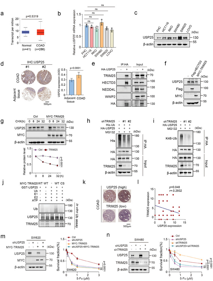
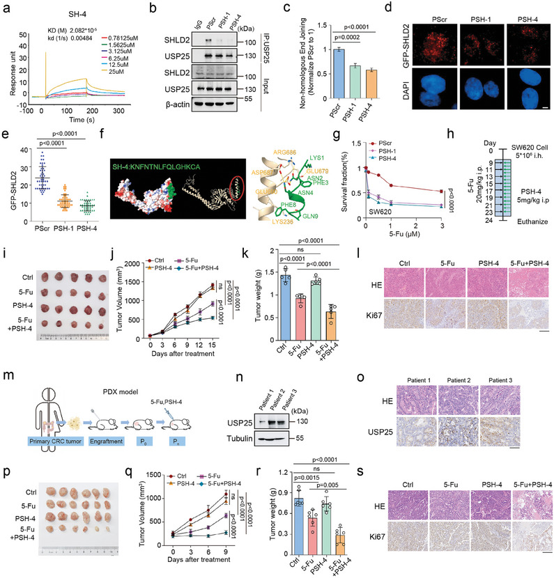
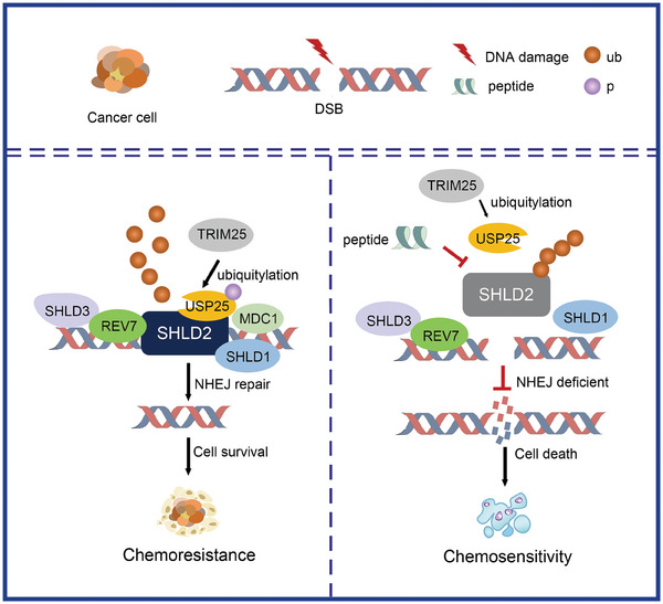
DNA damage plays a significant role in the tumorigenesis and progression of the disease. Abnormal DNA repair affects the therapy and prognosis of cancer. In this study, it is demonstrated that the deubiquitinase USP25 promotes non-homologous end joining (NHEJ), which in turn contributes to chemoresistance in cancer. It is shown that USP25 deubiquitinates SHLD2 at the K64 site, which enhances its binding with REV7 and promotes NHEJ. Furthermore, USP25 deficiency impairs NHEJ-mediated DNA repair and reduces class switch recombination (CSR) in USP25-deficient mice. USP25 is overexpressed in a subset of colon cancers. Depletion of USP25 sensitizes colon cancer cells to IR, 5-Fu, and cisplatin. TRIM25 is also identified, an E3 ligase, as the enzyme responsible for degrading USP25. Downregulation of TRIM25 leads to an increase in USP25 levels, which in turn induces chemoresistance in colon cancer cells. Finally, a peptide that disrupts the USP25-SHLD2 interaction is successfully identified, impairing NHEJ and increasing sensitivity to chemotherapy in PDX model. Overall, these findings reveal USP25 as a critical effector of SHLD2 in regulating the NHEJ repair pathway and suggest its potential as a therapeutic target for cancer therapy.

Keywords: DNA repair pathways; SHLD2; cancer therapy; deubiquitinase USP25; peptides.


Conflict of interest statement

The authors declare no conflict of interest.

Figures

Figure 1
Figure 1
USP25 interacts with and deubiquitinates SHLD2. a) Co‐immunoprecipitation (Co‐IP) analysis was performed to investigate the interaction between USP25 and SHLD2 in HEK293T cells. Cell lysates were subjected to HA resin and subsequently immunoblotted with indicated antibodies to detect the precipitated proteins. b) Co‐IP analysis was additionally performed to examine the interaction between SHLD2 and USP25 in HEK293T cells. Cell lysates were subjected to MYC resin, and the immunoprecipitations were then probed using the indicated antibodies. c) Co‐IP assay of the interaction between USP25 and SHLD2 using antibodies to USP25 in HEK293T cells. Lysates from cells were prepared for co‐IP experiments with USP25 antibody and then blotted with the indicated antibodies. d) The SHLD2 protein levels in Usp25 deleted mouse embryonic fibroblasts (MEFs) or e) USP25 knockdown HEK293T, RKO, SW620 cells were assessed by immunoblotting. The cells were lysed and subjected to Western blot analysis using the indicated antibodies. f) Control and USP25 knockdown cells were treated with MG132 for 4 h prior to harvest. MYC was immunoprecipitated and blots were then probed with the indicated antibodies. g) Control cells and USP25 knockdown cells reconstituted with USP25WT and USP25C178S mutant were treated to MG132 for 4 h before harvest. MYC was immunoprecipitated and blots were then probed with the indicated antibodies. h) Deubiquitination of SHLD2 in vitro by USP25. Ubiquitinated MYC‐SHLD2 was incubated with purified USP25WT and USP25C178S mutant in vitro and then probed with the indicated antibodies. i) HA‐Ublysine‐specific mutant constructs were transfected into control or USP25 knockdown cells and cells were treated to MG132 for 4 h before harvest. Blots were probed with the indicated antibodies. j) Control and USP25 knockdown cells were treated with MG132 for 4 h prior to harvest. MYC was immunoprecipitated and blots were then probed with the indicated antibody.
Figure 2
Figure 2
USP25 deubiquitinates SHLD2 to regulate DNA damage repair. a)Immunoblotting was performed to detect USP25 expression in U2OS cells stably expressing either control or USP25 shRNA. b) U2OS cells stably expressing control and USP25 shRNA were left irradiated with or without 2 Gy and probed with γH2AX foci at the indicated time point. Representative micrographs and the quantification of γH2AX foci were shown. n > 80 in each group. c) Immunoblotting was performed to detect USP25 expression in HEK293T cells stably expressing either control or USP25 shRNA. d) NHEJ or e) HR repair capacity of control and USP25 knockdown cells were subjected using a reporter assay. f) Immunoblot of USP25 in HEK293T cells stably expressing control, HA‐USP25WT or HA‐USP25C178S mutant. g) NHEJ or h) HR repair capacity of control, HA‐USP25WT, and HA‐USP25C178S mutant cells were assessed using a reporter assay. i) Control and USP25 knockdown HEK293T cells transfected short interfering RNAs (siRNA) against SHLD2 were blotted with the indicated antibodies and j) were then subjected to NHEJ assay. Statistical analysis was performed using two‐way ANOVA followed by a Turkey's multiple comparison test (b) or one‐way ANOVA followed by a Turkey's multiple comparison test (d,e,g,h,j).
Figure 3
Figure 3
K64 as the major deubiquitination site of SHLD2 for NHEJ.a‐c) U2OS cells stably expressing control or USP25 shRNA were irradiated with 5 Gy and probed with GFP‐SHLD1, GFP‐SHLD2, and GFP‐SHLD3 foci. Cells were stained with anti‐GFP antibodies. Representative micrographs and the quantification of a) GFP‐SHLD1, b) GFP‐SHLD2, and c) GFP‐SHLD3 foci were shown. n>80 in each group. d) Myc‐SHLD2 were transfected into RKO cells and cells were left untreated or treated with IR (10 Gy), 5‐Fu (20 um), or cisplatin (20 um), and cell lysates were incubated with the indicated antibody. e) USP25 knockdown RKO cells were transfected with MYC‐SHLD2 and left untreated or treated with cisplatin(20 um). MYC was immunoprecipitated, and blots were probed with the indicated antibodies. f) Cells were treated with IR and added MG132 for 4 h prior to harvest. MYC was immunoprecipitated in chromatin fraction samples or nonchromatin fraction samples. Blots were then probed with the indicated antibodies. g) Myc‐SHLD2 were transfected into control or USP25 knockdown cells. MYC was immunoprecipitated. Blots were probed with the indicated antibodies. h) Schematic representation of MYC‐SHLD2 full length or truncated MYC‐SHLD2. HEK293T cells were transfected with these deletion mutants subjected to anti‐MYC‐affinity gel, and blots were probed with indicated antibodies. i) MYC‐SHLD2 (aa1‐200) were transfected in control or USP25 knockdown cells. MYC was immunoprecipitated. Blots were probed with the indicated antibodies. j) SHLD2 (WT or KR mutants) constructs were transfected into control or USP25 knockdown cells. Myc‐SHLD2 was immunoprecipitated. Blots were probed with the indicated antibodies. k) Myc‐SHLD2 (WT or K64R) constructs were transfected into control or USP25 knockdown cells. Myc was immunoprecipitated. Blots were probed with the indicated antibodies. l) Control or USP25 knockdown HEK293T cells transfected short interfering RNAs (siRNA) against SHLD2 together with wild‐type or K64R Myc‐SHLD2 were subjected to NHEJ assay. Statistical analysis was performed using a t‐test (a–c) or one‐way ANOVA followed by Turkey's multiple comparison test (l).
Figure 4
Figure 4
Regulation of the DDR signaling by USP25. a) HEK293T cells were left untreated or treated with IR (10 Gy) or 5‐Fu (20 um). USP25 was immunoprecipitated and immunoblotted with phospho‐SQ/TQ (p‐SQ/TQ). b) HEK293T cells were pretreated with Ku55933 (25 µm) for 2 h followed by treatment with IR(10 Gy). After 1 h, USP25 was immunoprecipitated, left untreated, or treated with phosphatase and immunoblotted with p‐SQ/TQ. c) HEK293T cells transfected with USP25WT and indicated constructs were left treated with IR(10 Gy). USP25 was immunoprecipitated and immunoblotted with p‐SQ/TQ. d) USP25 knockdowm U2OS cells rescued with USP25WT or USP25T523A were treated with IR (2 Gy), and co‐localization USP25 with γH2AX was detected by immunofluorescence. n>80 in each group. e) HEK293T cells were left untreated or treated with 5‐Fu (20 um). MDC1 was immunoprecipitated and immunoblotted with USP25. f) HEK293T cells transfected with USP25 were treated with 5‐Fu (20 um), and cell lysates were incubated with GST or GST‐MDC1 deletions in vitro. The interaction USP25 and MDC1 deletions was detected. g) HEK293T cells transfected with USP25WT or USP25T523A were treated with IR, and cell lysates were incubated with GST or GST‐MDC1‐BRCT in vitro. The interaction USP25 and MDC1‐BRCT was detected. h) Non‐phosphorylated or phosphorylated Thr523 peptide was conjugated to beads and incubated with purified GST‐MDC1‐BRCT domain in buffer. Proteins bound to beads were blotted with the indicated antibodies. i) USP25 knockdown cells rescued USP25WT or USP25T523A cells were left untreated or treated with IR(10 Gy). MYC was immunoprecipitated, and blots were probed with the indicated antibodies. j) NHEJ repair capacity of control, USP25 knockdown, and USP25 knockdown rescued USP25WT or USP25T523A cells were subjected using a reporter assay. Statistical analysis was performed using t‐test (d) or one‐way ANOVA followed by Turkey's multiple comparison test (j).
Figure 5
Figure 5
USP25 promotes CSR through NHEJ.a) Serum immunoglobulin was measured in Usp25+/+ and Usp25−/− mouse. b) B cells isolated from Usp25+/+ and Usp25−/− mice were treated with LPS or LPS/IL4 for 72 h. Cells were lysed and AID expression levels were plotted for the indicated genotypes. c–e) CSR levels to IgG3 or IgE were measured in Usp25+/+ and Usp25−/− B cells on day 3 after LPS or LPS/IL4 stimulation. Representative c) growth curve, d,e) flow cytometry blot and quantification of data of indicated B cells. n  =  3. f) Apoptotic cell percentages (Annexin V+/PI) were assessed on Day 3 after stimulation with LPS or LPS/IL4 and plotted. n  =  3. A representative flow cytometry blot and quantitative analysis were presented for the respective B cells. g) CD19+ B220+ B cell population in spleen of Usp25+/+ and Usp25−/− mice were shown with flow cytometry and summary graphs. n  =  3. h) CD4+CD8+ T cell population in thymus of Usp25+/+ and Usp25−/− mice were shown with flow cytometry and summary graphs. n  =  3. Statistical analysis was performed using a t‐test (a,d,e,f,g,h) or two‐way ANOVA followed by Turkey's multiple comparison test (c).
Figure 6
Figure 6
TRIM25 ubiquitinates and inactivates USP25. a) The mRNA expression levels of USP25 in human normal colon tissue samples (n = 41) and colorectal adenocarcinoma tissue samples (n = 286 COAD patients) from the Ualcan database. b) The mRNA expression level of USP25 in human colon cell lines and colon carcinoma cell lines through qPCR. c) The protein expression level of USP25 in human colon cell lines and colon carcinoma cell lines through western blot. d) Tissue microarray with representative IHC images showing USP25 protein expression in COAD tumors and adjacent tissues. (n = 48). e) Co‐IP analysis was performed to detect the interaction between USP25 and TRIM25, HECTD3, NEDDL4, and WWP2. Cell lysates were subjected to HA resin, and the immunoprecipitants were subsequently blotted using the indicated antibodies. f) The protein expression levels of USP25 were evaluated in HEK293T cells transfected with MYC‐TRIM25 or Flag‐HECTD3. g) The effect of cycloheximide (CHX) treatment on USP25 expression levels was analyzed in HEK293T cells transfected with TRIM25 for an indicated duration of time. h) In vivo ubiquitination assays were performed in control and TRIM25 knockdown cells, transfected with indicated plasmids, and treated with MG132 for 4 h before harvesting. Western blot analysis was performed using indicated antibodies. i) Control and USP25 knockdown cells were treated with MG132 for 4 h prior to harvest. HA was immunoprecipitated and blots were then probed with k48‐ub antibodies. j) In vitro ubiquitination assays were conducted by incubating purified GST‐USP25 and MYC‐TRIM25 proteins with recombinant E1, UbcH5a, ubiquitin (Ub), and ATP buffer at 37 °C for 1 h. Samples were immunoblotted using the indicated antibodies. k‐l) Tissue microarray with representative IHC images showing USP25 or TRIM25 protein expression in COAD (n = 48 pairs of tumors). Representative images of two different specimens k), and l) the Pearson correlations of staining intensity between USP25 and TRIM25. m) Survival assays for control, USP25 knockdown, MYC‐TRIM25, and USP25 knockdown stably expressing MYC‐TRIM25 SW620 cells for CCK8 assay in response to the indicated concentration of 5‐Fu (0, 0.1, 0.5, 1, 3 µm) for 72 h. n) Survival assays for control, TRIM25 knockdown, USP25 knockdown, and double knockdown SW480 cells for CCK8 assay in response to the indicated concentration of 5‐Fu (0, 0.1, 0.5, 1, 3 µm) for 72 h. Statistical analysis was performed using t‐test (d) or one‐way ANOVA followed by Turkey's multiple comparison test or two‐way ANOVA followed by Turkey's multiple comparison test (g,m,n) or Pearson's correlation test (l).
Figure 7
Figure 7
Disturbing the USP25 and SHLD2 interaction counteracts colon cancer progression. a) The kinetic interaction of SH‐4 and USP25 was assessed using surface plasmon resonance (SPR) analyses. b) Co‐immunoprecipitation assays (Co‐IP) were performed to detect the interaction between USP25 and SHLD2 in HEK293T cells treated with indicated peptides. c) NHEJ repair capacity of HEK293T cells treated with indicated peptides was subjected using a reporter assay. d) Representative micrographs of GFP‐SHLD2 foci and e) quantification were shown following irradiation and indicated peptide treatment. Cells were stained with anti‐GFP antibodies. f) The highest‐scoring Dock model of the SH‐4 and USP25 complex is shown. Left: the surface of SH‐4 (green) and USP25 complex. Right: the 3D structure of SH‐4 (green) and USP25 complex. g) The effect of indicated peptides(20 µm) in combination with 5‐Fu (0, 0.1, 0.5, 1, 3 µm) on the chemosensitivity of SW620 cells was determined. h‐l) Xenograft tumor formation by SW620 cells in mice that were untreated or treated with PSH‐4 and 5‐Fu or each treatment alone was analyzed. A h) schematic model, i) representative tumor images, j) tumor volume over time, k) tumor weight, and l) HE or Ki67 staining assay were presented (n = 5 animals). m) A schematic model for generating patient‐derived tumor xenografts of colorectal adenocarcinoma (COAD) was provided. n) Western blots showing USP25 levels in COAD patient tumors (patients 1–3) were presented. o) Representative immunohistochemistry (IHC) micrographs showing USP25 expression in COAD patient‐derived tumors were provided. p–s) The PDX xenograft in mice that were untreated or treated with PSH‐4(5 mg k−1 ) and 5‐Fu (20 mg k−1 ) or each treatment alone were analyzed. Representative p) tumor images, q) tumor volume over time, r) tumor weight, and s) HE or Ki67 staining assay were presented (n = 6 animals). Statistical analysis was performed using one‐way ANOVA followed by a Turkey's multiple comparison test (c,e,k,r) or two‐way ANOVA followed by a Turkey's multiple comparison test (g,j,q).
Figure 8
Figure 8
Schematic model. We proposed a model in which USP25 deubiquitinates and modifies SHLD2 by K63‐linked polyubiquitin chains at the K64 site, promoting NHEJ repair. The phosphorylation of USP25 by ATM regulates the ubiquitination of SHLD2, enhances NHEJ repair, and contributes to chemoresistance. Peptide PSH‐4 was identified and found to disrupt the interaction between USP25 and SHLD2. Treatment of PDX mice in combination with PSH‐4 peptide and 5‐Fu significantly increased tumor chemosensitivity.

Similar articles

References

    1. Katsuki Y., Jeggo P. A., Uchihara Y., Takata M., Shibata A., Genome Instab Dis. 2020, 1, 155.
    1. Jackson S. P., Bartek J., Nature 2009, 461, 1071. - PMC - PubMed
    1. Mao Z., Bozzella M., Seluanov A., Gorbunova V., DNA Repair (Amst) 2008, 7, 1765. - PMC - PubMed
    1. Dev H., Chiang T. W., Lescale C., de Krijger I., Martin A. G., Pilger D., Coates J., Sczaniecka‐Clift M., Wei W., Ostermaier M., Herzog M., Lam J., Shea A., Demir M., Wu Q., Yang F., Fu B., Lai Z., Balmus G., Belotserkovskaya R., Serra V., O'Connor M. J., Bruna A., Beli P., Pellegrini L., Caldas C., Deriano L., Jacobs J. J. L., Galanty Y., Jackson S. P., Nat. Cell Biol. 2018, 20, 954. - PMC - PubMed
    1. Gupta R., Somyajit K., Narita T., Maskey E., Stanlie A., Kremer M., Typas D., Lammers M., Mailand N., Nussenzweig A., Lukas J., Choudhary C., Cell 2018, 173, 972. - PMC - PubMed

MeSH terms

LinkOut - more resources