

Expectant management for umbilical artery thrombosis in the third trimester of pregnancy: a case report
- PMID: 38803431
- PMCID: PMC11129344
- DOI: 10.3389/fphar.2024.1395344
Expectant management for umbilical artery thrombosis in the third trimester of pregnancy: a case report
Abstract
Background: Umbilical artery thrombosis (UAT) is a rare complication of pregnancy and is associated with adverse pregnancy outcomes, including fetal intrauterine distress, intrauterine growth restriction, and still birth. UAT is unpredictable, and prenatal diagnosis is challenging. There is no consensus on the treatment strategy of UAT, especially for patients with prenatal detection of one of the umbilical artery embolisms. In most previous cases, an emergency cesarean section was performed, or intrauterine fetal death occurred at the time of UAT diagnosis.
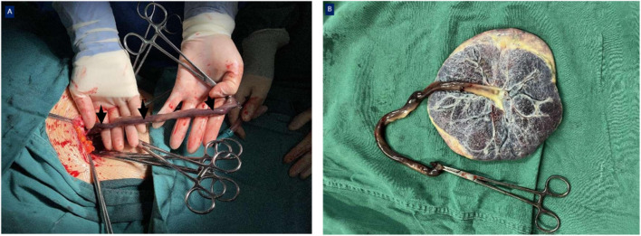
Case presentation: In this report, we describe a case of thrombosis in one of the umbilical arteries detected by routine ultrasonography at 31+3 weeks of gestation in a 34-year-old woman. Following expectant management with intensive monitoring for 4 four days, an emergency cesarean section was performed because of abnormal fetal umbilical cord blood flow and middle cerebral artery blood flow; the newborn was in good condition at birth. The final umbilical cord histopathology revealed thrombosis in one of the umbilical arteries. Both mother and newborn described in this case underwent long-term follow-up for nearly 2 two years and are currently in good health without any complications.
Conclusions: Based on our experience, obstetricians should comprehensively consider the current gestational age and fetal intrauterine status when UAT is suspected to determine the best delivery time. The appropriate gestational age should be prolonged as long as the mother and fetus are stable when the fetus is immature, trying our best to complete the corticosteroid treatment to promote fetal lung maturity and magnesium sulfate to protect fetal brain. During expectant management, ultrasound monitoring, electronic fetal heart monitoring, and fetal movement counting should be strengthened. Clinicians should ensure that the patients and their families are informed about all potential risks of expectant management for UAT.
Keywords: case report; fetal distress; third trimester; ultrasound monitoring; umbilical artery thrombosis.
Copyright © 2024 Gong, Zhang and Wang.
Conflict of interest statement
The authors declare that the research was conducted in the absence of any commercial or financial relationships that could be construed as a potential conflict of interest.
Figures


Similar articles
-
Expectant management for umbilical artery thrombosis in monochorionic diamniotic twin pregnancies: a case report.BMC Pregnancy Childbirth. 2023 Jul 14;23(1):515. doi: 10.1186/s12884-023-05834-9. BMC Pregnancy Childbirth. 2023. PMID: 37452280 Free PMC article.
-
[Clinical analysis of 31 cases of fetal umbilical artery thrombosis].Zhonghua Fu Chan Ke Za Zhi. 2023 Jul 25;58(7):495-500. doi: 10.3760/cma.j.cn112141-20230106-00008. Zhonghua Fu Chan Ke Za Zhi. 2023. PMID: 37474322 Chinese.
-
Fetal umbilical artery thrombosis: prenatal diagnosis, treatment and follow-up.Orphanet J Rare Dis. 2022 Nov 12;17(1):414. doi: 10.1186/s13023-022-02563-8. Orphanet J Rare Dis. 2022. PMID: 36371215 Free PMC article.
-
Expectant management for umbilical artery thrombosis: a report of two cases and literature review.J Matern Fetal Neonatal Med. 2022 Dec;35(25):9296-9298. doi: 10.1080/14767058.2022.2029398. Epub 2022 Jan 27. J Matern Fetal Neonatal Med. 2022. PMID: 35086411 Review.
-
Application of low molecular weight heparins in umbilical artery thrombosis: A case series and review of the literature.Medicine (Baltimore). 2023 Apr 14;102(15):e33501. doi: 10.1097/MD.0000000000033501. Medicine (Baltimore). 2023. PMID: 37058068 Free PMC article. Review.
Cited by
-
Umbilical Artery Thrombosis Masquerading as Single Umbilical Artery in a Stillbirth.Diagnostics (Basel). 2025 Jan 3;15(1):94. doi: 10.3390/diagnostics15010094. Diagnostics (Basel). 2025. PMID: 39795622 Free PMC article.
References
-
- Guocai H., Zhaoqin L., Qing B., Qiuqun X. (2019). Detection and clinical significance of antiphospholipid antibody in pregnant women. Laboratory Med. Clin. 16 (14), 2044–2046.
-
- Jieqiong L., Wen Z. (2018). Diagnosis, treatment and pathogenesis of antiphospholipid syndrome. Chin. Clin. Immunol. allergy complex 12 (04), 423–429.
Publication types
LinkOut - more resources
Full Text Sources