

Kimura Disease: A Detailed Analysis of Clinical and Radiological Manifestations in a Retrospective Case Series
- PMID: 38803691
- PMCID: PMC11129738
- DOI: 10.2147/JIR.S462098
Kimura Disease: A Detailed Analysis of Clinical and Radiological Manifestations in a Retrospective Case Series
Abstract
Background: Kimura disease (KD) is a rare chronic inflammatory disease that affects mainly young Asian men and is characterized by painless subcutaneous masses, lymphadenopathy, and elevated serum IgE levels. Despite its benign nature, KD poses a diagnostic and therapeutic challenge due to its rarity and clinical variability.
Objective: This study aimed to provide a comprehensive analysis of the clinical and radiological features of KD in a retrospective case series, to assess treatment outcomes, and to discuss the implications for diagnosis and management.
Methods: We retrospectively analyzed four histologically confirmed cases of KD admitted to Zhejiang Provincial People's Hospital from January 2018 to October 2023. Clinical and radiological data were retrospectively analyzed, and imaging findings were analyzed by two neuroradiologists to determine lesion characteristics and contrast enhancement patterns.
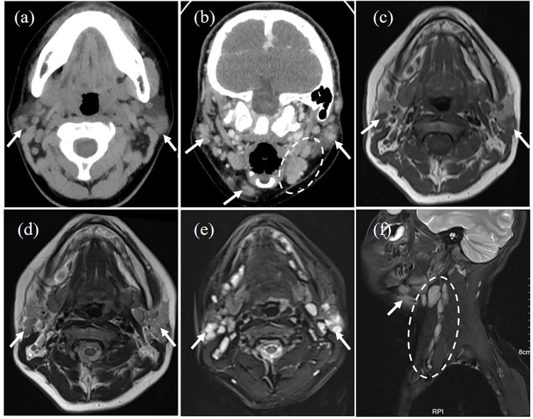
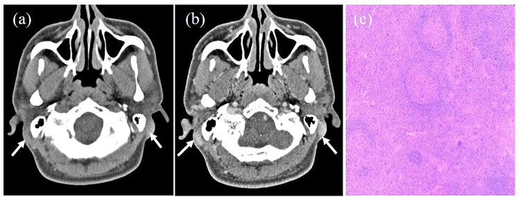
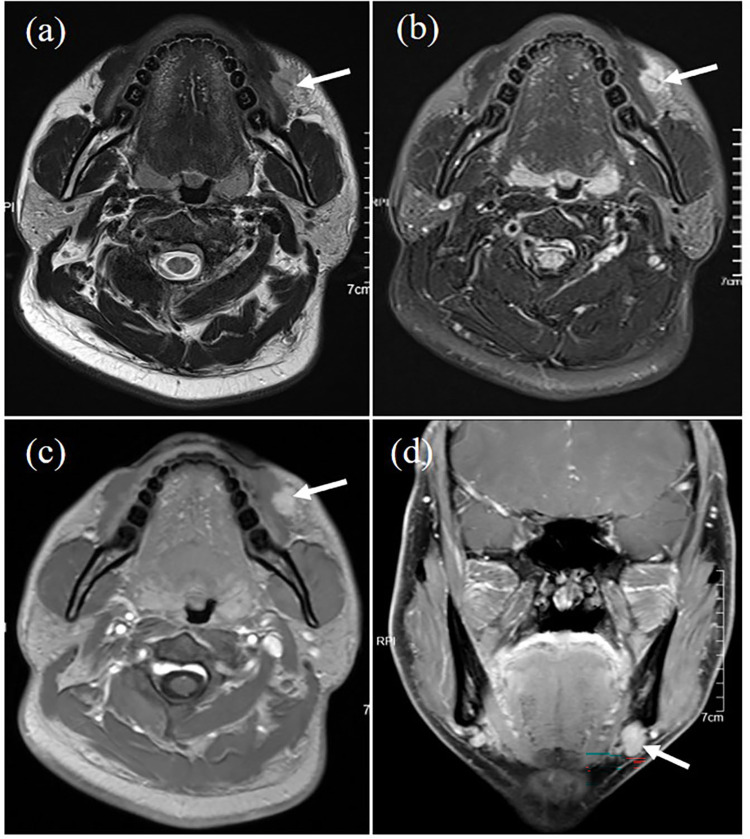
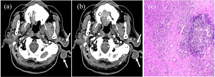
Results: Our findings showed that the patients were predominantly male, with a mean age of 43 years and an age range of 13-71 years. All patients presented with painless subcutaneous masses and three of them had peripheral blood eosinophilia and elevated serum IgE levels. Radiographically, the lesions were predominantly ill-defined with heterogeneous enhancement, accompanied by subcutaneous fat atrophy. Complete surgical excision and oral corticosteroids were effective treatments, and no recurrence was noted during follow-up.
Conclusion: KD should be considered in the differential diagnosis of painless subcutaneous masses in the head and neck region, especially in the presence of eosinophilia and elevated IgE levels. Our findings contribute to the understanding of KD's clinical and radiological spectrum and highlight the need for long-term follow-up due to the risk of recurrence.
Keywords: CT; Kimura disease; MRI; computed tomography; eosinophilia; magnetic resonance imaging.
© 2024 Zhao et al.
Conflict of interest statement
All authors declare that the research was conducted in the absence of any commercial or financial relationships that could be construed as a potential conflict of interest.
Figures
Similar articles
-
Kimura disease: A rare case affecting the inguinal lymph node.Int J Surg Case Rep. 2025 Jun;131:111332. doi: 10.1016/j.ijscr.2025.111332. Epub 2025 Apr 21. Int J Surg Case Rep. 2025. PMID: 40334445 Free PMC article.
-
Kimura disease: A rare case in Vietnamese woman.Asia Pac Allergy. 2024 Aug;14(3):143-147. doi: 10.5415/apallergy.0000000000000134. Epub 2024 Feb 6. Asia Pac Allergy. 2024. PMID: 39220575 Free PMC article.
-
The clinicopathological characteristics of Kimura disease in Chinese patients.Clin Rheumatol. 2019 Dec;38(12):3661-3667. doi: 10.1007/s10067-019-04752-6. Epub 2019 Aug 22. Clin Rheumatol. 2019. PMID: 31440918
-
Kimura Disease: A Case Series and Systematic Review of Clinico-radiological Features.Curr Probl Diagn Radiol. 2022 Jan-Feb;51(1):130-142. doi: 10.1067/j.cpradiol.2020.10.003. Epub 2020 Nov 17. Curr Probl Diagn Radiol. 2022. PMID: 33250297
-
Kimura's Disease without Peripheral Eosinophilia: An Unusual and Challenging Case Simulating Venous Malformation on Imaging Studies-Case Report and Review of literature.J Clin Diagn Res. 2017 Jun;11(6):ME01-ME04. doi: 10.7860/JCDR/2017/28603.10063. Epub 2017 Jun 1. J Clin Diagn Res. 2017. PMID: 28764210 Free PMC article. Review.
Cited by
-
Giant Kimura Disease of the Parotid Region Managed by Modified Rhytidectomy: A Case Report and Review of Surgical Treatment Strategies.Cureus. 2025 Jul 8;17(7):e87498. doi: 10.7759/cureus.87498. eCollection 2025 Jul. Cureus. 2025. PMID: 40786271 Free PMC article.
References
-
- Kim HT, Szeto C. Eosinophilic hyperplastic lymphogranuloma: comparison with Mikulicz’s disease. Chin Med J. 1937;23:699–700. in Chinese.
-
- Kimura T, Yoshimura S, Ishikawa E. Unusual granuloma combined with hyperplastic changes in lymphatic tissues. Trans Soc Path Jpn. 1948;13:179–180.
LinkOut - more resources
Full Text Sources
