. 2024 Sep 10;150(11):867-883.
doi: 10.1161/CIRCULATIONAHA.124.068624. Epub 2024 May 28.

Skeletal Muscle SIRT3 Deficiency Contributes to Pulmonary Vascular Remodeling in Pulmonary Hypertension Due to Heart Failure With Preserved Ejection Fraction

Affiliations

Skeletal Muscle SIRT3 Deficiency Contributes to Pulmonary Vascular Remodeling in Pulmonary Hypertension Due to Heart Failure With Preserved Ejection Fraction

Jia-Rong Jheng et al. Circulation. .

Abstract

Background: Pulmonary hypertension (PH) is a major complication linked to adverse outcomes in heart failure with preserved ejection fraction (HFpEF), yet no specific therapies exist for PH associated with HFpEF (PH-HFpEF). We have recently reported on the role of skeletal muscle SIRT3 (sirtuin-3) in modulation of PH-HFpEF, suggesting a novel endocrine signaling pathway for skeletal muscle modulation of pulmonary vascular remodeling.

Methods: Using skeletal muscle-specific Sirt3 knockout mice (Sirt3skm-/-) and mass spectrometry-based comparative secretome analysis, we attempted to define the processes by which skeletal muscle SIRT3 defects affect pulmonary vascular health in PH-HFpEF.

Results: Sirt3skm-/- mice exhibited reduced pulmonary vascular density accompanied by pulmonary vascular proliferative remodeling and elevated pulmonary pressures. Comparative analysis of secretome by mass spectrometry revealed elevated secretion levels of LOXL2 (lysyl oxidase homolog 2) in SIRT3-deficient skeletal muscle cells. Elevated circulation and protein expression levels of LOXL2 were also observed in plasma and skeletal muscle of Sirt3skm-/- mice, a rat model of PH-HFpEF, and humans with PH-HFpEF. In addition, expression levels of CNPY2 (canopy fibroblast growth factor signaling regulator 2), a known proliferative and angiogenic factor, were increased in pulmonary artery endothelial cells and pulmonary artery smooth muscle cells of Sirt3skm-/- mice and animal models of PH-HFpEF. CNPY2 levels were also higher in pulmonary artery smooth muscle cells of subjects with obesity compared with nonobese subjects. Moreover, treatment with recombinant LOXL2 protein promoted pulmonary artery endothelial cell migration/proliferation and pulmonary artery smooth muscle cell proliferation through regulation of CNPY2-p53 signaling. Last, skeletal muscle-specific Loxl2 deletion decreased pulmonary artery endothelial cell and pulmonary artery smooth muscle cell expression of CNPY2 and improved pulmonary pressures in mice with high-fat diet-induced PH-HFpEF.

Conclusions: This study demonstrates a systemic pathogenic impact of skeletal muscle SIRT3 deficiency in remote pulmonary vascular remodeling and PH-HFpEF. This study suggests a new endocrine signaling axis that links skeletal muscle health and SIRT3 deficiency to remote CNPY2 regulation in the pulmonary vasculature through myokine LOXL2. Our data also identify skeletal muscle SIRT3, myokine LOXL2, and CNPY2 as potential targets for the treatment of PH-HFpEF.

Keywords: Sirtuin 3; heart failure, diastolic; musculokeletal abnormalities; pulmonary heart disease.


Conflict of interest statement

M.T.G. is a coinventor of patents and patent applications directed to the use of recombinant neuroglobin and heme-based molecules as antidotes for carbon monoxide poisoning, which have been licensed by Globin Solutions, Inc. M.T.G. is a shareholder, advisor, and director in Globin Solutions, Inc. M.T.G. is also coinventor on patents directed to the use of nitrite salts in cardiovascular diseases, which were previously licensed to United Therapeutics, and are now licensed to Globin Solutions and Hope Pharmaceuticals. M.T.G. is a principal investigator in an investigator-initiated research study with Bayer Pharmaceuticals to evaluate riociguat as a treatment for patients with sickle cell disease.

Figures

Figure 1.
Figure 1.
Absence of SIRT3 in skeletal muscle leads to reduced pulmonary vascular density, increased pulmonary vascular remodeling, and elevated pulmonary pressures. A, SIRT3 protein levels in soleus, liver, pulmonary artery smooth muscle cells (PASMCs), and left ventricle (LV) of WT and Sirt3skm−/− mice. B, Representative micro-CT images of lungs infused with Mercox II resin from WT and Sirt3skm−/− mice. Reduction in vascular density, with narrowed vascular diameter (yellow square), and decreased peripheral vasculature (green arrowheads), was observed in Sirt3skm−/− mice. C, Volumetric analysis of the pulmonary vasculature. D, Representative images of lung sections stained with α-smooth muscle actin (α-SMA, 20X). E-G, Right ventricular systolic pressure (RVSP; E), left ventricular end-diastolic pressure (LVEDP; F), and LV ejection fraction (LVEF, G) were measured in WT and Sirt3skm−/− mice. H and I, LV and RV mass normalized to tibial length. J-K, Body weight (J) and glucose tolerant abilities (K) were measured. All mice were fed on regular diet starting at age of 8 weeks for 16 weeks. Data are mean ± SEM; n = 6–12 mice/group. Statistical comparisons were performed using Mann-Whitney U-test. For Figure 1K, two-way ANOVA followed by Bonferroni’s post hoc test was performed. *P < 0.05.
Figure 2.
Figure 2.
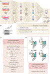
Secreted proteins associated with skeletal muscle SIRT3 deficiency identified by mass spectrometry. A, Schematic overview of comparative secretome analysis. Briefly, each sample (condition/replicate) was labeled with a unique TMT reagent tag at the peptide level. While these tags are isobaric, they are different with respect to the position of isotopically heavy nuclei in their structural backbone. After labeling, the individual samples are combined to produce a single multiplexed sample. Upon fragmentation in the mass spectrometer, a sample-specific fragment, a reporter ion, is released and recorded in the mass spectrum. Labeled peptides derived from different samples will have a unique reporter ion whose abundance is related to the abundance of the peptide in the given sample. Relative quantification is performed by measuring the relative abundances of the reporter ions released during fragmentation of “protein-specific” tryptic peptides. B, Efficiency of siRNA-mediated knock-down of SIRT3 in C2C12 myotubes. C, Proteins identified as differentially regulated by SIRT3. D, Abundance ratio of lysyl oxidases (LOX) and LOX-like family members (LOXL1, LOXL2 and LOXL3) for 3 conditions (with 3 biological replicates each). Data are mean ± SEM. Comparisons of parameters were performed using unpaired Student’s t-test after testing for normality with Shapiro-Wilk test.
Figure 3.
Figure 3.
Increased LOXL2 in plasma and skeletal muscle of Sirt3skm−/− mice, Ob-Su rats and patients with PH-HFpEF. Plasma levels of LOXL2 (A), periostin (B), and Grem1 (C) were measured in WT and Sirt3skm−/− mice. D, Representative Western blots for LOXL2 protein expression levels in soleus of Sirt3skm−/− and WT mice. E, Plasma levels of LOXL2 were measured in Ob-Su rat model of PH-HFpEF. F, Correlation between RVSP and LOXL2 in Ob-Su rats. Spearman r is shown. G, Circulating levels of LOXL2 were measured in plasma of control subjects (n = 12) and patients with PH-HFpEF [n = 14; Age: 70.4 ± 7.9; male gender: 8; BMI: 37.2 ± 10.2; mPAP: 39.9 ± 8.6 mmHg; PCWP: 20 ± 7.8 mmHg; WHO function class II: 2 (14%), III: 11 (79%), and IV: 1 (7%)]. H and I, Representative Western blots of LOXL2 (H) and SIRT3 (I) in vastus lateralis muscle obtained from human patients with HFpEF or PH-HFpEF. Data are mean ± SEM. Statistical comparisons were performed using Mann-Whitney U-test.
Figure 4.
Figure 4.
Treatment with LOXL2 increases CNPY2 protein expression in PCLS, PAECs, and PASMCs. A, PCLS from C57B6 mice were treated with recombinant LOXL2 protein (500 ng/ml, labeled as “+”) or vehicle control (labeled as “-“) for 7 days. Representative images of Western blots for p50-ATF6, ATF4, CHOP, and CNPY2 protein expression levels in PCLS (n = 3). B, Representative images of PCLS stained with CNPY2 (red), α-smooth muscle actin (α-SMA, green), CD31 (light blue), and counterstained with DAPI (40X). C, Representative Western blots for CNPY2 protein expression levels in lungs of Ob-Su rats compared with lean rats ± SU5416 exposure (Ln and Ln-Su). D and E, Representative images of Western blots for CNPY2 protein expression levels in cultured human PAECs (D) and PASMCs (E) treated with recombinant LOXL2 protein (250 ng/ml, serum-free medium) for 2 days. F and G, C2C12 cells were differentiated to 70% confluence and transiently transfected with siRNA targeting SIRT3 or scrambled control for 72 hours. Conditioned media from scrambled control (labeled as “Ci”) or SIRT3-deficient (labeled as “Si”) C2C12 cells were treated to PAECs (F) or PASMCs (G) isolated from C57B6 mice for 2 days. CNPY2 protein levels were measured. Data are mean ± SEM. n = 3–4/group. Statistical comparisons were performed using unpaired Student’s t-test after testing for normality with Shapiro-Wilk test. For Figure 4C, one-way ANOVA followed by Tukey’s post hoc test was performed.
Figure 5.
Figure 5.
Elevated CNPY2 expression in PAECs and PASMCs of Sirt3skm−/− mice and Ob-Su rats, and PASMCs of human subjects with obesity/diabetes. A and B, Representative Western blots for CNPY2 protein expression levels in PAECs of Sirt3skm−/− mice (A) and Ob-Su rats (B). C and D, Representative images of CNPY2 levels in PASMCs of Sirt3skm−/− mice (C) and Ob-Su rats (D). E, Clinical information for human PASMCs. F, Representative images of Western blots for CNPY2 protein expression levels in PASMCs obtained from humans with or without obesity (using BMI of 29 as a cut-off to define obesity). Data are mean ± SEM. Statistical comparisons were performed using Mann-Whitney U-test.
Figure 6.
Figure 6.
Elevated CNPY2, either by overexpression or LOXL2 stimulation, promotes cell migration and proliferation. A-C, Cultured human PAECs were transfected with empty or CNPY2-expressing vector for 30 hours. Efficiency of CNPY2 overexpression and its effect on p53 were measured by Western blots (A). Representative images of wound closure and related quantitative data (B). Cell proliferation assessed by manual cell counts (C). D, Human PAECs treated with recombinant LOXL2 protein (250 ng/ml, serum-free medium) for 2 days. p53 expression levels were measured and quantified. E and F, Human PAECs were transiently transfected with siRNA targeting CNPY2 or scramble control before treatment with recombinant LOXL2 protein. Migration (E) and proliferation (F) of PAECs were measured. G-I, Human PASMCs were overexpressed with CNPY2 for 30 hours. Representative Western blots for CNPY2 and p53 protein levels (G). Cell proliferations measured by cell counts (H) and colorimetric BrdU incorporation (I). J, Human PASMCs treated with recombinant LOXL2 protein (250 ng/ml, serum-free medium) for 2 days. p53 expression levels were measured and quantified. K and L, Human PASMCs were transiently transfected with siRNA targeting CNPY2 or scramble control for 24 hours before stimulation with recombinant LOXL2 protein (500 ng/ml, complete medium) for 5 days. Cell proliferation assessed by manual cell counts (K) and colorimetric BrdU incorporation (L). Data are mean ± SEM; n = 3–5/group. Statistical comparisons were performed using Mann-Whitney U-test, two-way ANOVA followed by Tukey’s post hoc test, or unpaired Student’s t-test after testing for normality with Shapiro-Wilk test.
Figure 7.
Figure 7.
Skeletal muscle-specific LOXL2 deletion reduces PAECs/PASMCs expression of CNPY2 and improves pulmonary pressures in mice with HFD-induced PH-HFpEF. A, Eight-week-old male Loxl2skm−/− and WT mice were fed with a HFD (60% lipids/kcal) or RD (10% lipids/kcal) for 16 weeks. B-D, Body weight (B), glucose tolerant abilities (C), and right ventricular systolic pressures (RVSP, D) were measured. E, Representative images of lungs stained with α-smooth muscle actin (α-SMA, green), CNPY2 (red), CD31 (light blue), and counterstained with DAPI (40X). Scale bar, 30 μm. Medial index was calculated. F-G, Representative Western blots and quantification of CNPY2, p53, or PCNA in PAECs (F) and PASMCs (G) isolated from WT and Loxl2skm−/− mice. H, Schema summarizing results relating skeletal muscle secretome to pulmonary vascular remodeling in PH-HFpEF. Statistical comparisons were performed using two-way ANOVA followed by Tukey’s post hoc test. For Figure 7C, two-way ANOVA followed by Bonferroni’s post hoc test was performed.

References

    1. Oktay AA, Rich JD, Shah SJ. The emerging epidemic of heart failure with preserved ejection fraction. Current heart failure reports. 2013;10:401–410. doi: 10.1007/s11897-013-0155-7 - DOI - PMC - PubMed
    1. Lloyd-Jones D, Adams R, Carnethon M, De Simone G, Ferguson TB, Flegal K, Ford E, Furie K, Go A, Greenlund K, et al. Heart disease and stroke statistics−-2009 update: a report from the American Heart Association Statistics Committee and Stroke Statistics Subcommittee. Circulation. 2009;119:e21–181. doi: 10.1161/CIRCULATIONAHA.108.191261 - DOI - PubMed
    1. Thenappan T, Shah SJ, Gomberg-Maitland M, Collander B, Vallakati A, Shroff P, Rich S. Clinical characteristics of pulmonary hypertension in patients with heart failure and preserved ejection fraction. Circ Heart Fail. 2011;4:257–265. doi: 10.1161/CIRCHEARTFAILURE.110.958801 - DOI - PubMed
    1. Klapholz M, Maurer M, Lowe AM, Messineo F, Meisner JS, Mitchell J, Kalman J, Phillips RA, Steingart R, Brown EJ, Jr., et al. Hospitalization for heart failure in the presence of a normal left ventricular ejection fraction: results of the New York Heart Failure Registry. Journal of the American College of Cardiology. 2004;43:1432–1438. doi: 10.1016/j.jacc.2003.11.040 - DOI - PubMed
    1. Simon MA, Vanderpool RR, Nouraie M, Bachman TN, White PM, Sugahara M, Gorcsan J, Parsley EL, Gladwin MT. Acute hemodynamic effects of inhaled sodium nitrite in pulmonary hypertension associated with heart failure with preserved ejection fraction. JCI Insight. 2016;1:e89620. doi: 10.1172/jci.insight.89620 - DOI - PMC - PubMed

MeSH terms