. 2024 Apr 30;8(2):17.
doi: 10.3390/epigenomes8020017.

Blood Vitamin C Levels of Patients Receiving Immunotherapy and Relationship to Monocyte Subtype and Epigenetic Modification

Affiliations

Blood Vitamin C Levels of Patients Receiving Immunotherapy and Relationship to Monocyte Subtype and Epigenetic Modification

Ben Topham et al. Epigenomes. .

Abstract

The treatment of metastatic melanoma has been revolutionised by immunotherapy, yet a significant number of patients do not respond, and many experience autoimmune adverse events. Associations have been reported between patient outcome and monocyte subsets, whereas vitamin C (ascorbate) has been shown to mediate changes in cancer-stimulated monocytes in vitro. We therefore investigated the relationship of ascorbate with monocyte subsets and epigenetic modifications in patients with metastatic melanoma receiving immunotherapy. Patients receiving immunotherapy were compared to other cancer cohorts and age-matched healthy controls. Ascorbate levels in plasma and peripheral blood-derived mononuclear cells (PBMCs), monocyte subtype and epigenetic markers were measured, and adverse events, tumour response and survival were recorded. A quarter of the immunotherapy cohort had hypovitaminosis C, with plasma and PBMC ascorbate levels significantly lower than those from other cancer patients or healthy controls. PBMCs from the immunotherapy cohort contained similar frequencies of non-classical and classical monocytes. DNA methylation markers and intracellular ascorbate concentration were correlated with monocyte subset frequency in healthy controls, but correlation was lost in immunotherapy patients. No associations between ascorbate status and immune-related adverse events or tumour response or overall survival were apparent.

Keywords: DNA methylation; ascorbate; metastatic melanoma; monocytes.


Conflict of interest statement

The authors declare no conflicts of interest. The funders had no role in the design of the study; in the collection, analyses or interpretation of data; in the writing of the manuscript; or in the decision to publish the results.

Figures

Figure 1
Figure 1
Ascorbate intake and plasma concentrations in patients with cancer and healthy controls. The cohorts included patients receiving immunotherapy (Im) or chemotherapy (Ch), or patients prior to their surgery to remove cancer (PS), or healthy controls (HC). (A) Estimated daily intake of ascorbate according to 24 h dietary recall, and (B) plasma concentrations of ascorbate, measured by HPLC-EC. Im n = 63 filled circles, Ch n = 103 open squares, PS n = 82 open triangles, HC n = 16 open circles, compared by one-way ANOVA, * p ≤ 0.05, median shown in red. ns—not significant.
Figure 2
Figure 2
Ascorbate levels in healthy controls and patients with cancer receiving immunotherapy. No relationship between plasma ascorbate and intracellular ascorbate in (A) healthy controls or (B) patients receiving immunotherapy; Pearson correlation. (C) Comparison of plasma levels between patients and controls. (D) Comparison of intracellular ascorbate levels between patients and controls; Mann–Whitney test. Intracellular ascorbate was measured by HPLC-EC in peripheral blood-derived mononuclear cells. Patients with metastatic melanoma receiving immunotherapy n = 25, healthy controls n = 16; ns—not significant, * p ≤ 0.05; median shown in red.
Figure 3
Figure 3
Immune cell and epigenetic markers on PBMCs from patients with metastatic melanoma receiving immunotherapy and healthy volunteers. Comparisons between controls and patients with respect to frequency of (A) CD14+CD16+, (B) CD14+CD16 and (C) HLADR mean fluorescence intensity (MFI). Surface markers were measured by flow cytometry; patients with metastatic melanoma receiving immunotherapy n = 20, healthy controls n = 14. Comparison between PBMCs from controls and patients with respect to (D) 5-hmC, (E) 5-mC and (F) cytidine content. Epigenetic markers were measured by mass spectrometry; patients with metastatic melanoma receiving immunotherapy n = 20, healthy controls n = 9; ns—not significant, * p ≤ 0.05; median shown in red.
Figure 4
Figure 4
Associations between ascorbate and immune cell and epigenetic markers in PBMCs from controls and patients with melanoma. Correlations between intracellular ascorbate and (A) percentage of classical monocytes (CD14+16), (B) 5-mC and (C) cytidine. Correlations between percentage of non-classical monocytes (CD14+CD16+) and epigenetic markers (D) 5-hmC, (E) 5-mC and (F) cytidine. Patient data are shown as filled circles with solid lines, and those of controls as open circles with dashed lines; bold lines indicate significance. Relationships were tested using Pearson’s correlation; ns—not significant, significance * p < 0.05, ** p < 0.01.
Figure 5
Figure 5
Relationship of ascorbate status and patient or tumour response to immunotherapy. Plasma ascorbate levels vs. (A) grade of side effects in patients with metastatic melanoma receiving immunotherapy, and (B) tumour response based on RECIST criteria. Intracellular ascorbate in PBMCs vs. (C) grade of side effects and (D) RECIST criteria. PD—progressive disease, SD—stable disease, PR—partial response, CR—complete response; plasma data for side effects available from n = 45 and for RECIST for n = 33 patients; intracellular ascorbate data available for n = 25 patients; ns – not significant; median shown in red.
Figure 6
Figure 6
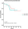
Survival probability of patients with metastatic melanoma receiving immunotherapy according to ascorbate status. The cohort was divided by median ascorbate (48 μM). Data are presented as a Kaplan–Meier curve; n = 44.

References

    1. Dzwierzynski W.W. Melanoma Risk Factors and Prevention. Clin. Plast. Surg. 2021;48:543–550. doi: 10.1016/j.cps.2021.05.001. - DOI - PubMed
    1. Cancer Society Melanoma Survival Rates|Melanoma Survival Statistics. [(accessed on 7 December 2023)]. Available online: https://www.cancer.org/cancer/melanoma-skin-cancer/detection-diagnosis-s....
    1. Ahmadi F., Karamitanha F., Ramezanpour A. Clustering trends of melanoma incidence and mortality: A worldwide assessment from 1995 to 2019. Australas. J. Dermatol. 2022;63:e206–e217. doi: 10.1111/ajd.13882. - DOI - PubMed
    1. Huang J., Chan S.C., Ko S., Lok V., Zhang L., Lin X., Lucero-Prisno D.E., III, Xu W., Zheng Z.J., Elcarte E., et al. Global incidence, mortality, risk factors and trends of melanoma: A systematic analysis of registries. Am. J. Clin. Dermatol. 2023;24:965–975. doi: 10.1007/s40257-023-00795-3. - DOI - PubMed
    1. Villani A., Scalvenzi M., Micali G., Lacarrubba F., Fornaro L., Martora F., Potestio L. Management of Advanced Invasive Melanoma: New Strategies. Adv. Ther. 2023;40:3381–3394. doi: 10.1007/s12325-023-02555-5. - DOI - PMC - PubMed

LinkOut - more resources