. 2024 Jun;14(6):e1692.
doi: 10.1002/ctm2.1692.

Inhibition of HDAC2 sensitises antitumour therapy by promoting NLRP3/GSDMD-mediated pyroptosis in colorectal cancer

Affiliations

Inhibition of HDAC2 sensitises antitumour therapy by promoting NLRP3/GSDMD-mediated pyroptosis in colorectal cancer

Xin Guan et al. Clin Transl Med. 2024 Jun.

Abstract

Background: Although numerous studies have indicated that activated pyroptosis can enhance the efficacy of antitumour therapy in several tumours, the precise mechanism of pyroptosis in colorectal cancer (CRC) remains unclear.

Methods: Pyroptosis in CRC cells treated with antitumour agents was assessed using various techniques, including Western blotting, lactate dehydrogenase release assay and microscopy analysis. To uncover the epigenetic mechanisms that regulate NLRP3, chromatin changes and NLRP3 promoter histone modifications were assessed using Assay for Transposase-Accessible Chromatin using sequencing and RNA sequencing. Chromatin immunoprecipitation‒quantitative polymerase chain reaction was used to investigate the NLRP3 transcriptional regulatory mechanism. Additionally, xenograft and patient-derived xenograft models were constructed to validate the effects of the drug combinations.

Results: As the core molecule of the inflammasome, NLRP3 expression was silenced in CRC, thereby limiting gasdermin D (GSDMD)-mediated pyroptosis. Supplementation with NLRP3 can rescue pyroptosis induced by antitumour therapy. Overexpression of HDAC2 in CRC silences NLRP3 via epigenetic regulation. Mechanistically, HDAC2 suppressed chromatin accessibility by eliminating H3K27 acetylation. HDAC2 knockout promotes H3K27ac-mediated recruitment of the BRD4-p-P65 complex to enhance NLRP3 transcription. Inhibiting HDAC2 by Santacruzamate A in combination with classic antitumour agents (5-fluorouracil or regorafenib) in CRC xenograft-bearing animals markedly activated pyroptosis and achieved a significant therapeutic effect. Clinically, HDAC2 is inversely correlated with H3K27ac/p-P65/NLRP3 and is a prognostic factor for CRC patients.

Conclusion: Collectively, our data revealed a crucial role for HDAC2 in inhibiting NLRP3/GSDMD-mediated pyroptosis in CRC cells and highlighted HDAC2 as a potential therapeutic target for antitumour therapy.

Highlights: Silencing of NLRP3 limits the GSDMD-dependent pyroptosis in colorectal cancer. HDAC2-mediated histone deacetylation leads to epigenetic silencing of NLRP3. HDAC2 suppresses the NLRP3 transcription by inhibiting the formation of H3K27ac/BRD4/p-P65 complex. Targeting HDAC2 activates pyroptosis and enhances therapeutic effect.

Keywords: H3K27ac; HDAC2; NLRP3; colorectal cancer; pyroptosis.


Conflict of interest statement

The authors declare they have no conflicts of interest.

Figures

FIGURE 1
FIGURE 1
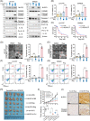
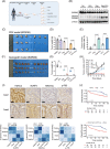
Loss of NLRP3 expression in colorectal cancer (CRC) limits gasdermin D (GSDMD)‐mediated pyroptosis during antitumour therapy. (A and B) Representative immunohistochemistry (IHC) staining images showing NLRP3 and GSDMD expression in CRC tissue (tumour) and corresponding adjacent normal colorectal tissue (normal) from 82 patients. Scale bar: 50 µm. (C) RT‐qPCR analysis demonstrates reduced NLRP3 expression in 30 CRC tissue samples compared to matched adjacent normal tissue samples collected at our institution. (D) Western blot analysis demonstrated the expression levels of the NLRP3 and GSDMD protein in both HIEC‐6 and cancerous cell lines. (E) CRC cells were evaluated for pyroptosis 24 h post‐treatment with regorafenib, as assessed by Western blot. (F) Cell death by regorafenib in CRC cancer cells was morphologically assessed via microscopy. Red arrowheads indicate large bubbles and pores emerging from the plasma membrane. Scale bar: 50 µm. (G and H) Flow cytometry analysis for activated Caspase‐1/propidium iodide (PI). (I) CRC cells were evaluated for pyroptosis 24 h post‐treatment with regorafenib, as assessed by lactate dehydrogenase (LDH) release. Statistical significance is indicated (* p < .05, ** p < .01, *** p < .001, **** p < .0001).
FIGURE 2
FIGURE 2
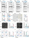
Supplementation of NLRP3 expression in colorectal cancer (CRC) cells rescues gasdermin D (GSDMD)‐mediated pyroptosis in vivo and in vitro. (A and B) Cells stably expressing NLRP3 and control cells were treated with 10 µM regorafenib (Reg) for 24 h. Pyroptosis‐related proteins were assessed by Western blot analysis (A). Lactate dehydrogenase (LDH) release was measured to evaluate pyroptosis levels (B). (C) LS174T and SW620 cells were treated with regorafenib (0, 5, 10, 20 and 40 µM) for 48 h. Cell viability was assessed by CCK‐8 assay. (D and E) Transmission electron microscopy (scale bar: 5 µm) of cells incubated with regorafenib or the corresponding control. Red arrowheads indicate pyroptosis. (F and G) Flow cytometry analysis for activated Caspase‐1/propidium iodide (PI). (H) SW620 xenografts were established in nude mice and treated as shown (n = 5/group). (I) Tumour growth curves were generated from volumes recorded every 2 days. Tumour weight was measured on day 12 after drug treatment. (J) The expression of NLRP3 and cleaved GSDMD in xenografts from different treatment groups was analysed by immunohistochemistry (IHC). Scale bar: 60 µm. Statistical significance is indicated (* p < .05, ** p < .01, *** p < .001, **** p < .0001).
FIGURE 3
FIGURE 3
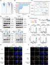
HDAC2‐mediated histone deacetylation leads to epigenetic silencing of NLRP3 in colorectal cancer (CRC). (A) Overview of the general mechanisms and canonical target sites for various epigenetic drugs. (B and C) Multiple epigenetic drugs were applied to LS174T and SW620 cells. RT‐qPCR and Western blotting were used to analyse the changes in the mRNA and protein levels of NLRP3, respectively. (D and E) RT‐qPCR and Western blot analyses were conducted to assess the expression levels of NLRP3 in LS174T and SW620 cells after exposure to escalating doses of histone deacetylase (HDAC) pan‐inhibitors. Two distinct small molecule HDAC pan‐inhibitors were utilised independently. (F) Profiling of HDAC enzymes influencing NLRP3 transcription. Spearman correlation analysis was performed between 15 deacetylases (DACs) and NLRP3 using The Cancer Genome Atlas (TCGA) data. RNA interference was used to individually knockdown 15 DACs, and RT‐qPCR was performed to assess the effects on NLRP3 gene expression. (G) Western blot analysis of NLRP3 and HDAC2 protein levels in 14 matched pairs of CRC tissues and neighbouring normal tissues. (H) Western blot analysis demonstrated the expression levels of the NLRP3 and HDAC2 protein in both HIEC‐6 and CRC cells. (I) A joint analysis of HDAC2 expression in CRC cancer tissues and their corresponding normal tissues was conducted using data from the TCGA and GTEx databases. (J) RT‐qPCR and Western blot analyses were conducted to assess the expression levels of NLRP3 in CRC cells after exposure to escalating doses of Santacruzamate A (SCA). (K) NLRP3 protein expression in LS174T and SW620 cells was detected by Western blot following HDAC2 knockdown. Statistical significance is indicated (* p < .05, ** p < .01, *** p < .001, **** p < .0001).
FIGURE 4
FIGURE 4
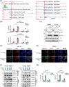
Knocking out HDAC2 activates gasdermin D (GSDMD)‐mediated pyroptosis by upregulating NLRP3. (A‒D) LS174T and SW620 HDAC2 knockout lines were generated with CRISPR/Cas9. Treated with 10 µM regorafenib, and transfected with NLRP3 siRNA in knockout cells for rescue experiments. Pyroptosis pathway proteins were analysed by Western blotting (A and B), and pyroptosis was evaluated by measuring lactate dehydrogenase (LDH) release (C and D). (E) Organoids were treated with regorafenib (10 µM), and the percentage of organoids with changed morphology was quantified using light microscopy. Scale bar: 50 µm. (F and G) Typical bright‐field microscopy images of LS174T cells are shown. Large bubbles protruding from the plasma membrane are highlighted by red arrows. Scale bar: 50 µm. Dead cells were tallied and quantified based on five separate images. (H and I) Flow cytometry analysis for activated Caspase‐1/propidium iodide (PI). Statistical significance is indicated (* p < .05, ** p < .01, *** p < .001, **** p < .0001).
FIGURE 5
FIGURE 5
HDAC2 blocks the transcriptional effect of p‐P65 on NLRP3 by inhibiting chromatin accessibility. (A)  Heatmap showing the average ATAC‐Seq signal centred on the transcription start site (TSS) of the genes in HDAC2 knockout (KO) or wild‐type SW620. The regions of enrichment were expanded by 3 kb on either side of their central point. (B) Gene set enrichment analysis (GSEA) software was used to perform functional enrichment analysis on ranked gene sets of accessibility changes related to various forms of programmed cell death. (C) Genome browser visualisation of ATAC‐Seq data for the NLRP3, gasdermin D (GSDMD), Caspase‐1 and PYCARD Loci in SW620 cells following HDAC2 silencing. (D) The levels of total GSK3β, p‐GSK3β (Ser9), total P65 and p‐P65 (Ser536) were analysed by Western blotting in LS174T or SW620 cells treated with 10 µM regorafenib at the indicated time points. (E) Schematic representation illustrating the positions of potential P65 binding sites in the promoter region of the NLRP3 gene. (F) Chromatin immunoprecipitation (ChIP)‒qPCR validated the interaction between p‐P65 and the NLRP3 promoter in LS174T and SW620 cells under conditions with or without HDAC2 knockdown, as well as with or without Santacruzamate A (SCA) treatment. (G) Expression of NLRP3, HDAC2, P65 and p‐P65 was analysed by Western blotting in LS174T and SW620 cells treated with 10 µM regorafenib in the presence or absence of HDAC2 knockdown. (H and I) The subcellular localisation of p‐P65 and HDAC2 was assessed by double immunofluorescence staining using their respective antibodies. Images were acquired using a confocal microscope. Scale bar: 20 µm. Statistical significance is indicated (* p < .05, ** p < .01, *** p < .001, **** p < .0001).
FIGURE 6
FIGURE 6
Inhibition of HDAC2 activates NLRP3 transcription by facilitating the formation of the H3K27ac‐BRD4‒p‐P65 complex. (A) Chromatin immunoprecipitation (ChIP)‐seq analysis of H3K27ac binding to the NLRP3 promoter region in various cells as predicted by Cistrome DataBrowser and visualised by UCSC Genome Browser. H3K27ac marks active chromatin. (B) ChIP‒qPCR analysis of the binding between the indicated histone proteins (H3K9ac and H3K27ac) and the NLRP3 promoter in colorectal cancer (CRC) wild‐type or HDAC2 knockout (KO) cells. Total genomic DNA and nonspecific immunoglobulin G were the input and negative antibody controls, respectively. (C) Protein levels of NLRP3 and H3K27ac were detected in LS174T cells and SW620 cells after HDAC2 KO. Two independent HDAC2 KO cell lines were provided each for either LS174T cells or SW620 cells. (D and E) The subcellular localisation of p‐P65 and HDAC2 was examined by double immunofluorescence staining with their corresponding antibodies. Images were obtained using confocal microscopy. Scale bar: 20 µm. (F) Co‐immunoprecipitation (Co‐IP) and immunoblotting showing the effect of HDAC2 knockdown on p‐P65 binding with BRD4/H3K27ac and BRD4 binding with p‐P65/H3K27ac in regorafenib‐treated CRC cells. (G) ChIP‒qPCR analysis of the binding between p‐P65 and the NLRP3 promoter in HDAC2 KO LS174T and SW620 cells under normal or BRD4 knockdown conditions. Statistical significance is indicated (* p < .05, ** p < .01, *** p < .001, **** p < .0001).
FIGURE 7
FIGURE 7
HDAC2 correlates with H3K27ac/p‐P65/NLRP3, predicts prognosis and represents a promising target in colorectal cancer (CRC). (A) A schematic diagram outlines the workflow for establishing patient‐derived xenograft (PDX) models of CRC. To evaluate therapeutic strategies, mice bearing established PDX tumours were then randomly divided into six treatment groups as depicted in the schematic. (B) Expression of gasdermin D (GSDMD) and Cleaved GSDMD in tumour tissues from BP0036 mice under different treatment conditions was analysed by Western blotting. (C and D) In the CRC PDX model BP0036, which exhibits high expression of HDAC2, tumour size (C) and weight (D) are shown after grouping and 12 days of treatment. (E) Tumour growth inhibition rate of each PDX model under different treatment regimens. (F and G) The xenograft tumour size (F) and weight (G) were assessed approximately 12 days after initiating treatment. (H) Tumour volumes were measured every 2 days. (I) Representative images of immunohistochemistry (IHC) staining for HDAC2, NLRP3, H3K27ac and p‐P65 in clinical CRC specimens. Scale bar: 50 µm. Correlations (Fisher's exact test) between HDAC2 IHC scores and IHC scores of three proteins (NLRP3, H3K27ac, p‐P65) in CRC tissues were displayed for our cohort (n = 82). (J) Analysis of overall survival of CRC patients in our institution categorised by HDAC2 and NLRP3 status using Kaplan‒Meier survival curves, n = 200, statistical significance was evaluated by the log‐rank test. Statistical significance is indicated (* p < .05, ** p < .01, *** p < .001, **** p < .0001).
FIGURE 8
FIGURE 8
Schematic illustrating inhibition of HDAC2 can reverse the epigenetic silencing of NLRP3 to induce pyroptosis in colorectal cancer.

References

    1. Morgan E, Arnold M, Gini A, et al. Global burden of colorectal cancer in 2020 and 2040: incidence and mortality estimates from GLOBOCAN. Gut. 2023;72:338‐344. - PubMed
    1. Ciardiello F, Ciardiello D, Martini G, Napolitano S, Tabernero J, Cervantes A. Clinical management of metastatic colorectal cancer in the era of precision medicine. CA Cancer J Clin. 2022;72:372‐401. - PubMed
    1. Cervantes A, Adam R, Roselló S, et al. Metastatic colorectal cancer: ESMO Clinical Practice Guideline for diagnosis, treatment and follow‐up. Ann Oncol. 2023;34:10‐32. - PubMed
    1. Grothey A, Blay JY, Pavlakis N, Yoshino T, Bruix J. Evolving role of regorafenib for the treatment of advanced cancers. Cancer Treat Rev. 2020;86:101993. - PubMed
    1. Azwar S, Seow HF, Abdullah M, Faisal Jabar M, Mohtarrudin N. Recent updates on mechanisms of resistance to 5‐fluorouracil and reversal strategies in colon cancer treatment. Biology (Basel). 2021;10:854. - PMC - PubMed

MeSH terms

Substances