

Survival in dogs with meningoencephalomyelitis of unknown etiology with and without lesions detected by magnetic resonance imaging
- PMID: 38804716
- PMCID: PMC11256124
- DOI: 10.1111/jvim.17109
Survival in dogs with meningoencephalomyelitis of unknown etiology with and without lesions detected by magnetic resonance imaging
Abstract
Background: The prognosis of individual dogs with meningoencephalomyelitis of unknown etiology (MUE) remains difficult to predict. MUE cases with no lesions detected by magnetic resonance imaging (MRI) occur, but it is unknown whether this finding is associated with prognosis.
Hypothesis: MUE cases without detectable lesions on MRI have a better outcome than cases with detectable lesions.
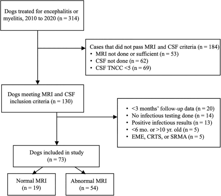
Animals: Study included 73 client-owned dogs with MUE presenting to Purdue University Veterinary Hospital from 2010 to 2020.
Methods: Retrospective study. Dogs with a clinical diagnosis of MUE were identified by medical record search. MRI reports were reviewed for presence or absence of lesions consistent with MUE. Clinical findings at presentation, treatment, disease-specific survival, and outcomes including rates of remission and relapse were compared between cases with normal MRI or abnormal MRI.
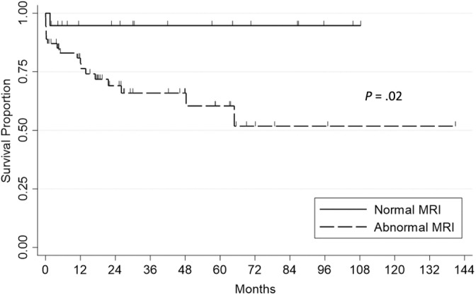
Results: Overall, 54 dogs (74%) were classified as abnormal MRI, and 19 dogs (26%) were classified as normal MRI cases. Death caused by MUE occurred in 1/19 (5%) normal MRI dogs and 18/54 (33%) abnormal MRI dogs (P = .016). Median survival was >107 months in both groups, but survival was significantly longer in the normal MRI group (P = .019). On multivariate analysis, abnormal MRI was significantly related to death (hazard ratio, 7.71; 95% confidence interval 1.03-58.00, P = .0470), whereas significant relationships with death were not identified for either the use of secondary immunosuppressive medications or cerebrospinal fluid nucleated cell count.
Conclusions: MUE dogs with no detectable lesions on MRI have reduced disease-related death compared with dogs with abnormal MRI. The presence or absence of MRI lesions in MUE dogs is prognostically relevant.
Keywords: GME; brain; canine; encephalitis; meningitis; necrotizing encephalitis.
© 2024 The Author(s). Journal of Veterinary Internal Medicine published by Wiley Periodicals LLC on behalf of American College of Veterinary Internal Medicine.
Conflict of interest statement
Georg E. Moore serves as Consulting Editor for Experimental Design and Statistics for the Journal of Veterinary Internal Medicine. He was not involved in review of this manuscript. No other authors declare a conflict of interest.
Figures
References
-
- Cornelis I, Van Ham L, Gielen I, De Decker S, Bhatti SFM. Clinical presentation, diagnostic findings, prognostic factors, treatment and outcome in dogs with meningoencephalomyelitis of unknown origin: a review. Vet J. 2019;244:37‐44. - PubMed
-
- Lowrie M, Smith PM, Garosi L. Meningoencephalitis of unknown origin: investigation of prognostic factors and outcome using a standard treatment protocol. Vet Rec. 2013;172(20):527. - PubMed
-
- Jia Y, Wang H, Zhang M. LGI1 antibody‐associated encephalitis without evidence of inflammation in CSF and brain MRI. Acta Neurol Belg. 2023;123(3):849‐856. - PubMed
MeSH terms
LinkOut - more resources
Full Text Sources
Medical