. 2024 Jul 23;8(14):3798-3809.
doi: 10.1182/bloodadvances.2024012835.

Packaging of supplemented urokinase into alpha granules of in vitro-grown megakaryocytes for targeted nascent clot lysis

Affiliations

Packaging of supplemented urokinase into alpha granules of in vitro-grown megakaryocytes for targeted nascent clot lysis

Mortimer Poncz et al. Blood Adv. .

Abstract

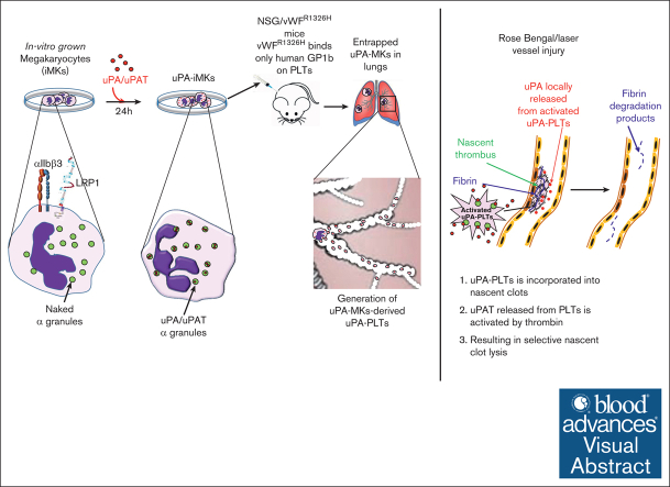
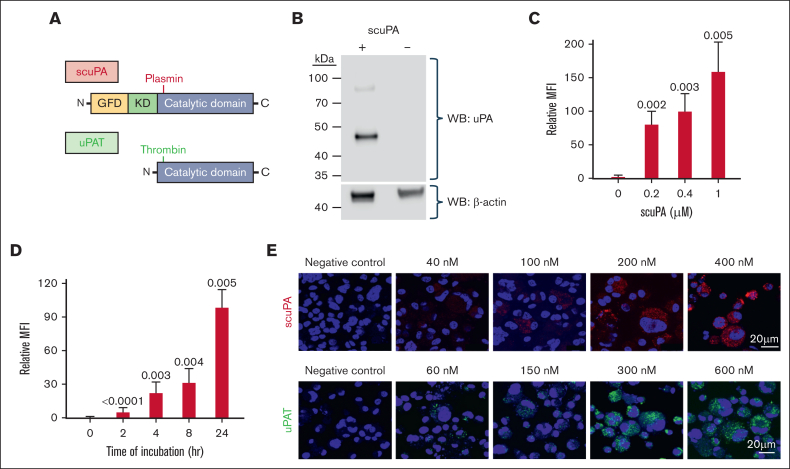
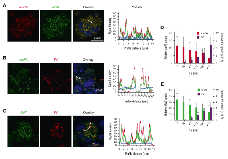
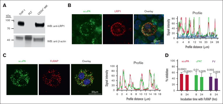
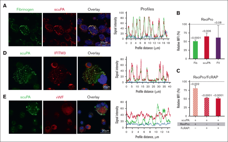
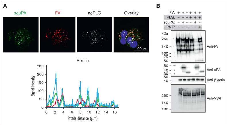
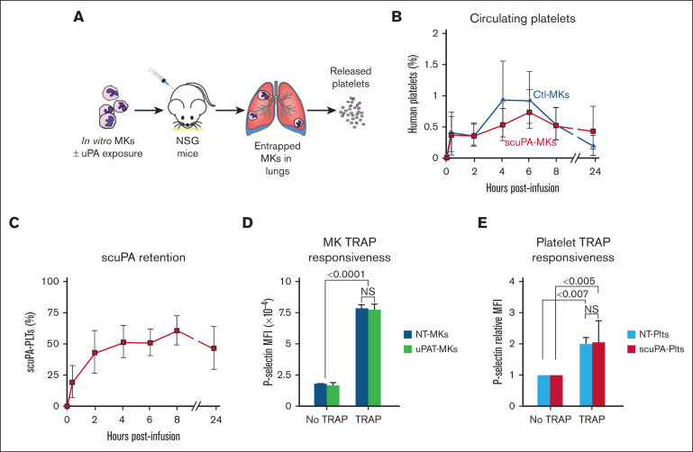
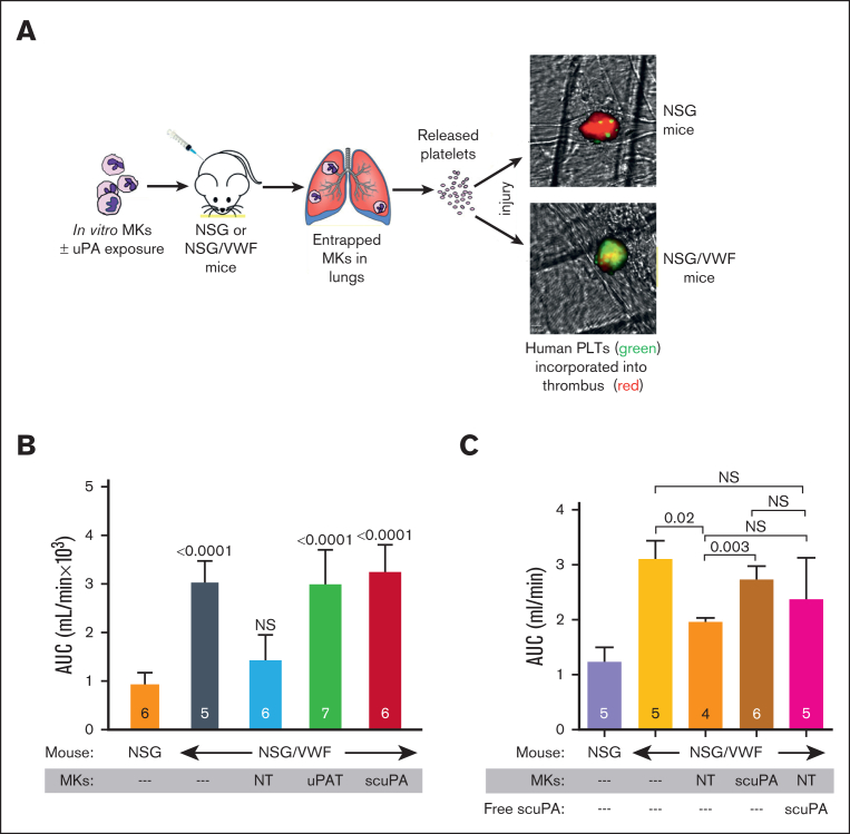
Fibrinolytics delivered into the general circulation lack selectivity for nascent thrombi, reducing efficacy and increasing the risk of bleeding. Urokinase-type plasminogen activator (uPA) transgenically expressed within murine platelets provided targeted thromboprophylaxis without causing bleeding but is not clinically feasible. Recent advances in generating megakaryocytes prompted us to develop a potentially clinically relevant means to produce "antithrombotic" platelets from CD34+ hematopoietic stem cell-derived in vitro-grown megakaryocytes. CD34+ megakaryocytes internalize and store in alpha granules (α-granules) single-chain uPA (scuPA) and a plasmin-resistant thrombin-activatable variant (uPAT). Both uPAs colocalized with internalized factor V (FV), fibrinogen and plasminogen, low-density lipoprotein receptor-related protein 1 (LRP1), and interferon-induced transmembrane protein 3, but not with endogenous von Willebrand factor (VWF). Endocytosis of uPA by CD34+ megakaryocytes was mediated, in part, via LRP1 and αIIbβ3. scuPA-containing megakaryocytes degraded endocytosed intragranular FV but not endogenous VWF in the presence of internalized plasminogen, whereas uPAT-megakaryocytes did not significantly degrade either protein. We used a carotid artery injury model in nonobese diabetic-severe combined immunodeficiency IL2rγnull (NSG) mice homozygous for VWFR1326H (a mutation switching binding VWF specificity from mouse to human glycoprotein Ibα) to test whether platelets derived from scuPA- or uPAT-megakaryocytes would prevent thrombus formation. NSG/VWFR1326H mice exhibited a lower thrombotic burden after carotid artery injury compared with NSG mice unless infused with human platelets or megakaryocytes, whereas intravenous injection of uPA-megakaryocytes generated sufficient uPA-containing human platelets to lyse nascent thrombi. These studies describe the use of in vitro-generated megakaryocytes as a potential platform for delivering uPA or other ectopic proteins within platelet α-granules to sites of vascular injury.


Conflict of interest statement

Conflict-of-interest disclosure: The authors declare no competing financial interests.

Figures

None
Graphical abstract
Figure 1.
Figure 1.
uPA is endocytosed by in vitrogrown MKs and stored in granules. (A) Schematics of the structures of scuPA (top) and uPAT (bottom). Full-length scuPA is composed of (1) an N-terminal growth factor-like domain (GFD, yellow) that binds uPAR; (2) a Kringle domain (K, green) that mediates LRP1-dependent intracellular uptake, binding to integrins, and nuclear translocation; and (3) the protease domain (catalytic domain, gray). Site of activation by plasmin is shown in red. uPAT is composed of the protease domain in which 157F158K has been deleted to create a thrombin cleavage/activation site (shown in green). (B) Representative WB of lysates of in vitro–grown day-11 MKs in the presence (+) or absence (−) of scuPA (400 nM) added on day 10. WB at the top was with anti-human uPA mouse monoclonal antibody followed by horseradish peroxidase (HRP)-conjugated goat anti-mouse antibody, and bottom shows HPR-conjugated anti–β-actin antibody as a loading control. Size marker is shown to the left of the blot. (C) Dose-dependent uptake of Alexa-568 scuPA by CD34+ MKs. y-axes denote mean fluorescence intensity (MFI) measured by flow cytometry. Mean ± 1 standard deviation (SD) of relative uptake of scuPA compared with the values in its absence. n = 4 independent studies. P values were determined by ordinary 1-way analysis of variance (ANOVA). (D) Same as in panel C but for time course of Alexa-568 scuPA (400 nM) uptake by the CD34+ MKs. (E) Visualization using confocal microscopy of Alexa-568 scuPA (top, red) or Alexa-488 uPAT (bottom, green) endocytosed by day-11 CD34+ MKs for 24 hours starting on day 10 initiation of culture. DAPI (4′,6-diamidino-2-phenylindole; blue) depicts the nuclei. Scale bar is shown.
Figure 2.
Figure 2.
scuPA, uPAT, and FV share an endocytic pathway in CD34+ MKs. (A) Representative confocal images of day-10 CD34+ MKs loaded simultaneously by preincubation with Alexa-568 scuPA (red) and Alexa-488 uPA-T (green) for 24 hours. Nuclear staining by DAPI is in blue. Overlap is in yellow and is shown to the right. Scale bar is shown. Quantitative analysis of overlap is shown in Table 1. (B-C) Similar confocal image studies as in panel A but of CD34+ MKs preincubated with (B) Alexa-488 scuPA or (C) Alexa-488 uPAT for 24 hours and then incubated with Alexa-568 FV for 2 hours (red) on day 11. White segmented lines in overlay images represent profiles along which the intensity of the fluorescence signal in each channel was measured using the ImageJ software. The plotted profiles are presented in right panels. Abscissa indicates the length of the profile in μm. Ordinate indicates relative fluorescence intensity. Coincidence of the peaks might provide clear evidence for colocalization of the signal in indicated channels. (D-E) Uptake of (D) Alexa-488 scuPA or (E) Alexa-488 uPAT, each at 400 nM, by day-11 CD34+ MKs in the absence or presence of Alexa-568-FV (0-400 nM). Abscissa denotes MFI measured by flow cytometry. Mean ± 1 SD are shown. n = 4 independent experiments.
Figure 3.
Figure 3.
LRP1 mediates uptake of scuPA, uPA-T, and FV by CD34+ MKs. (A) Representative WB, done as in Figure 1B, of lysates from THP-1 cell line as the positive control, donor-derived PLTs, and day-11 CD34+ MKs using anti-LRP light chain rabbit polyclonal antibody followed by HRP-conjugated goat anti-mouse antibody and HRP-conjugated anti–β-actin antibody as the loading control. (B) Confocal images of day-11 CD34+ MKs preloaded with scuPA (1 μM) for 24 hours and stained with mouse anti-uPA monoclonal antibody and anti-LRP1 rabbit polyclonal antibody followed by Alexa-488 goat anti-mouse polyclonal antibody (green) and Alexa-555 goat anti-rabbit polyclonal antibody (red) and DAPI nuclear stain (blue). (C) Confocal images of day-11 CD34+ MKs preloaded with Alexa-488 scuPA for 24 hours (green) followed by incubation with Alexa-555 FcRAP for 2 hours (red) on day 11. Scale bar is shown. Profile measurements in panels C-D are as in Figure 2A. (D) Inhibition of scuPA, uPA-T, and FV uptake by FcRAP. Day-11 CD34+ MKs were incubated with Alexa-488 scuPA or Alexa-488 uPAT or Alexa-568 FV (200 nM) in the absence or presence of FcRAP (2 μM) for either 8 or 24 hours, and MFI was measured using flow cytometry. Ordinate denotes percent uptake in the presence of FcRAP relative to in its absence. Mean ± 1 SD is shown. n = 3 to 4 independent studies. Data were analyzed using an ordinary 1-way ANOVA.
Figure 4.
Figure 4.
αIIbβ3 mediates uptake of scuPA by CD34+ MKs. (A) Confocal images of day-11 CD34+ MKs loaded with Alexa-488 fibrinogen and Alexa-586 scuPA for 24 hours. Colocalization of scuPA and fibrinogen is depicted in yellow in the overlay panel (right). DAPI (blue) depicts the nuclei. Scale bar is shown. For quantitation of colocalization, see also Table 1. Profile measurements are as in Figure 2A. (B) Inhibition of scuPA, fibrinogen, and FV (200 nM each) uptake by ReoPro monoclonal antibody (200 μg/mL). (C) A similar study as in panel B but for scuPA alone and inhibition was by ReoPro (200 μg/mL) and/or FcRAP (200 μg/mL). In both, ordinate denotes percent of uptake inhibition by ReoPro and/or FcRAP. Data were analyzed using the 2-way ANOVA. Mean ± 1 SD is shown. n = 3 to 4 independent studies. Data were analyzed using a 2-way Student t test. (D-E) Confocal studies of MKs loaded with scuPA (600 nM) and stained with Alexa-488 mouse anti-uPA monoclonal antibody, and (D) anti-IFITM3 rabbit polyclonal antibody, (E) anti-VWF antibody rabbit polyclonal antibody, followed by Alexa-555 or Alexa-647 goat anti-rabbit polyclonal antibody (red). Nuclei were stained blue by DAPI. For quantitation of overlap, see also Table 1. Profile measurements in panels D-E are as in Figure 2A.
Figure 5.
Figure 5.
Effects of PLG on uPA-MKs. (A) Confocal images of day-11 CD34+ MKs preincubated for 24 hours with Alexa-488 scuPA (green), Alexa-568 FV (red), and Alexa-647 ncPLG (white). Nuclei were stained blue by DAPI. For quantitation of overlap, see also Table 1. Profile measurements are as in Figure 2A. (B) WB analysis of lysates prepared from day-11 CD34+ MKs with or without loading with enzymatically active PLG (20 μg/mL) on day 10 for 18 hours followed by a wash and incubation with scuPA or uPAT (200 nM each) and/or FV (400 nM) for 24 hours. WB membranes were probed with mouse monoclonal antibodies recognizing reduced FV, nonreduced uPA, and rabbit polyclonal antibody recognizing reduced VWF and β-actin (nonconjugated and HRP-conjugated, respectively).
Figure 6.
Figure 6.
Release of human uPA-PLTs in vivo after infusion of not treated (NT) or scuPA-loaded CD34+ MKs in NSG mice. (A) Schematic representation of intrapulmonary generation in NSG mice of human PLTs from infused CD34+ MKs that had or had not been incubated with exogenous uPA variant. (B) Day-12 CD34+ MKs (6 × 106) that had or had not been loaded with Alexa488-scuPA (400 nM) for 24 hours were injected into NSG mice. At each time point, peripheral blood sample was withdrawn and stained with APC-human CD41 and BUV395-mouse CD41 antibodies to measure circulating human PLTs numbers relative to murine PLTs. MFI was measured using flow cytometry. Ordinate denotes percent human PLTs of the total number of human and mouse PLTs measured in the blood sample. CD34+ MKs (blue) or uPA-MKs (red) infused into mice. Mean ± 1 SD is shown. n = 8 (control MKs) and n = 6 (scuPA-MKs). (C) Same experiment as in panel B but scuPA retention was determined as the percent human PLTs that remained Alexa-488 scuPA positive at indicated time points. Mean ± 1 SD is shown. n = 6. (D) Studies of agonist responsiveness for the control MKs vs uPAT-loaded MKs. Flow cytometry studies of the control MKs vs uPAT-loaded MKs at 24 hours after adding uPAT (400 nM) to the medium. P-selectin exposure after activation of the control and uPAT-loaded MKs by human-specific, thrombin receptor activating peptide 6 (TRAP6; 50 μg/mL) was measured using fluorescein isothiocyanate (FITC)-conjugated anti-human CD41 and allophycocyanin (APC)-conjugated anti–P-selectin monoclonal antibody. Mean ± 1 SD is shown. n = 3 per arm. ∗P ≤ .0001 by 1-way ANOVA comparing TRAP responsiveness of NT-MKs vs uPAT-MKs. (E) Studies of agonist responsiveness for the PLTs released in vivo after infusion of NT-MKs vs scuPA-MKs. Flow cytometry studies 6 hours after infusion of the NT-MKs vs uPAT-MKs are shown. P-selectin exposure after activation of the non-treated (NT)-human (h) PLTs and uPAT-hPLTs by (TRAP6, 50 μg/mL) was measured in whole mouse blood using FITC anti-human CD41 and APC anti–P-selectin monoclonal antibody. Mean ± 1 SD is shown. n = 3 per arm. ∗P ≤ .0001 by 1-way ANOVA comparing TRAP responsiveness of NT-MK–derived PLTs vs uPAT-MK–derived PLTs.
Figure 7.
Figure 7.
Antithrombotic effects of uPA-PLTs. (A) Schematic representation of the in vivo thrombosis model. Day-12 CD34+ MKs that had been loaded with scuPA or uPAT for 24 hour were injected in NSG or NSG/VWF mice. Infused MKs are trapped in the lung vasculature in which they release PLTs. Photochemical intravascular injury is induced by injection of Rose Bengal and exposure to a 540 nm laser. Human PLT released from CD34+ MKs (green) are incorporated into the nascent thrombus also containing murine PLTs (red). (B) Area under the curve (AUC) of blood flow over the first 40 minutes after injury. The genotype of the recipient mice and the infusion of CD34+ MKs are shown in the abscissa as well whether the MKs had been incubated with scuPA or uPAT. The mean ± 1 SD and the number of independent experiments are shown in each bar. P values show outcomes in NSG/VWF studies compared with NSG control studies (orange bar) as determined by ordinary 1-way ANOVA analysis. (C) AUC of blood flow over the first 40 minutes after injury. The genotype of the recipient mice and the infusion of CD34+ MKs without or along with scuPA (2 mg/kg) are shown in the abscissa as well as whether the MKs had been incubated with scuPA. The mean ± 1 SD and the number of independent experiments are shown in each bar. P values show outcomes in NSG/VWF studies compared with NSG control mouse (purple bar) as determined by ordinary 1-way ANOVA analysis.

Update of

Similar articles

Cited by

  • The winding road to platelet α-granules.
    Ambrosio AL, Di Pietro SM. Ambrosio AL, et al. Front Cell Dev Biol. 2025 Apr 16;13:1584059. doi: 10.3389/fcell.2025.1584059. eCollection 2025. Front Cell Dev Biol. 2025. PMID: 40309239 Free PMC article. Review.

References

    1. Abdul Rahim P, Rengaswamy D. Fibrinolytic enzyme - an overview. Curr Pharm Biotechnol. 2022;23(11):1336–1345. - PubMed
    1. Altaf F, Wu S, Kasim V. Role of fibrinolytic enzymes in anti-thrombosis therapy. Front Mol Biosci. 2021;8 - PMC - PubMed
    1. Risman RA, Kirby NC, Bannish BE, Hudson NE, Tutwiler V. Fibrinolysis: an illustrated review. Res Pract Thromb Haemost. 2023;7(2) - PMC - PubMed
    1. Tang M, Hu C, Lin H, Yan H. Fibrinolytic drugs induced hemorrhage: mechanisms and solutions. Blood Coagul Fibrinolysis. 2023;34(5):263–271. - PubMed
    1. Kaufman C, Kinney T, Quencer K. Practice trends of fibrinogen monitoring in thrombolysis. J Clin Med. 2018;7(5) - PMC - PubMed

MeSH terms

Substances