. 2024 Jul:73:103214.
doi: 10.1016/j.redox.2024.103214. Epub 2024 May 24.

An essential role for EROS in redox-dependent endothelial signal transduction

Affiliations

An essential role for EROS in redox-dependent endothelial signal transduction

Markus Waldeck-Weiermair et al. Redox Biol. 2024 Jul.

Abstract

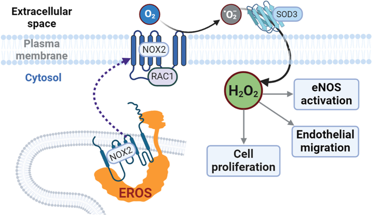
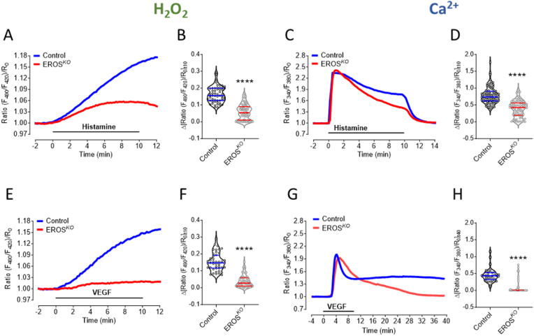
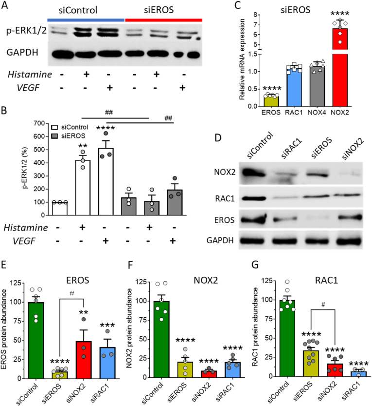
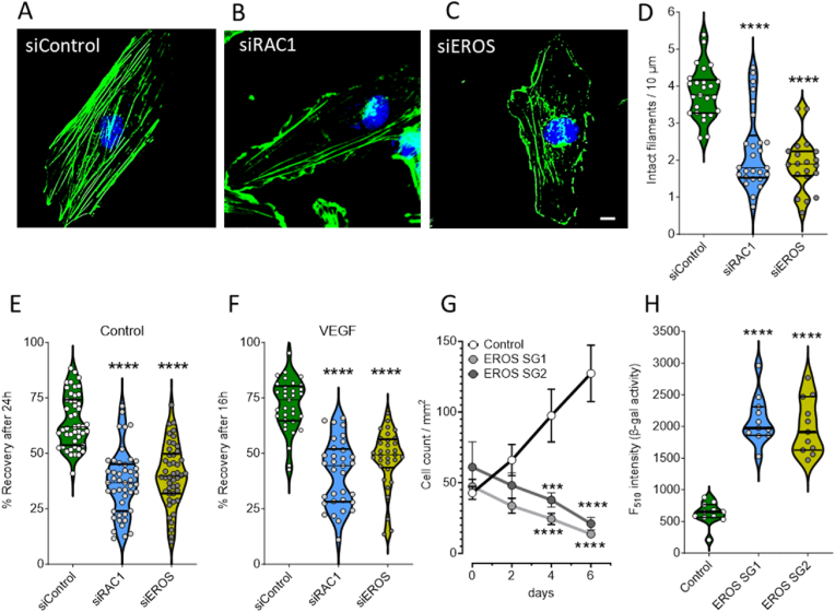
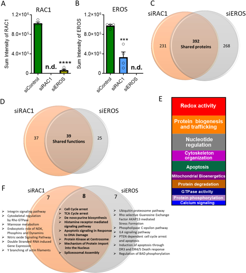
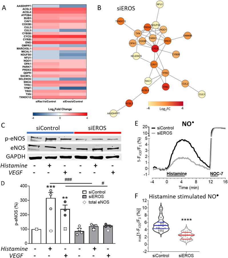
The chaperone protein EROS ("Essential for Reactive Oxygen Species") was recently discovered in phagocytes. EROS was shown to regulate the abundance of the ROS-producing enzyme NADPH oxidase isoform 2 (NOX2) and to control ROS-mediated cell killing. Reactive oxygen species are important not only in immune surveillance, but also modulate physiological signaling responses in multiple tissues. The roles of EROS have not been previously explored in the context of oxidant-modulated cell signaling. Here we show that EROS plays a key role in ROS-dependent signal transduction in vascular endothelial cells. We used siRNA-mediated knockdown and developed CRISPR/Cas9 knockout of EROS in human umbilical vein endothelial cells (HUVEC), both of which cause a significant decrease in the abundance of NOX2 protein, associated with a marked decrease in RAC1, a small G protein that activates NOX2. Loss of EROS also attenuates receptor-mediated hydrogen peroxide (H2O2) and Ca2+ signaling, disrupts cytoskeleton organization, decreases cell migration, and promotes cellular senescence. EROS knockdown blocks agonist-modulated eNOS phosphorylation and nitric oxide (NO) generation. These effects of EROS knockdown are strikingly similar to the alterations in endothelial cell responses that we previously observed following RAC1 knockdown. Proteomic analyses following EROS or RAC1 knockdown in endothelial cells showed that reduced abundance of these two distinct proteins led to largely overlapping effects on endothelial biological processes, including oxidoreductase, protein phosphorylation, and endothelial nitric oxide synthase (eNOS) pathways. These studies demonstrate that EROS plays a central role in oxidant-modulated endothelial cell signaling by modulating NOX2 and RAC1.

Keywords: EROS; Endothelial cells; NOX2; RAC1; Redox signaling; Reductive stress.


Conflict of interest statement

Declaration of competing interest The authors declare no competing financial interests.

Figures

Image 1
Graphical abstract
Fig. 1
Fig. 1
EROS knockout modulates agonist-induced H2O2and Ca2+signaling A. Real time imaging of H2O2 level in response to histamine was measured in CRISPR/Cas9 transduced EROS knockout (EROS SG, red curve) vs. lentiviral CAS9 transduced HUVEC (Control, blue curve). B. Detected H2O2 levels are significantly lower in EROS SG cells (n = 74) compared to Control (n = 55) 10 min after histamine stimulation. C. Ca2+ measurements under same experimental condition as shown in A. D. Histamine treated EROS SG cells (n = 78) show lower Ca2+ levels than Control (n = 93) 10 min after histamine stimulation. E. Average curves of HyPer7 signals in response to VEGF in Control (blue curve) and EROS SG cells (red curve). F. Values of EROS SG (n = 53) and Control (n = 44) 10 min after VEGF treatment. G. Average curves of Ca2+ imaging experiments in Control (blue curve) and EROS SG (red curve) cells stimulated for 10 min with VEGF. H. Statistical analysis of Control (n = 38) and EROS SG (n = 52) indicates decreased Ca2+ levels in EROS SG cells 30 min after VEGF treatment. Violin plots are marked with median and first and third quartiles for the graphed data, ****P < 0.0001 using unpaired t-test. (For interpretation of the references to color in this figure legend, the reader is referred to the Web version of this article.)
Fig. 2
Fig. 2
Effect of EROS knockdown on ERK1/2 phosphorylation and abundance of EROS, NOX2 and RAC1 A. Representative immunoblot shows ERK1/2 phosphorylation in response to histamine and VEGF stimulation in siControl transfected HUVEC, which is strongly attenuated in EROS knockdown cells. B. Statistical analysis of ERK1/2 phosphorylation (n = 3 for all conditions). C. Quantitative RT-PCR of HUVEC mRNA downregulating EROS reveals low EROS (yellow bar, n = 6), unchanged RAC1 (blue bar, n = 6) as well as NOX4 (gray bar, n = 6) and high NOX2 transcripts (red bar, n = 6). D. Western blots of protein lysates from HUVEC transfected with either siControl, siRAC1, siEROS or siNOX2 probed with antibodies for NOX2, RAC1, EROS and GAPDH. E-G. EROS, NOX2 and RAC1 are significantly downregulated within all single-guided siRNA samples, siEROS (yellow bars), siNOX2 (red bars) and siRAC1 (blue bars) compared to siControl (green bars). All values are presented as means ± SEM, **P < 0.01, ***P < 0.001 and ****P < 0.0001 compared to untreated siControl HUVEC, #P < 0.05 of EROS siRNA vs. NOX2 siRNA and ##P < 0.01 compared to same treatments, either histamine or VEGF, following siRNA knockdown of RAC1 vs. Control siRNA using 1-way ANOVA. (For interpretation of the references to color in this figure legend, the reader is referred to the Web version of this article.)
Fig. 3
Fig. 3
Loss of EROS disrupts endothelial cell function A-C. Representative images of siRNA transfected HUVEC with either siControl, siRAC1 or siEROS show staining of F-actin binding fluorescent dye conjugated phalloidin (green) and nuclear counterstain with DAPI (blue). Scale bar is 10 μm. D. Quantitative analysis of intact actin filaments as performed shown in Figs. S5A and B indicates disorganized cytoskeleton in HUVEC downregulating EROS (yellow plot, n = 20) as well as RAC1 (blue plot, n = 26) versus siControl (green plot, n = 25). E-F. Statistical evaluation of scratch area recovery in Control (E) or under VEGF-treated conditions (F) indicates lower migratory activities in HUVEC following knockdown of RAC1 (blue plots) as well as EROS (yellow plots) versus siControl (green plots). G. CAS9 (Control) and EROS SG1 or SG2 transduced HUVEC were seeded to ∼20 % confluence in endothelial cell growth medium and monitored at day 0, 2, 4 and 6. Graph shows progression of cell proliferation in Control cells (black line, n = 8), while both EROS SG populations (dark and light gray lines, n = 8 each) over time even decreased in cell number counted per mm2. H. Statistical analysis of fluorescence-based β-galactosidase activity indicates similar degree of senescence progression in EROS SG1 (blue plot, n = 11) as well as EROS SG2 (yellow plot, n = 11) compared to CAS9 infected cells (Control, green plot, n = 11). For all panels when indicated, ***P < 0.001 and ****P < 0.0001 (1way ANOVA for equal variance). (For interpretation of the references to color in this figure legend, the reader is referred to the Web version of this article.)
Fig. 4
Fig. 4
EROS and RAC1 knockdown show comparable endothelial proteomic regulation A. Detected sum intensities of RAC1 peptides by MS in HUVEC transfected with either siControl, siEROS or siRAC1. B. Detected sum intensities of EROS peptides by mass spectrometry in HUVEC transfected with either Control, EROS or RAC1 siRNA transfection. C. Venn diagram shows numbers of altered protein expression in siRAC1 and siEROS samples. Expression levels equal or lower than 0.5-fold were considered as downregulated proteins and levels equal or higher than 2-fold as upregulated protein changes compared with siControl. Changes in the proteome of individual knockdown largely overlaps (expressed as shared proteins). All altered proteins following siRNA-medicated knockdown of RAC1 or EROS were regulated in the same direction (detailed regulation of individual protein expression levels is provided in Data S1). D. Gene ontology annotated biological processes following siRNA-mediated knockdown of EROS or RAC1 indicates similar overlap in functional changes (for detailed lists of shared and unique functions see Data S2). E. Vertical slice chart presents Gene ontology analysis of shared processes within EROS or RAC1 siRNA-mediated downregulation distributed according to their different functional categories (Data S3). F. Gene ontology pathway analysis using R studio highlights most affected shared and unique pathways (Data S4 lists unique and shared top hits of pathways, protein interactions, phosphatases and kinome enrichments). ****P < 0.0001 (1way ANOVA for equal variance).
Fig. 5
Fig. 5
EROS knockdown induces reductive stress associated protein regulation resulting in diminished nitric oxide formation A. Heat map of critical siEROS and siRAC1 regulated shared proteins identified in functional analysis. B. Network analysis shows most significant protein interactions. The network has been refined with a highest confidence cut off of 0.900. Nodes are colored based on calculated fold change. C. Representative immunoblots show abundances of eNOS phosphorylation (p-eNOS, Ser1177), total eNOS (eNOS) and GAPDH following transfection with Control or EROS siRNA in HUVEC after stimulation with histamine or VEGF vs. untreated. D. Analysis of p-eNOS blots reveals p-eNOS Ser1177 phosphorylation in siControl HUVEC stimulated with histamine or VEGF for 10 min, which is blocked in EROS downregulated HUVEC. Total eNOS (gray open rhombic characters) is unchanged among samples. All values are presented as mean ± SEM, **P < 0.01 and ***P < 0.001 compared to untreated Control siRNA and #P < 0.05 or ###P < 0.001 compared to same treatments, either histamine or VEGF, of siEROS vs. siControl cells using 1way ANOVA. E. Average curves of NO imaging experiments in Control (black curve) and EROS (gray curve) siRNA-transfected cells stimulated with histamine and NOC-7 as indicated. F. Statistical analysis of Control (n = 27) and EROS siRNA-transfected (n = 27) cells indicates decreased NO levels following siRNA-mediated EROS knockdown, ****P < 0.0001 using unpaired t-test.

References

    1. Sies H. Hydrogen peroxide as a central redox signaling molecule in physiological oxidative stress: oxidative eustress. Redox Biol. 2017;11:613–619. doi: 10.1016/j.redox.2016.12.035. - DOI - PMC - PubMed
    1. Thomas D.C., Clare S., Sowerby J.M., Pardo M., Juss J.K., Goulding D.A., van der Weyden L., Storisteanu D., Prakash A., Espéli M., Flint S., Lee J.C., Hoenderdos K., Kane L., Harcourt K., Mukhopadhyay S., Umrania Y., Antrobus R., Nathan J.A., Adams D.J., Bateman A., Choudhary J.S., Lyons P.A., Condliffe A.M., Chilvers E.R., Dougan G., Smith K.G.C. Eros is a novel transmembrane protein that controls the phagocyte respiratory burst and is essential for innate immunity. J. Exp. Med. 2017;214:1111–1128. doi: 10.1084/jem.20161382. - DOI - PMC - PubMed
    1. Randzavola L.O., Mortimer P.M., Garside E., Dufficy E.R., Schejtman A., Roumelioti G., Yu L., Pardo M., Spirohn K., Tolley C., Brandt C., Harcourt K., Nichols E., Nahorski M., Woods G., Williamson J.C., Suresh S., Sowerby J.M., Matsumoto M., Santos C.X.C., Kiar C.S., Mukhopadhyay S., Rae W.M., Dougan G.J., Grainger J., Lehner P.J., Calderwood M.A., Choudhary J., Clare S., Speak A., Santilli G., Bateman A., Smith K.G.C., Magnani F., Thomas D.C. EROS is a selective chaperone regulating the phagocyte NADPH oxidase and purinergic signalling. Elife. 2022;11:1–31. doi: 10.7554/eLife.76387. - DOI - PMC - PubMed
    1. Thomas D.C., Charbonnier L.M., Schejtman A., Aldhekri H., Coomber E.L., Dufficy E.R., Beenken A.E., Lee J.C., Clare S., Speak A.O., Thrasher A.J., Santilli G., Al-Mousa H., Alkuraya F.S., Chatila T.A., Smith K.G.C. EROS/CYBC1 mutations: decreased NADPH oxidase function and chronic granulomatous disease. J. Allergy Clin. Immunol. 2019;143:782–785.e1. doi: 10.1016/j.jaci.2018.09.019. - DOI - PMC - PubMed
    1. Arnadottir G.A., Norddahl G.L., Gudmundsdottir S., Agustsdottir A.B., Sigurdsson S., Jensson B.O., Bjarnadottir K., Theodors F., Benonisdottir S., Ivarsdottir E.V., Oddsson A., Kristjansson R.P., Sulem G., Alexandersson K.F., Juliusdottir T., Gudmundsson K.R., Saemundsdottir J., Jonasdottir A., Jonasdottir A., Sigurdsson A., Manzanillo P., Gudjonsson S.A., Thorisson G.A., Magnusson O.T., Masson G., Orvar K.B., Holm H., Bjornsson S., Arngrimsson R., Gudbjartsson D.F., Thorsteinsdottir U., Jonsdottir I., Haraldsson A., Sulem P., Stefansson K. A homozygous loss-of-function mutation leading to CYBC1 deficiency causes chronic granulomatous disease. Nat. Commun. 2018;9:1–9. doi: 10.1038/s41467-018-06964-x. - DOI - PMC - PubMed

MeSH terms

LinkOut - more resources