. 2024 Jun 18;5(6):101589.
doi: 10.1016/j.xcrm.2024.101589. Epub 2024 May 27.

Distinctive multicellular immunosuppressive hubs confer different intervention strategies for left- and right-sided colon cancers

Affiliations

Distinctive multicellular immunosuppressive hubs confer different intervention strategies for left- and right-sided colon cancers

Bingxin Liu et al. Cell Rep Med. .

Abstract

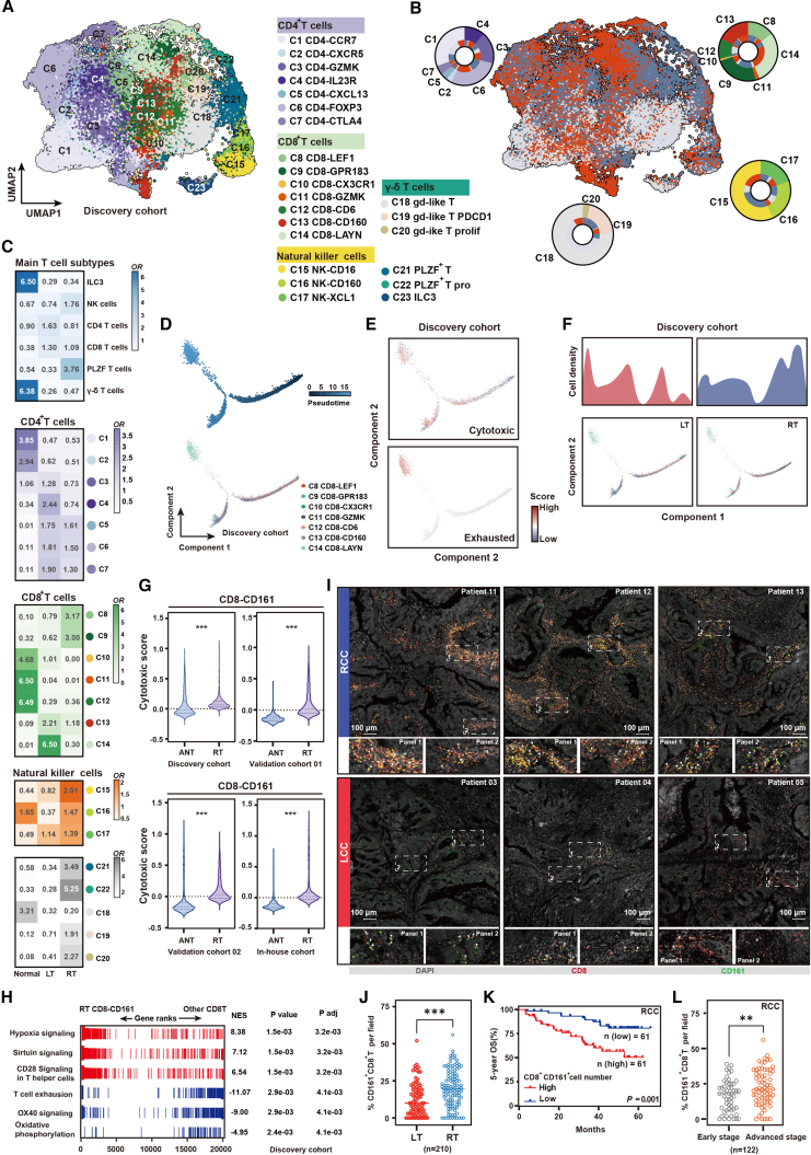
Primary colon cancers arising from the left and right sides exhibit distinct clinical and molecular characteristics. Sidedness-associated heterogeneity relies intricately on the oncogenic properties of cancer cells and multicellular interactions in tumor microenvironments. Here, combining transcriptomic profiling of 426,863 single cells from 105 colon cancer patients and validation with spatial transcriptomics and large-scale histological analysis, we capture common transcriptional heterogeneity patterns between left- and right-sided malignant epithelia through delineating two side-specific expression meta-programs. The proliferation stemness meta-program is notably enriched in left-sided malignant epithelia that colocalize with Mph-PLTP cells, activated regulatory T cells (Tregs), and exhausted CD8-LAYN cells, constituting the glucose metabolism reprogramming niche. The immune secretory (IS) meta-program exhibits specific enrichment in right-sided malignant epithelia, especially in smoking patients with right-sided colon cancer. The IShigh malignant epithelia spatially localize in hypoxic regions and facilitate immune evasion through attenuating Mph-SPP1 cell antigen presentation and recruiting innate-like cytotoxicity-reduced CD8-CD161 cells.

Keywords: expression meta-programs; immune microenvironment; left- and right-sided colon cancers; malignant epithelium; single-cell RNA sequencing; spatial transcriptome.


Conflict of interest statement

Declaration of interests The authors declare no competing interests.

Figures

None
Graphical abstract
Figure 1
Figure 1
Single-cell transcriptome atlas of LCCs and RCCs (A) Sample collection and analytic process of this study. (B) Uniform manifold approximation and projection (UMAP) plot of 93,146 CD45 cells and 96,807 CD45+ cells, colored by cell type or cancer type. (C) Proportions of the major cell types in normal mucosa and colon tumor tissues in individual samples, grouped by left- (LCC) and right-sided colon cancer (RCC). (D) Heatmaps showing the odds ratios (ORs) of major cell types occurring in each tissue. OR > 1.5 indicates distribution preference. (E) Multiplex immunohistochemistry (mIHC) staining of EPCAM (epithelia), CD68 (myeloid cells), CD3 (T cells), VIM (stromal cells), CD79 (B cells), and MZB1 (plasma cells) in left- and right-sided tumor tissues (validation cohort 5).
Figure 2
Figure 2
Identification of expression meta-programs within malignant epithelium (A) Heatmap showing the chromosomal map of large-scale CNVs for individual cells (rows) from representative patients (C125 for LCC and C109 for RCC) inferred by scRNA-seq using immune and stromal cells as reference (bottom) and epithelia as observed cells (top). The color represents log2 CNV. (B) UMAP plot of 84,975 epithelial cells, colored by malignant and non-malignant cells. Bar plots showing the proportion of malignant and non-malignant epithelial cells in LCC and RCC. (C) UMAP plots of malignant epithelium clustering colored by patients (left) or cancer types (right). The numbers on the UMAP plots represent different clusters. (D) Stacked histograms showing proportions of patients (top) or cancer type (bottom) for each malignant epithelium cluster, colored by patients or cancer types as in (C). (E) Heatmap depicting shared expression meta-programs across all patients. (F) PS and IS meta-programs in malignant epithelium with significantly different activities in left- versus right-sided colon cancers. For each program, the top genes are shown, and circles indicate the relative weight of each gene in the program. UMAP plots show program activity across all cell types (global UMAP); location of epithelium is indicated on the left in blue. Mann-Whitney-Wilcoxon test, ∗∗p < 0.01. (G) Representative immunohistochemistry (IHC) staining of the proteins produced by the signature genes for PS (top) and IS (bottom) meta-programs in validation cohort 5. (H) The H-score of the expression of PS and IS signature genes between LT and RT. Two-sided Wilcoxon test, ∗∗∗p < 0.001. (I) The distribution of the PS and IS staining score in early-stage and advanced-stage colon tumor tissues. Mat/Ent, mature enterocyte.
Figure 3
Figure 3
Deciphering side-specific expression meta-programs of the malignant epithelium (A) Spike plot showing PS AUCell score distribution in left-sided malignant colonic epithelium. The red dotted line represents the optimal cutoff (AUC > 0.15). (B) Top 5 enrichment pathways from GSEA for PShigh vs. PSlow left-sided malignant colonic epithelium. (C) IHC staining of Ki-67 (top) and LDHA (bottom) in validation cohort 5. (D) The distribution of Ki67 and LDHA staining score in left- and right-sided colon tumor tissues. (E) Spearman’s correlations between PS score of left malignant colonic epithelium and aerobic glycolysis score (top) and MCT4 (bottom) expression, respectively. (F) Representative mIHC staining of LT (validation cohort 5). ALDH1 (green), MCT4 (red), DAPI (blue), and merged channels are shown. (G) Spike plot showing IS AUCell score distribution in right-sided malignant colonic epithelium. The red dotted line represents the optimal cutoff (AUC > 0.15). (H) Top 5 enrichment pathways from GSEA for IShigh vs. ISlow right-sided malignant colonic epithelium. (I) IHC staining of CAIX in LT and RT (validation cohort 5). (J) The distribution of CAIX staining score in left- and right-sided colon tumor tissues (validation cohort 5). (K) Spearman’s correlations between the IS score and the hypoxia score in right-sided malignant colonic epithelium. (L) Spike plots showing standardized log rank statistic for different PS and IS cutoffs in LCC patients with bulk RNA-seq data (TCGA-COAD). The red arrow represents the optimal cutoff. Stacked barplot showing the chi-squared test of CMS for each PS subtype. Kaplan-Meier plots show the 5-year OS of PS and IS subgroups in LCC (left); 5-year OS for PS/CMS2 subgroups (right). (M) Spike plots showing standardized log rank statistic for different PS and IS cutoffs in RCC patients with bulk RNA-seq data (TCGA-COAD). The red arrow represents the optimal cutoff. Stacked barplot showing the chi-squared test of CMS for each IS subtype. Kaplan-Meier plots show the 5-year OS of PS and IS subgroups in RCC; 5-year OS for IS/CMS1 subgroups (middle). p < 0.05 is considered as a statistically significant difference. ∗∗p < 0.01, ∗p < 0.05, >0.05 is non-significant. Two-sided log rank test was used to assess statistical significance in (L) and (M). Two-sided Wilcoxon test was used to assess statistical significance in (D) and (J).
Figure 4
Figure 4
Characteristics of myeloid cells (A) UMAP plot of myeloid cells, color coded by cell type. (B) Bubble heatmap showing marker genes across myeloid clusters. Dot size indicates fraction of expressing cells, color indicating normalized expression levels. (C) UMAP plot colored by sample type (normal, LT, or RT). Pie chart of myeloid clusters and their distribution in normal, LT, and RT sections. (D) Heatmap showing the ORs of myeloid clusters occurring in each tissue. (E) GSEA enrichment for gene sets in Mph-SPP1 cells comparing to other macrophage subpopulations in RT. (F) Representative mIHC staining of SPP1+ macrophages in LT and RT (validation cohort 5). (G) Scatterplot showing the density of SPP1+ macrophages in left- and right-sided colon tumors based on mIHC staining results. Each sample on the scatterplot represents individual patient data. (H) The Kaplan-Meier curves showing RCC patients OS with different SPP1+ macrophage infiltration. Significance was calculated using the log rank test. (I) The density of SPP1+ macrophages in tumor sections from early-stage and advanced-stage RCC patients. Two-sided Wilcoxon test was used to assess statistical significance in (G) and (I); ∗∗∗p < 0.001, ∗∗p < 0.01.
Figure 5
Figure 5
Characterization of T cell states (A) UMAP plot of T cells, color coded by cell type. (B) UMAP plot colored by sample type (normal, LT, or RT). Pie chart of T cell clusters and their distribution in normal, LT, and RT sections. (C) Heatmap showing the ORs of T cell subpopulations occurring in each tissue. (D) Pseudotime-ordered analysis of CD8+ T cells. T cell subtypes are labeled by colors. (E) 2D pseudotime plot showing the dynamics of cytotoxic (upper panel) or exhausted signals (lower panel) in CD8+ T cells from LT and RT samples. (F) 2D graph of the pseudotime-ordered CD8+ T cells from LT (left panel) and RT (right panel) samples. The cell density distribution, by cell subtypes, is shown at the top of the figure. (G) Violin plots showing the cytotoxic scores of CD8-CD161 cells from adjacent normal tissues and RT samples. The p values were calculated by Student’s t test; ∗∗∗p < 0.001. (H) GSEA enrichment for gene sets in CD8-CD161 cells compared to other T subpopulations in RT. (I) Representative mIHC staining of CD161+CD8+ T cells in LT and RT (validation cohort 5). (J) Scatterplot showing the density of CD161+CD8+ T cells in left- and right-sided colon tumors based on mIHC staining results. (K) The Kaplan-Meier OS curves of RCC patients stratified by CD161+ CD8+ T cell infiltration. Significance was calculated using the log rank test. (L) The density of CD161+ CD8+ T cells in tumor sections from early-stage and advanced-stage RCC patients. Two-sided Wilcoxon test was used to assess statistical significance in (J) and (L); ∗∗∗p < 0.001, ∗∗p < 0.01.
Figure 6
Figure 6
Distinct cell-cell interaction patterns in left-sided versus right-sided colon cancer microenvironments (A) Bar charts showing the intensity of communication between PShigh and PSlow malignant epithelium, respectively, with immune cells in LT. Circle plots showing the change in cell communication intensity between PShigh malignant epithelium and immune cell subpopulations in LT compared to PSlow malignant epithelium and immune cell subpopulations, where the red line represents an increase in signal intensity, while the blue line represents signal strength decreases. The thickness of the line is directly proportional to the strength of the interaction (discovery cohort). (B) Bar charts showing the intensity of communication between IShigh and ISlow malignant epithelium, respectively, with immune cells in RT. Circle plots showing the change in cell communication intensity between IShigh malignant epithelium and immune cell subpopulations in RT compared to ISlow malignant epithelium and immune cell subpopulations (discovery cohort). (C) Spatial feature plots of PS signature score, Mph-PLTP cells, CD8-LAYN cells, and Tregs in LCC tissue sections (top). Spatial feature plots of IS signature score, Mph-SPP1 cells, CD8-CD161 cells, and NK-XCL1 cells in RCC tissue sections (bottom). (D) Representative mIHC staining of ALDH1+ malignant epithelium and CD163+ macrophages in LT (validation cohort 5). (E) The upregulated signaling ligand-receptor pairs between PShigh malignant epithelium with Mph-PLTP cells in LT compared to PSlow malignant epithelium. Dot sizes represent p value (only p < 0.05 shown). (F) Top-ranked ligands inferred to regulate PShigh malignant epithelium by Mph-PLTP cells according to NicheNet. Dot plots show the expression percentage (dot size) and intensity (dot intensity) of top-ranked ligands in Mph-PLTP cells. Heatmap showing regulatory potential of top-ranked ligand and the downstream target genes in PShigh malignant epithelium. (G and H) Representative images of mIHC staining indicating CD74+ malignant epithelium, SPP1+ macrophages (G), and CD161+ CD8+ T cells (H) in the hypoxic area of RT samples (validation cohort 5). (I) The upregulated signaling ligand-receptor pairs between IShigh malignant epithelium with Mph-SPP1 cells and CD8-CD161 cells in RT compared to ISlow malignant epithelium. Dot sizes represent p value (only p < 0.05 shown). Arrows show the direction of regulatory relationships between cell populations. (J) Representative mIHC staining showing the crosstalk between PD-L1+ IShigh malignant epithelium and CD80+SPP1+ macrophages via the ligand-receptor of PD-L1-CD80 in the hypoxic area of RT.
Figure 7
Figure 7
Resolution of IS meta-program in malignant epithelium correlates with immune checkpoint inhibitor therapy (A) The treatment-related information on colon cancer patients. (B) UMAP plot of broad cell types from all samples (left). Cell cluster frequency shown as a fraction of total cells for each sample (right). (C) UMAP plots showing malignant epithelium origins by color, and the expression of IS score in malignant epithelium. Quantification of IS score in malignant epithelium representation among patient groups. Stacked barplot shows the chi-squared test of IShigh/ISlow malignant epithelium for −ICI and +ICI/pCR. (D) GSEA enrichment for gene sets in all macrophages comparing −ICI vs. +ICI/pCR. NES, normalized enrichment score. The p values were determined by fast preranked GSEA. (E) UMAP plot of myeloid cells and heatmap showing the ORs of cell subpopulations occurring in each group. (F) Representative images of mIHC staining indicating SPP1+ macrophages in pre-treatment and post-treatment tumors of +ICI/pCR groups. Each dot in the scatterplot presents the fraction of cells in each sample based on mIHC staining image. Two-sided Wilcoxon test; ∗p < 0.05. (G) GSEA enrichment for gene sets in all CD8+ T cells comparing −ICI vs. +ICI/pCR (top) and −ICI vs. +ICI/non-pCR (bottom). (H) UMAP plot of CD8+ T cells and heatmap showing the ORs of cell subpopulations occurring in each group. (I) Expression of HLA-DRA, CD74, and HLA-DQA1 in the scRNA-seq dataset. The pie charts showing the percentage of gene-expressing cells (counts > 0) and the violin and scatterplots showing the normalized counts in the CD8-CD161 cells from −ICI and +ICI/pCR group. (J) Pseudotime-ordered analysis of CD8+ T cell clusters from RT samples. CD8+ T cell subsets are labeled by color.

References

    1. Tie J., Cohen J.D., Lahouel K., Lo S.N., Wang Y., Kosmider S., Wong R., Shapiro J., Lee M., Harris S., et al. Circulating Tumor DNA Analysis Guiding Adjuvant Therapy in Stage II Colon Cancer. N. Engl. J. Med. 2022;386:2261–2272. doi: 10.1056/NEJMoa2200075. - DOI - PMC - PubMed
    1. Arnold D., Lueza B., Douillard J.Y., Peeters M., Lenz H.J., Venook A., Heinemann V., Van Cutsem E., Pignon J.P., Tabernero J., et al. Prognostic and predictive value of primary tumour side in patients with RAS wild-type metastatic colorectal cancer treated with chemotherapy and EGFR directed antibodies in six randomized trials. Ann. Oncol. 2017;28:1713–1729. doi: 10.1093/annonc/mdx175. - DOI - PMC - PubMed
    1. Normanno N., Rachiglio A.M., Lambiase M., Martinelli E., Fenizia F., Esposito C., Roma C., Troiani T., Rizzi D., Tatangelo F., et al. Heterogeneity of KRAS, NRAS, BRAF and PIK3CA mutations in metastatic colorectal cancer and potential effects on therapy in the CAPRI GOIM trial. Ann. Oncol. 2015;26:1710–1714. doi: 10.1093/annonc/mdv176. - DOI - PubMed
    1. Petrelli F., Tomasello G., Borgonovo K., Ghidini M., Turati L., Dallera P., Passalacqua R., Sgroi G., Barni S. Prognostic Survival Associated With Left-Sided vs Right-Sided Colon Cancer: A Systematic Review and Meta-analysis. JAMA Oncol. 2017;3:211–219. doi: 10.1001/jamaoncol.2016.4227. - DOI - PubMed
    1. Yahagi M., Okabayashi K., Hasegawa H., Tsuruta M., Kitagawa Y. The Worse Prognosis of Right-Sided Compared with Left-Sided Colon Cancers: a Systematic Review and Meta-analysis. J. Gastrointest. Surg. 2016;20:648–655. doi: 10.1007/s11605-015-3026-6. - DOI - PubMed