. 1985 Jan;144(1):127-33.
doi: 10.2214/ajr.144.1.127.

Cholangiography and interventional biliary radiology in adult liver transplantation

Cholangiography and interventional biliary radiology in adult liver transplantation

A B Zajko et al. AJR Am J Roentgenol. 1985 Jan.

Abstract

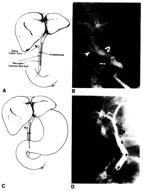
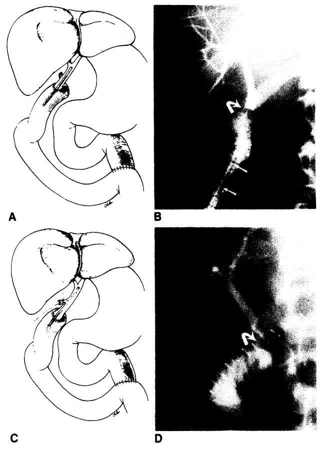
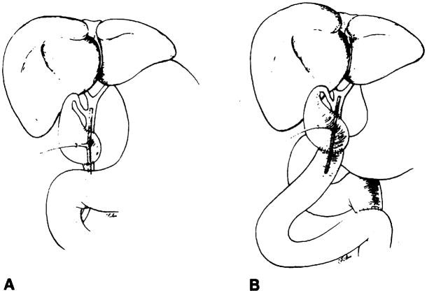
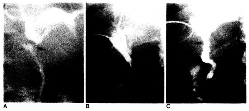
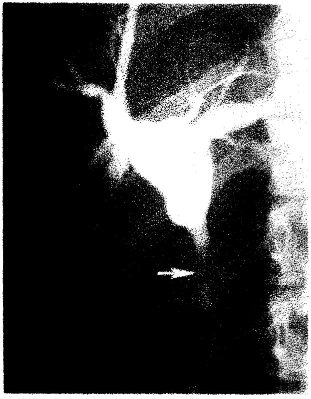
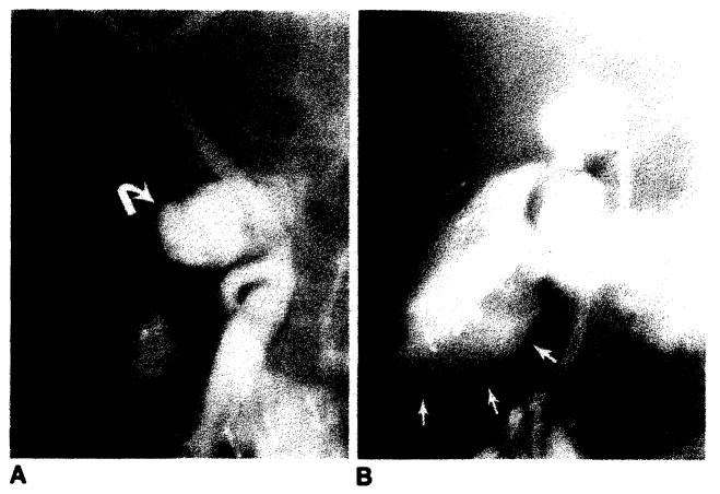
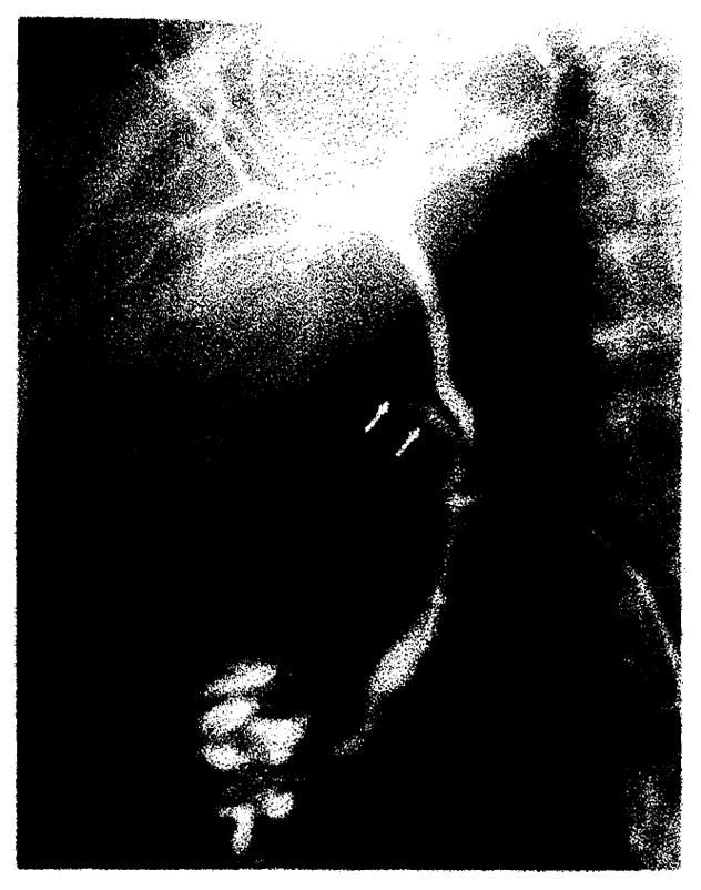
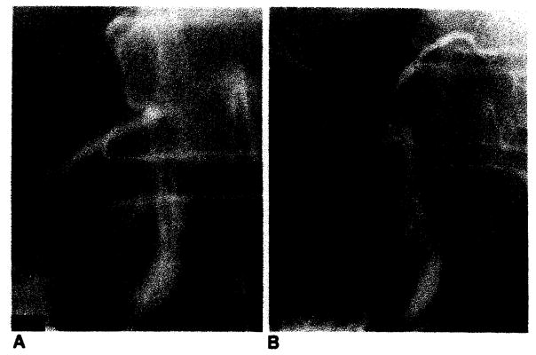
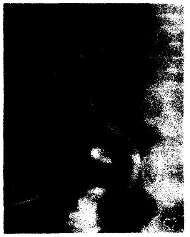
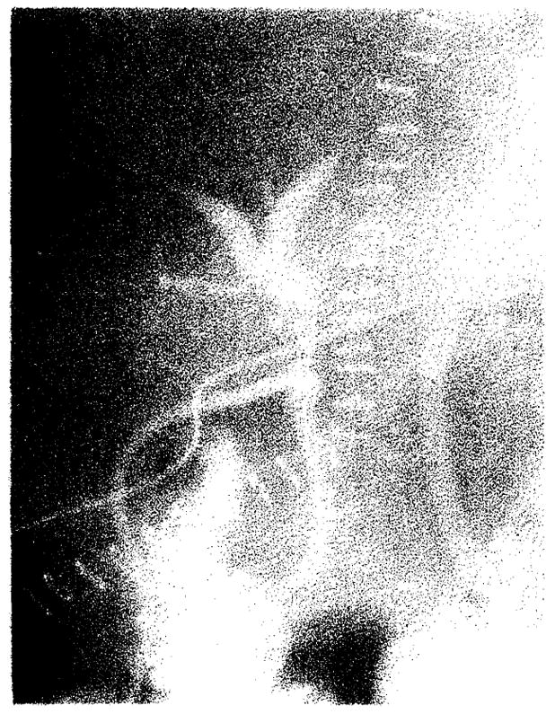
Radiographic assessment of the biliary tract is often essential in patients who have undergone liver transplantation. T- or straight-tube cholangiography, percutaneous transhepatic cholangiography, and endoscopic retrograde cholangiography all may be used. A total of 264 cholangiograms in 79 adult liver transplant patients (96 transplants) was reviewed. Normal radiographic features of biliary reconstructive procedures, including choledochocholedochostomy and choledochojejunostomy, are demonstrated. Complications diagnosed by cholangiography included obstruction, bile leaks, and tube problems, seen in eight, 24, and 12 transplants respectively. Stretching and incomplete filling of intrahepatic biliary ducts were frequently noted and may be associated with rejection and other conditions. Transhepatic biliary drainage, balloon catheter dilatation of strictures, replacement of dislodged T-tubes, and restoring patency of obstructed T-tubes using interventional radiologic techniques were important in avoiding complications and additional surgery in selected patients.


Figures

Fig. 1
Fig. 1
Normal choleclochochotedochostomy. A, Schematic diagram demonstrates T-tube stent, donor cystic duct remnant, and anastomosis. T-tube enters choledochotomy in recipient’s common bile duct. B, T-tube cholangiogram demonstrates donor’s (arrowhead) and recipient’s (arrow) cystic duct remnants and anastomosis (curved arrow). This anastomosis between donor’s common bile duct and recipient’s common hepatic duct is less frequently performed than common bile duct-to-common bite duct anastomosis shown in A. C, Schematic diagram demonstrates straight internal stent across biliary anastomosis. D, PTC demonstrates anastomosis (curved arrow) and position of stent (arrows).
Fig. 2
Fig. 2
Normal choledochojejunostomy in Roux-en-Y. A, Schematic diagram shows straight internal stent across biliary anastomosis. B, PTC shows biliary anastomosis (curved arrow). Note migration of stent (arrows) into Roux limb of jejunum. C, Schematic diagram shows T-tube stent which enters donors cystic duct remnant. D, T-tube cholangiogram shows biliary anastomosis (curved arrow).
Fig. 3
Fig. 3
Schematic diagrams demonstrate biliary reconstruction using donor’s gallbladder as pedicle graft. There are two anastomoses. In both A and B. proximal anastomosis is between Hartmann’s pouch and donor’s common bile duct. Distal anastomosis is between fundus of gallbladder and either recipient’s common bile duct (A) or recipient’s jejunum in Roux-en-Y (B).
Fig. 4
Fig. 4
Treatment of common hepatic duct stricture by transhepatic balloon dilatation. A. T-tube cholangiogram 3 weeks after transplantation demonstrates biliary obstruction due to stricture in donor’s common hepatic duct (arrow). Note donor’s and recipient’s cystic duct remnants (arrowheads). B, Transhepatic dilatation with 8-mm balloon. C. Catheter cholangiogram 6 weeks after dilatation demonstrates patent donor’s common hepatic duct (arrow). Catheter was removed. Patient was asymptomatic in 20-month follow-up.
Fig. 5
Fig. 5
Anastomotic stricture (arrow) at choledochocholedochostomy, which proved to be cholangiocarcinoma by transcatheter brush biopsy. PTC 16 months after liver transplantation. Patient was treated by iridium-192 wire through transhepatic drainage catheter. Another patient (not shown) with benign anastomotic stricture had similar cholangiography findings and was treated by transhepatic balloon dilatation.
Fig. 6
Fig. 6
Bile leak from choledochocholedochostomy resulting in both periductal (A, curved arrow) and subhepatic bile collections (B, arrows). B is later film of same patient shown in A.
Fig. 7
Fig. 7
Abnormal position of proximal limb of T-tube in donor’s cystic duct remnant (arrows) demonstrated on operative cholangiogram.
Fig. 8
Fig. 8
Biliary tube replacement after distodgment of T-tube. A, T-tube cholangiogram demonstrates T-tube outside choledochotomy. B, Cholangiogram after straight tube was replaced into common hepatic duct for external biliary drainage.
Fig. 9
Fig. 9
Clinical rejection. Poor filling, stretching, and mild attenuation of intrahepatic biliary tree.
Fig. 10
Fig. 10
Clinical rejection. Marked attenuation and essentially no filling of intrahepatic biliary tree.

Similar articles

Cited by

References

    1. Starzl TE, Marchioro TL, Von Kaulla KN, Hermann G, Brittain RS, Waddell WR. Homotransplantation of the liver in humans. Surg Gynecol Obstet. 1963;117:659–676. - PMC - PubMed
    1. Iwatsuki S, Klintmalm GBG, Starzl TE. Total hepatectomy and liver replacement (orthotopic liver transplantation) for primary hepatic malignancy. World J Surg. 1982;6:81–85. - PMC - PubMed
    1. Starzl TE. Liver transplantation. Johns Hopkins Med J. 1978;143:73–83. - PubMed
    1. Martineau G, Porter KA, Corman J, et al. Delayed biliary duct obstruction after orthotopic liver transplantation. Surgery. 1972;72:604–610. - PMC - PubMed
    1. Starzl TE, Koep U, Halgrimson CG, et al. Fifteen years of clinical liver transplantation. Gastroenterology. 1979;77:375–388. - PMC - PubMed