. 2024 May 28;14(1):12249.
doi: 10.1038/s41598-024-61115-1.

Genomic and morphological characterization of Knufia obscura isolated from the Mars 2020 spacecraft assembly facility

Affiliations

Genomic and morphological characterization of Knufia obscura isolated from the Mars 2020 spacecraft assembly facility

Atul Munish Chander et al. Sci Rep. .

Abstract

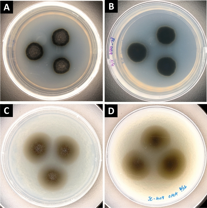
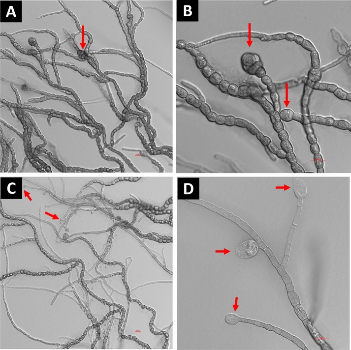
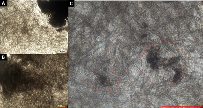
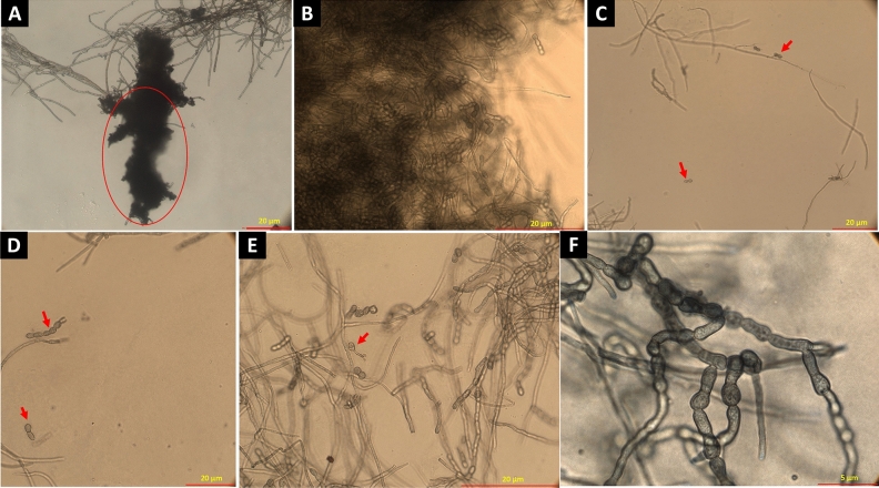
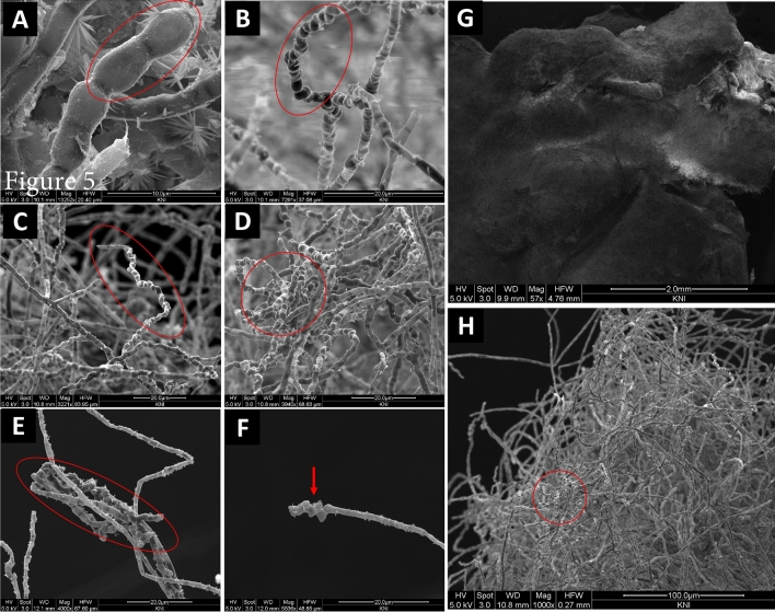
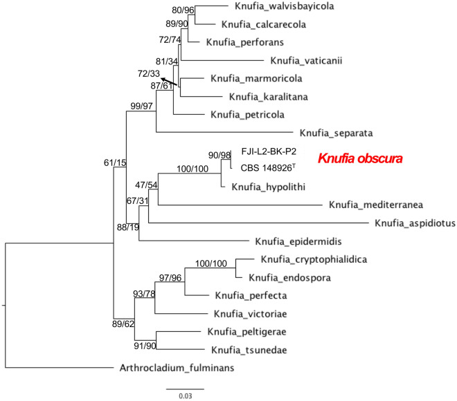
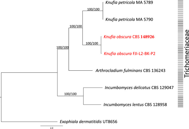
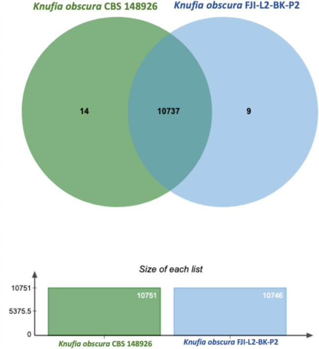
Members of the family Trichomeriaceae, belonging to the Chaetothyriales order and the Ascomycota phylum, are known for their capability to inhabit hostile environments characterized by extreme temperatures, oligotrophic conditions, drought, or presence of toxic compounds. The genus Knufia encompasses many polyextremophilic species. In this report, the genomic and morphological features of the strain FJI-L2-BK-P2 presented, which was isolated from the Mars 2020 mission spacecraft assembly facility located at the Jet Propulsion Laboratory in Pasadena, California. The identification is based on sequence alignment for marker genes, multi-locus sequence analysis, and whole genome sequence phylogeny. The morphological features were studied using a diverse range of microscopic techniques (bright field, phase contrast, differential interference contrast and scanning electron microscopy). The phylogenetic marker genes of the strain FJI-L2-BK-P2 exhibited highest similarities with type strain of Knufia obscura (CBS 148926T) that was isolated from the gas tank of a car in Italy. To validate the species identity, whole genomes of both strains (FJI-L2-BK-P2 and CBS 148926T) were sequenced, annotated, and strain FJI-L2-BK-P2 was confirmed as K. obscura. The morphological analysis and description of the genomic characteristics of K. obscura FJI-L2-BK-P2 may contribute to refining the taxonomy of Knufia species. Key morphological features are reported in this K. obscura strain, resembling microsclerotia and chlamydospore-like propagules. These features known to be characteristic features in black fungi which could potentially facilitate their adaptation to harsh environments.

Keywords: Chaetothyriales; Extremophile; Trichomeriaceae; Black fungi; Genomics.


Conflict of interest statement

The authors declare no competing interests.

Figures

Figure 1
Figure 1
Colony morphology of Knufia obscura FJI-L2-BK-P2. Colonies after incubation at 25 °C on PDA medium (14 days after inoculation; A,B) and on OMA medium (21 days after inoculation; C,D). (A) and (C) are front side of the plates whereas (B) and (D) are back side of the plates.
Figure 2
Figure 2
Microscopic features shown by DIC microscopy for Knufia obscura FJI-L2-BK-P2. (A,B). Mature hyphae with multicellular globose apex containing large pear shaped chlamydospores. (C,D) Conidia and conidiophores (arrows). Image (A) and (C) are at ×400 total magnification and image (B) and (D) are taken at ×1000 total magnification.
Figure 3
Figure 3
Bright field (A,B) and phase contrast microscopy (C) provides evidences for presence of microsclerotia in Knufia obscura FJI-L2-BK-P2: (A,B) Extensive hyphal branching and hyphae intermingle to form dense microsclerotia type network. (C) Closely attached multi-celled chlamydospores intermingled form microsclerotia indicated by using phase contrast microscopy. Images (A) and (B) are at ×200 and image (C) is at ×1000 total magnification.
Figure 4
Figure 4
Bright field microscopy revealed chlamydospore featured mature microsclerotia. (A) Microsclerotium forms a dense clump of biomass (encircled). (B) Microsclerotia containing intermingled arthroconidia and chlamydospores. (C,D) Arthroconidia released (arrow). (E) Germinating conidia (arrow). (F,G) Arthroconidia. Images (A) and (C) are taken at ×200, (B), (D) and (E) are taken at ×400 and image (F) is taken at ×1000 total magnification.
Figure 5
Figure 5
SEM images. (AC) Arthroconidia (encircled). (D,E) Microsclerotia. (F) Chlamydospore/chlamydospore like propagules. (G) Microsclerotia network creates dense surface layer over the fungal colony. (H) Microsclerotia.
Figure 6
Figure 6
The MLSA of Knufia obscura. The genes ITS, LSU, tef1, rpb1 and ß-tubulin were used to investigate phylogenetic placement of the K. obscura via ML tree on the IQTREE2 software.
Figure 7
Figure 7
The WGS based phylogenomic analysis for K. obscura. Phylogenomic tree was constructed for Knufia strains whose genomes were available. Maximum-likelihood tree was constructed using the RAxML and ASTRAL software packages. Exophiala dermatitidis was set as the outgroup and the branches are proportional to the number of mutations. Branch fidelity used posterior probabilities, which were added next to the corresponding branches.
Figure 8
Figure 8
Shared and Unique Genomic Clusters between Knufia obscura strains. Venn diagram depicting the shared and unique genomic clusters between Knufia obscura FJI-L2-BK-P2 and CBS 148926T strains. The figure presents a Venn diagram that illustrates the comparative genomic analysis of Knufia obscura strains, highlighting the shared and unique clusters of each.

Similar articles

Cited by

References

    1. Muggia L, Ametrano CG, Sterflinger K, Tesei D. An overview of genomics, phylogenomics and proteomics approaches in Ascomycota. Life. 2020 doi: 10.3390/life10120356. - DOI - PMC - PubMed
    1. Chander AM, et al. Genome insight and comparative pathogenomic analysis of Nesterenkonia jeotgali strain CD08_7 isolated from duodenal mucosa of celiac disease patient. Front. Microbiol. 2017;8:129. doi: 10.3389/fmicb.2017.00129. - DOI - PMC - PubMed
    1. Min K, Neiman AM, Konopka JB. Fungal pathogens: Shape-shifting invaders. Trends Microbiol. 2020;28:922–933. doi: 10.1016/j.tim.2020.05.001. - DOI - PMC - PubMed
    1. Rong M, et al. Phenotypic plasticity of Staphylococcus aureus in liquid medium containing vancomycin. Front. Microbiol. 2019;10:809. doi: 10.3389/fmicb.2019.00809. - DOI - PMC - PubMed
    1. Corona Ramirez A, et al. Assessment of fungal spores and spore-like diversity in environmental samples by targeted lysis. BMC Microbiol. 2023;23:68. doi: 10.1186/s12866-023-02809-w. - DOI - PMC - PubMed