Clinical Trial
. 2024 May 28;15(1):4210.
doi: 10.1038/s41467-024-48416-9.

L-RNA aptamer-based CXCL12 inhibition combined with radiotherapy in newly-diagnosed glioblastoma: dose escalation of the phase I/II GLORIA trial

Affiliations
Clinical Trial

L-RNA aptamer-based CXCL12 inhibition combined with radiotherapy in newly-diagnosed glioblastoma: dose escalation of the phase I/II GLORIA trial

Frank A Giordano et al. Nat Commun. .

Abstract

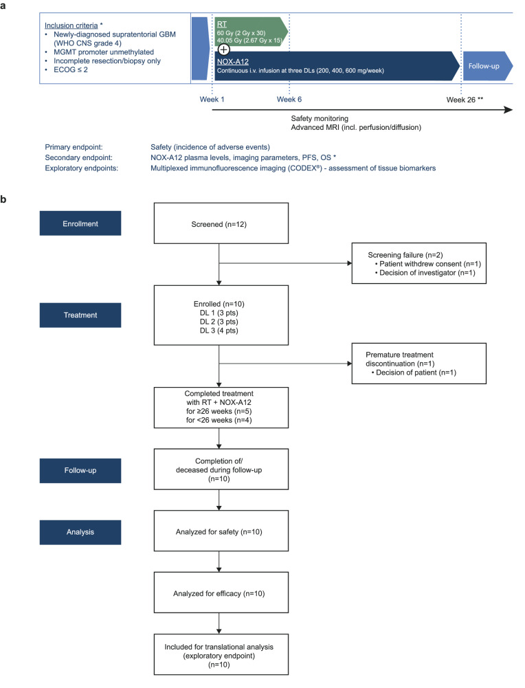
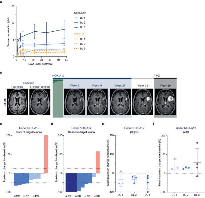
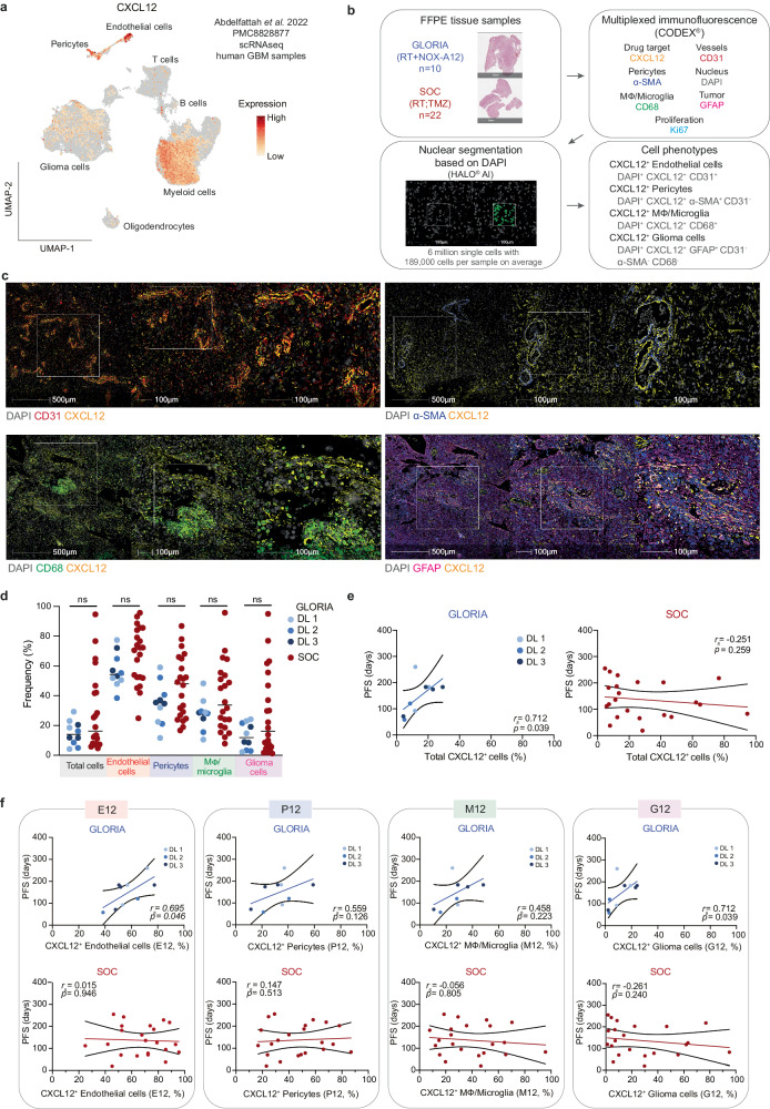
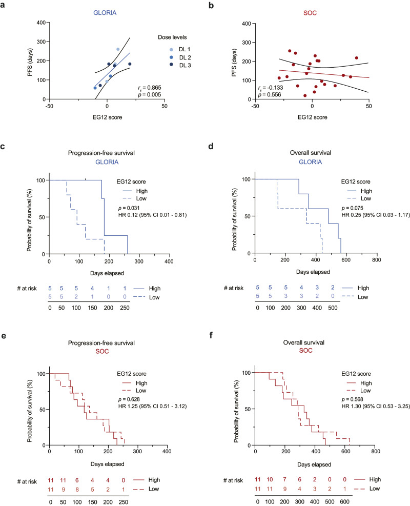
The chemokine CXCL12 promotes glioblastoma (GBM) recurrence after radiotherapy (RT) by facilitating vasculogenesis. Here we report outcomes of the dose-escalation part of GLORIA (NCT04121455), a phase I/II trial combining RT and the CXCL12-neutralizing aptamer olaptesed pegol (NOX-A12; 200/400/600 mg per week) in patients with incompletely resected, newly-diagnosed GBM lacking MGMT methylation. The primary endpoint was safety, secondary endpoints included maximum tolerable dose (MTD), recommended phase II dose (RP2D), NOX-A12 plasma levels, topography of recurrence, tumor vascularization, neurologic assessment in neuro-oncology (NANO), quality of life (QOL), median progression-free survival (PFS), 6-months PFS and overall survival (OS). Treatment was safe with no dose-limiting toxicities or treatment-related deaths. The MTD has not been reached and, thus, 600 mg per week of NOX-A12 was established as RP2D for the ongoing expansion part of the trial. With increasing NOX-A12 dose levels, a corresponding increase of NOX-A12 plasma levels was observed. Of ten patients enrolled, nine showed radiographic responses, four reached partial remission. All but one patient (90%) showed at best response reduced perfusion values in terms of relative cerebral blood volume (rCBV). The median PFS was 174 (range 58-260) days, 6-month PFS was 40.0% and the median OS 389 (144-562) days. In a post-hoc exploratory analysis of tumor tissue, higher frequency of CXCL12+ endothelial and glioma cells was significantly associated with longer PFS under NOX-A12. Our data imply safety of NOX-A12 and its efficacy signal warrants further investigation.


Conflict of interest statement

F.A.G. reports travel expenses, stocks and honoraria from TME Pharma AG related to this work; research grants and travel expenses from ELEKTA AB; grants, research grants, travel expenses and honoraria from Carl Zeiss Meditec AG; travel expenses and research grants from Varian Medical Systems, Inc.; travel expenses and/or honoraria from Bristol-Myers Squibb, Cureteq AG, Roche Pharma AG, MSD Sharp and Dohme GmbH, Siemens Healthineers AG, Varian Medical Systems, and AstraZeneca GmbH; non-financial support from Oncare GmbH and Opasca GmbH and patent US10857388B2 together with Carl Zeiss Meditec AG; all unrelated to this work. J.P.L. reports stocks and travel expenses from TME Pharma AG related to this work; travel expenses from Carl Zeiss Meditec AG, stocks and honoraria from Siemens Healthineers AG, and stocks from Bayer AG and BioNTech AG, all unrelated to this work. S.L. reports travel expenses from TME Pharma AG related to this work. C.S. has received speaker and/or advisory board honoraria from AbbVie, Bristol-Myers Squibb, HRA Pharma, Medac, Novocure, Roche, and Seagen not related to this work. E.S. reports travel expenses and honoraria for lectures from Carl Zeiss Meditec AG. C.O. reports travel support from Novocure; honoria by Horizon and Novocure and has received a Clinician Scientist Stipend of the University Medicine Essen Clinician Scientist Academy (UMEA) sponsored by the faculty of medicine and Deutsche Forschungsgemeinschaft (DFG). U.H. reports honoraria from Medac and Bayer AG, unrelated to this work. M.H. reports travel expenses, honoraria for webinars and research support (consumables) from TME Pharma AG related to this work. M.H. also reports honoraria from Bristol-Myers Squibb and Novartis unrelated to this work. A patent application related to biomarker identification has been filed by F.A.G., J.P.L., S.L., and M.H. (EP23000076.2; EP23000075.4). The remaining authors declare no competing interests.

Figures

Fig. 1
Fig. 1. Study outline of the GLORIA trial.
a Graphical overview of the study. GLORIA is a multicentric phase I/II trial conducted to assess the safety and efficacy of RT combined with escalating DLs of continuous i.v. treatment with NOX-A12 in newly diagnosed, incompletely resected, or biopsied GBM (CNS WHO grade 4) lacking MGMT promoter methylation (n = 10). *A complete and more detailed list of eligibility criteria and outcome measures is provided under ClinicalTrials.gov Identifier: NCT04121455. **End of treatment: 26 weeks as per protocol; treatment continuation beyond 26 weeks per investigator’s choice if the patient has clear clinical benefit. b Flow chart of the study. CODEX® CO-Detection by indEXing, DL dose level, ECOG Eastern Cooperative Oncology Group performance score, GBM glioblastoma, MGMT O6-methylguanine DNA methyltransferase, MRI magnetic resonance imaging, NOX-A12 olaptesed pegol, OS overall survival, PFS progression-free survival, RT radiotherapy.
Fig. 2
Fig. 2. Treatment with RT and NOX-A12 is safe and shows radiographic responses in conventional and advanced MRI.
a Serial plasma NOX-A12 (blue, full lines) and CXCL12 (orange, dashed lines) concentrations (µM) over treatment time (days) in respective GLORIA DLs (n = 10) indicated by color coding. Error bars indicate the standard error of the mean. b Representative illustration of the treatment course of a responding patient. Patient C1-003 was treated with RT (6 weeks; 2 Gy ad 60 Gy) and continuous NOX-A12 infusion for 26 weeks as per protocol, reaching partial remission in week 9. The patient relapsed at the end of NOX-A12 treatment (week 27) and deteriorated both before and after the initiation of TMZ. c, d Waterfall plots for best radiographic response as per mRANO under NOX-A12 (maximum change from baseline) of the sum of target lesion SPD (T1 Gd MRI) (c) and the best responding non-target lesion SPD (T1 Gd MRI) (d). Colors from blue to red indicate CR, PR, SD, and PD for each patient. As per mRANO, red dotted line indicates 25% increase (PD), blue dotted line indicates −50% decrease (PR). e, f Dot plots depicting mean maximum change from baseline under NOX-A12 for FTBhigh (e) and ADC (f) of patients in the respective DLs (color-labeling in blue; 200 (n = 3), 400 (n = 3), 600 mg/week (n = 4)). Error bars indicate mean and standard deviation. Source data are provided as a Source Data file. ADC apparent diffusion coefficient, CR complete response, DL dose level, FTBhigh high fractional tumor burden, GBM glioblastoma, mRANO modified Criteria for Radiographic Response, NOX-A12 olaptesed pegol, PD progressive disease, PR partial response, RT radiotherapy, SD stable disease, SPD sum of product of perpendicular diameters, TMZ temozolomide.
Fig. 3
Fig. 3. CXCL12 positivity in endothelial cells and glioma cells correlates with PFS in the GLORIA cohort.
a UMAP projection overlayed with CXCL12 mRNA expression in cell types from scRNAseq in human GBM samples (dataset from Abdelfattah et al.). b Experimental setup of mIF imaging and outline of analysis pipeline. FFPE tissue samples were used for 7-plex mIF imaging; GLORIA cohort (RT + NOX-A12) (n = 10) and SOC cohort (RT; TMZ) (n = 22). All tumor areas were confirmed independently by two neuropathologists. Cell types and CXCL12 positivity were identified as indicated. c Representative images of GBM tissue samples from GLORIA cohort patients (n = 10) showing CXCL12 (yellow) expression in the cell types of interest: CD31+ endothelial cells (red); α-SMA+ pericytes (blue); CD68+ M⏀/microglia (green) and GFAP+ glioma cells (magenta). d Frequency of CXCL12+ cells per cell type measured in the GLORIA cohort (in blue; different DLs as indicated; n = 10) and in the SOC cohort (in red; n = 22). Unpaired two-tailed Mann–Whitney U test; ns: not significant (p > 0.05). e Spearman’s rank correlation (rs) calculated between PFS (days) and total CXCL12+ cells (%) measured in the GLORIA cohort (left; in blue and with DLs as indicated; n = 10) and in the SOC cohort (right; in red; n = 22). rs- and p values (two-tailed) are depicted in the corresponding graphs. f Spearman’s rank correlation (rs) calculated between PFS (days) and CXCL12+ endothelial cells (%) out of total endothelial cells (E12), CXCL12+ pericytes (%) out of total pericytes (P12), CXCL12+ M⏀/Microglia (%) out of total M⏀/microglia (M12) and CXCL12+ glioma cells (%) out of total glioma cells (G12) measured in the GLORIA cohort (upper panels; in blue with DLs as indicated; n = 10) and in the SOC cohort (lower panels; in red; n = 22). rs- and p values (two-tailed) are depicted in the corresponding graphs. Source data are provided as a Source Data file. DL dose level, FFPE formalin-fixed paraffin-embedded, GBM glioblastoma, M⏀ macrophages, mIF multiplexed immunofluorescence, NOX-A12 olaptesed pegol, PFS progression-free survival, RT radiotherapy, scRNAseq single-cell RNA sequencing, SOC standard-of-care, TMZ temozolomide, UMAP uniform manifold approximation, and projection.
Fig. 4
Fig. 4. EG12 correlates with PFS and is associated with improved survival in the GLORIA, but not in a SOC cohort.
a, b Correlation analysis of progression-free survival (days) with EG12 score in corresponding tumor tissue of the GLORIA cohort (n = 9; colors depict DL as indicated) (a) and the SOC cohort (n = 22) (b). Spearman’s rank correlation (rs); rs- and p values (two-tailed) are depicted in the corresponding graphs. c, d Kaplan–Meier curves of progression-free (c) and overall survival (d) in days in the GLORIA cohort according to high (n = 5; continuous line) versus low (n = 5; dashed line) EG12 score. e, f Kaplan–Meier curves of progression-free (e) and overall survival (f) in days according to high (n = 11; continuous line) versus low (n = 11; dashed line) EG12 score in the SOC cohort. Log-rank test (two-tailed); p values are depicted in the corresponding graphs. Source data are provided as a Source Data file. CI confidence interval, DL dose level, HR hazard ratio, PFS progression-free survival, SOC standard-of-care.

References

    1. Ostrom QT, et al. CBTRUS statistical report: primary brain and other central nervous system tumors diagnosed in the United States in 2012–2016. Neuro-Oncol. 2019;21:v1–v100. doi: 10.1093/neuonc/noz150. - DOI - PMC - PubMed
    1. Stupp R, et al. Effects of radiotherapy with concomitant and adjuvant temozolomide versus radiotherapy alone on survival in glioblastoma in a randomised phase III study: 5-year analysis of the EORTC-NCIC trial. Lancet Oncol. 2009;10:459–466. doi: 10.1016/S1470-2045(09)70025-7. - DOI - PubMed
    1. Wen PY, et al. Glioblastoma in adults: a Society for Neuro-Oncology (SNO) and European Society of Neuro-Oncology (EANO) consensus review on current management and future directions. Neuro-Oncol. 2020;22:1073–1113. doi: 10.1093/neuonc/noaa106. - DOI - PMC - PubMed
    1. Hegi ME, et al. MGMT gene silencing and benefit from temozolomide in glioblastoma. N. Engl. J. Med. 2005;352:997–1003. doi: 10.1056/NEJMoa043331. - DOI - PubMed
    1. Kreth F-W, et al. Gross total but not incomplete resection of glioblastoma prolongs survival in the era of radiochemotherapy. Ann. Oncol. 2013;24:3117–3123. doi: 10.1093/annonc/mdt388. - DOI - PubMed

Publication types