. 2024 May 28;15(1):4542.
doi: 10.1038/s41467-024-48989-5.

The low-density lipoprotein receptor and apolipoprotein E associated with CCHFV particles mediate CCHFV entry into cells

Affiliations

The low-density lipoprotein receptor and apolipoprotein E associated with CCHFV particles mediate CCHFV entry into cells

Maureen Ritter et al. Nat Commun. .

Abstract

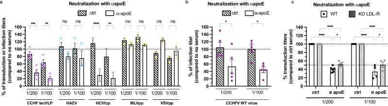
The Crimean-Congo hemorrhagic fever virus (CCHFV) is an emerging pathogen of the Orthonairovirus genus that can cause severe and often lethal hemorrhagic diseases in humans. CCHFV has a broad tropism and can infect a variety of species and tissues. Here, by using gene silencing, blocking antibodies or soluble receptor fragments, we identify the low-density lipoprotein receptor (LDL-R) as a CCHFV entry factor. The LDL-R facilitates binding of CCHFV particles but does not allow entry of Hazara virus (HAZV), another member of the genus. In addition, we show that apolipoprotein E (apoE), an exchangeable protein that mediates LDL/LDL-R interaction, is incorporated on CCHFV particles, though not on HAZV particles, and enhances their specific infectivity by promoting an LDL-R dependent entry. Finally, we show that molecules that decrease LDL-R from the surface of target cells could inhibit CCHFV infection. Our study highlights that CCHFV takes advantage of a lipoprotein receptor and recruits its natural ligand to promote entry into cells.


Conflict of interest statement

The authors declare no competing interests. A European patent application has been filed by Inserm-Transfert.

Figures

Fig. 1
Fig. 1. LDL-R is a cofactor of CCHFV infectivity.
a WT CCHFV, manipulated in BSL4, was produced in Huh-7.5 cells, whereas CCHF tecVLPs were produced in Huh-7.5 or HEK-293T cells. Infection or transduction assays were performed with serial dilutions. The level of infection was determined by RNA measurement in infected cells lysates. For tecVLP-GFP, target cells were pre-transfected with CCHFV NP and L expression vectors to amplify the GFP signal by minigenome replication and the level of transduction was assessed by flow cytometry. For tecVLP-NanoLuc, the level of transduction was assessed by measurement of nanoLuc levels. Created with Biorender.com. b Western blot analysis of cell lysates from Huh-7.5 cells transduced with lentiviral vectors allowing expression of control shRNA or shRNA targeting Lrp1 or LDL-R or SR-B1 (top). Representative image of 3 experiments. Quantification of the abundance of corresponding receptors (bottom). c Cells described in (b) were transduced with CCHF tecVLP-NanoLuc. Independent experiments: N = 5 SR-BI; N = 6 Lrp1; N = 7 LDL-R. Kruskal-Wallis test with Dunn’s multiple comparisons (ctrl vs. Lrp1: p = 0.0278, ctrl vs. LDL-R: p = 0.0498, ctrl vs. SR-BI: p > 0.9999). d Huh-7.5 cells were incubated with different concentration of LDL-R antibody (open bars) or control isotype (IgG goat, dashed bars) for 1 h at 37 °C before transduction with CCHF tecVLP-GFP (pink), MLVpp (green), and VSVpp (yellow) or infection with HAZV (blue). Independent experiments: N = 3 MLVpp; N = 5 CCHFV tecVLPs and VSVpp; N = 4 HAZV (0.25μg/mL and 4μg/mL) and N = 2 HAZV (1μg/mL). Two-way ANOVA test with Sidak’s multiple comparisons (αLDL-R vs. IgG: CCHF tecVLPs, 0.25μg/mL p = 0.0091, 1μg/mL p < 0.0001, 4μg/mL p < 0.0001; HAZV, 0.25μg/mL p > 0.9999, 1μg/mL p = 0.8951, 4μg/mL p > 0.9999; MLVpp, 0.25μg/mL p > 0.9999, 1μg/mL p = 0.8343, 4μg/mL p > 0.9999; VSVpp, 0.25μg/mL p = 0.0028, 1μg/mL p < 0.0001, 4μg/mL p < 0.0001). e Same experiment using WT CCHFV. N = 4 (1μg/mL) or N = 5 (4μg/mL) independent experiments. Two-way ANOVA test with Sidak’s multiple comparisons (αLDL-R vs. IgG: 1μg/mL p = 0.0042, 4μg/mL p = 0.0004). f Huh-7.5 cells stably expressing FLuc cells were transduced with a lentiviral vector allowing expression of VLDL-R. Surface expression of VLDL-R assessed by flow cytometry (left). Cells were then transduced with CCHF tecVLP-NanoLuc. N = 6 independent experiments. One sample t-test (two-tailed) p = 0.0467. Data are represented as means ± SEM.
Fig. 2
Fig. 2. LDL-R entry functions are conserved for infection of different human cells but not for bovine cells.
a Expression of LDL-R at the surface of Huh-7.5, A549, TE-671, EBL, MDBK, and PHH cells assessed by flow cytometry. b Huh-7.5 cells or PHH were incubated with 4μg/mL of LDL-R antibody (open bars) or IgG goat (dashed bars) for 1 h at 37 °C before transduction with CCHF tecVLP-NanoLuc. N = 4 independent experiments. Two-way ANOVA test with Sidak’s multiple comparisons (αLDL-R vs. IgG: Huh-7.5 p < 0.0001; PHH p < 0.0001). c Same as (b) with Huh-7.5, TE-671, A549 cells with harvesting at 48 h post-transduction (p.t.). N = 4 independent experiments. Two-way ANOVA test with Sidak’s multiple comparisons (αLDL-R vs. IgG: Huh-7.5 p < 0.0001; TE671 p < 0.0001; A549 p < 0.0001). d Same as (c) with Huh-7.5, EBL, MDBK cells. N = 3 independent experiments. Two-way ANOVA test with Sidak’s multiple comparisons (αLDL-R vs. IgG: Huh-7.5 p < 0.0001; EBL p = 0.8740; MDBK p = 0.0715). Data are represented as means ± SEM.
Fig. 3
Fig. 3. LDL-R promotes CCHFV entry.
a Huh-7.5 cells were incubated with 4 µg/mL of LDL-R antibody or control isotype before, during or after transduction with CCHF tecVLPs GFP as indicated in grey. b Percentage of transduction titers of CCHF tecVLP-GFP relative to control isotype as in the experimental set up described in (a). N = 3 independent experiments. Two-way ANOVA test with Dunnett’s multiple comparisons (αLDL-R vs. IgG: no Ab p > 0.9999; H-1 p = 0.0505; H-1 and H0 p = 0.0026; H0 p = 0.014; H + 2 p > 0.9999; H + 4 p = 0.9980; H + 6 p > 0.999). c CCHF tecVLP-GFP (pink), MLVpp (green), VSVpp (yellow) or HAZV (blue, N = 2) were incubated for 1 h at room temperature with soluble LDL-R (sLDL-R, open bars) or with soluble CD81 (CD81-LEL, dashed bars) at different concentrations before transduction or infection of Huh-7.5 cells. Independent experiments: N = 3 CCHF tecVLP and MLVpp; N = 4 VSVpp; N = 2 HAZV. Two-way ANOVA test with Sidak’s multiple comparisons (sLDL-R vs. CD81-LEL: CCHF tecVLPs, 0.5μg/mL p = 0.8354, 5μg/mL p < 0.0001, 10μg/mL p < 0.0001; HAZV, 0.5μg/mL p = 0.9998, 5μg/mL p > 0.999, 10μg/mL p = 0.8206; MLVpp, 0.5μg/mL p > 0.9999, 5μg/mL p > 0.9999, 10μg/mL p = 0.6061; VSVpp, 0.5μg/mL p < 0.0001, 5μg/mL p < 0.0001, 10μg/mL p < 0.0001). d Same experiment using WT CCHFV. Media was removed 1 h post-infection (p.i.) and cells were lysed 24 h p.i. The level of infectivity was quantified by RT-qPCR. N = 6 independent experiments. Two-way ANOVA test with Sidak’s multiple comparisons (sLDL-R vs. CD81-LEL: 0.5μg/mL p = 0.0125, 10μg/mL p = 0.0053). e CCHF tecVLPs (left) or CCHFV or HAZV (right) were incubated with sLDL-R or CD81-LEL (both expressing a 6xHis tag) for 1 h at RT before capture using magnetic beads. The levels of viral RNA co-captured were determined by RT-qPCR. One sample t-test (two-tailed) for CCHF tecVLPs (N = 5 independent experiments, p = 0.0227), two-way ANOVA test with Sidak’s multiple comparisons for HAZV and CCHFV (N = 3 independent experiments, HAZV p > 0.9999; CCHFV p = 0.025). Data are represented as means ± SEM.
Fig. 4
Fig. 4. LDL-R dependency of CCHFV entry is influenced by the virus producer cell type.
a Huh-7.5 cells were incubated with 0.25μg/mL or 4μg/mL of LDL-R antibody (open bars) or IgG goat (dashed bars) for 1 h at 37 °C before transduction with VSV-ΔG/GFP particles pseudotyped with VSV-G (yellow), CCHFV GPs (pink) or EBOV GP (blue). N = 3 independent experiments. Two-way ANOVA test with Sidak’s multiple comparisons (αLDL-R vs. IgG: VSV-G, 0.25μg/mL p = 0.0001, 4μg/mL p < 0.0001; CCHFV GPs, 0.25μg/mL p = 0.5799, 4μg/mL p = 0.0393; EBOV, 0.25μg/mL p = 0.7924, 4μg/mL p > 0.9988). b (left) Same as (a) with CCHF tecVLP-GFP particles produced in Huh-7.5 (pink) or HEK-293T (fuchsia) cells. N = 4 independent experiments. Two-way ANOVA test with Sidak’s multiple comparisons (αLDL-R vs. IgG: Huh-7.5, 0.25μg/mL p = 0.0106, 4μg/mL p < 0.0001; HEK-293T, 0.25μg/mL p = 0.1528, 4μg/mL p = 0.0159; Huh-7.5 vs. HEK-293T, αLDL-R 4μg/mL p = 0.0245, IgG 4μg/mL p = 0.9999). (right) HEK-293T cells were incubated with 0.25 or 4μg/mL of αLDL-R antibody (open bars) or IgG goat (dashed bars) for 1 h at 37 °C before transduction with CCHF tecVLP-NanoLuc particles produced in Huh7.5 (pink) or HEK-293T (fuchsia) cells. N = 3 independent experiments. Two-way ANOVA test with Sidak’s multiple comparisons (αLDL-R vs. IgG: Huh-7.5, 0.25μg/mL p = 0.1085, 4μg/mL p = 0.0004, HEK-293T; 0.25μg/mL p = 0.3004, 4μg/mL p = 0.4791; Huh-7.5 vs. HEK-293T, αLDL-R 4μg/mL p = 0.0545, IgG 4μg/mL p = 0.9999). c Intracellular levels of apoE in HEK-293T vs. HEK-239T stably expressing apoE as assessed by flow cytometry (left). CCHF tecVLPs produced in these cells were used for the experiment described as in (b) with 4μg/mL of αLDL-R antibody (open bars) or IgG goat (dashed bars) (right). N = 5 independent experiments. Two-way ANOVA test with Sidak’s multiple comparisons (HEK-293T: αLDL-R vs. IgG: 0.4196, HEK-293T + apoE: αLDL-R vs. IgG: 0.0152). Data are represented as means ± SEM.
Fig. 5
Fig. 5. ApoE promotes entry of CCHFV particles.
a CCHF tecVLPs (pink), MLVpp (light green), VSVGpp (yellow), HAZV (blue) or HCVtcp (green) were incubated for 1 h at room temperature with anti-apoE serum or control serum at different dilution before transduction or infection of Huh-7.5 cells. Independent experiments: N = 4 CCHF tecVLP; N = 3 MLVpp, VSVGpp, and HAZV; N = 2 HCVtcp. Two-way ANOVA test with Sidak’s multiple comparisons (αapoE vs. ctrl serum: CCHF tecVLP, 1/200 p = 0.0002, 1/100 p = 0.0018; HAZV, 1/200 p = 0.2582, 1/100 p = 0.3151; HCVtcp, 1/200 p < 0.0001, 1/100 p = 0.0032; MLVpp, 1/200 p = 0.9886, 1/100 p = 0.5832; VSVpp, 1/200 p = 0.0533, 1/100 p = 0.7327). b Same experiment as (a) using WT CCHFV particles. N = 5 independent experiments. Two-way ANOVA test with Sidak’s multiple comparisons (ctrl vs. αapoE: 1/100 p = 0.0273, 1/200 p = 0.0619). c CCHF tecVLP-NanoLuc particles were incubated for 1 h at room temperature with an apoE serum at different dilution before transduction of Huh-7.5 cells stably expressing FLuc and transduced with lentiviral vectors allowing expression of control shRNA or shRNA targeting LDL-R as described in Fig. 1. N = 6 independent experiments. Two-way ANOVA test with Sidak’s multiple comparisons (WT vs. KD LDL-R: 1/200 p = 0.012, 1/100 p = 0.0383; ctrl vs. αapoE: WT 1/200 p < 0.0001, WT 1/100 p < 0.0001, KD LDL-R 1/200 p < 0.0001, KD LDL-R 1/100 p < 0.0001). Data are represented as means ± SEM.
Fig. 6
Fig. 6. ApoE is associated with CCHFV particles and contribute to assembly/secretion and specific infectivity.
a Level of CCHFV minigenome RNA or HAZV RNA co-immunoprecipitated with an apoE serum vs. control IgGs and quantified by RT-qPCR. Results from N = 2 (HAZV) or N = 5 (CCHF tecVLP) independent experiments are presented as fold enrichment with apoE antibodies relative to control IgGs. Two-way ANOVA test with Sidak’s multiple comparisons (HAZV p = 0.9895, CCHF tecVLPs p = 0.0036). b Representative electron microscopy images of tecVLPs with simple negative stain (top) or with immunogold labelling with anti-Gn or anti-apoE antibodies or control antibodies (bottom). Scale bar represents 100 nm. Representative images from 2 experiments. c CCHFV particles were immunoprecipitated with an apoE serum vs. control IgGs. N = 4 independent experiments. Mann-Whitney test (two-tailed, p = 0.0286). d CCHFV particles were immunoprecipitated with an apoE serum vs. control IgGs and analyzed by western blot for apoE and Gn or Gc detection. Asterisks indicated unspecific bands from antibodies light chains. Representative image of 4 independent experiments. e Intracellular levels of apoE as assessed by flow cytometry and Western blot of cells transduced with shRNA control (NT) or targeting apoE. Representative image of 3 independent experiments. f Cells described in (e) were used for the production of CCHF tecVLPs or HAZV particles as described in Methods. Percentage of CCHFV NP transfected cells (top) or HAZV-eGFP expressing cells (bottom). Unpaired t-test (two-tailed, p = 0.8219) for CCHFV and Mann-Whitney test (two-tailed, p = 0.1) for HAZV. g Transduction efficiency of CCHF tecVLPs (top) or infectivity of HAZV (bottom) particles produced in cells described in (e) as assessed by flow cytometry. Unpaired t-test (two-tailed, p = 0.0003) for CCHFV and Mann-Whitney test (two-tailed, p = 0.4) for HAZV. h Level of secreted viral RNA of tecVLPs (top) or HAZV (bottom) assessed by RT-qPCR. Unpaired t-test (two-tailed, p = 0.0296) for CCHFV and Mann-Whitney test (two-tailed, p = 0.7) for HAZV. For (f–h), N = 5 independent experiments for CCHFV tecVLPs and N = 3 for HAZV. Data are represented as means ± SEM.
Fig. 7
Fig. 7. Molecules impairing LDL-R surface level impaired CCHFV infection.
a Cell surface expression of LDL-R of cells treated with 0 vs. 10μg/mL of PCSK9 for 3 h at 37 °C. Control isotypes are depicted in dotted lines. Representative image of N = 3 independent experiments. b Cell surface expression of LDL-R or CD81 of cells treated with 0 vs. 10μg/mL of PCSK9 for 3 h at 37 °C as assessed by confocal microscopy. The images provided are representative of two independent experiments. Scale bars represent 10 µm. c Cell viability of cells treated with PCSK9 relative to non-treated cells. N = 2 independent experiments. d Level of transduction or infection of cells treated with 0 vs. 10μg/mL of PCSK9 for 3 h at 37 °C. The media was replaced 3 h p.i. or p.t. and the cells were harvested at 48 h p.i. or p.t. for determination of the levels of transduction or infection by flow cytometry. Independent experiments: N = 5 CCHF tecVLP; N = 3 HAZV, VSVpp and MLVpp. Two-way ANOVA test with Sidak’s multiple comparisons (H20 vs. PCSK9: CCHF tecVLPs p < 0.0001, HAZV p = 0.9736, VSVpp p = 0.0023, MLVpp p = 0.5648). e Cell surface expression of LDL-R of cells treated with 0 vs. 75μM BBM for 2 h at 37 °C. Control isotypes are depicted in dotted lines. Representative image of 3 independent experiment. f Cell surface expression of LDL-R or CD81 of cells treated with 0 vs. 75μM BBM for 2 h at 37 °C as assessed by confocal microscopy. The images provided are representative of two independent experiments. Scale bars represent 10 µm. g Cell viability of cells treated with 0 vs. 75μM BBM for 2 h at 37 °C. N = 4 independent experiments. h Level of transduction or infection of cells treated with 0 vs. 75μM BBM for 2 h at 37 °C. The media was replaced 3 h p.i. or p.t. and the cells were harvested at 48 h p.i. or p.t. for determination of the levels of transduction or infection by flow cytometry. Independent experiments: N = 6 CCHF tecVLP, N = 3 HAZV and MLVpp, N = 4 VSVpp. Two-way ANOVA test with Sidak’s multiple comparisons (DMSO vs. BBM: CCHF tecVLPs p < 0.0001, HAZV p = 0.0286, VSVpp p = 0.0006, MLVpp p = 0.8694). Data are represented as means ± SEM.
Fig. 8
Fig. 8. Summary of the role of LDL-R in CCHFV entry.
CCHFV particles could incorporate apoE at their surface, which might contribute to the binding to LDL-R in addition to Gc. Followed this binding, CCHFV particles are endocytosed and then fuse with the late endosome membranes, allowing the release of viral genome. At this time, we cannot exclude the role of an additional factor (in blue) to promote endocytosis and/or fusion of CCHFV particles. In addition, an LDL-R-independent route of entry remains possible via a still unknown receptor (orange). Created with Biorender.com.

Similar articles

Cited by

References

    1. Bente DA, et al. Crimean-Congo hemorrhagic fever: history, epidemiology, pathogenesis, clinical syndrome and genetic diversity. Antivir. Res. 2013;100:159–189. doi: 10.1016/j.antiviral.2013.07.006. - DOI - PubMed
    1. Messina JP, et al. A global compendium of human Crimean-Congo haemorrhagic fever virus occurrence. Sci. Data. 2015;2:150016. doi: 10.1038/sdata.2015.16. - DOI - PMC - PubMed
    1. Freitas N, Legros V, Cosset FL. Crimean-Congo hemorrhagic fever: a growing threat to Europe. C. R. Biol. 2022;345:17–36. doi: 10.5802/crbiol.78. - DOI - PubMed
    1. Gargili A, et al. The role of ticks in the maintenance and transmission of Crimean-Congo hemorrhagic fever virus: A review of published field and laboratory studies. Antivir. Res. 2017;144:93–119. doi: 10.1016/j.antiviral.2017.05.010. - DOI - PMC - PubMed
    1. Hawman, D. W. & Feldmann, H. Crimean–Congo haemorrhagic fever virus. Nat. Rev. Microbiol.21, 463–477 (2023). - PMC - PubMed

MeSH terms