. 2024 May 28;14(1):12186.
doi: 10.1038/s41598-024-61941-3.

Polyetheretherketone bioactivity induced by farringtonite

Affiliations

Polyetheretherketone bioactivity induced by farringtonite

Martina Martínková et al. Sci Rep. .

Abstract

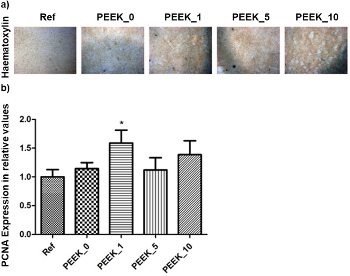
Polyetheretherketone (PEEK) is considered as an excellent biomaterial for bone grafting and connective tissue replacement. The clinical potential is, however, limited by its bioinertness, poor osteoconduction, and weak antibacterial activity. These disadvantages can be overcome by introducing suitable additives to produce mineral-polymer composites or coatings. In this work, a PEEK-based bioactive composite has been obtained by blending the polymer with magnesium phosphate (Mg3(PO4)2) particles in amounts ranging from 1 to 10 wt.% using the hot press technique. The obtained composite exhibited improved mechanical and physical properties, above the lower limits set for bone engineering applications. The tested grafts were found to not induce cytotoxicity. The presence of magnesium phosphate induced the mineralisation process with no adverse effects on the expression of the marker crucial for osteoblastic differentiation. The most promising results were observed in the grafts containing 1 wt.% of magnesium phosphate embedded within the PEEK matrix. The improved bioactivity of grafts, together with suitable physical-chemical and mechanical properties, indicate this composite as a promising orthopaedic implant material.


Conflict of interest statement

The authors declare no competing interests.

Figures

Figure 1
Figure 1
XRPD pattern of MP.
Figure 2
Figure 2
SEM observation of MP structure.
Figure 3
Figure 3
TGA-DTA curve for the employed PEEK.
Figure 4
Figure 4
SEM observation of the selected samples after osteoblast proliferation and differentiation tests, where (a) PEEK_0 and (b) PEEK_10.
Figure 5
Figure 5
FTIR microscope mapping illustrating the distribution of MP (green to red) in the PEEK matrix (blue) on the surface of the samples (left) and the cross-section (right). FTIR spectra of the two phases are shown at the bottom part of the figure.
Figure 6
Figure 6
The cytotoxicity of PEEK disks was evaluated by a decrease in cell viability of NIH/3T3 cells compared to the reference. The graphs show the averages of relative cell viability with standard deviation. Dashed lines mark the cytotoxicity threshold.
Figure 7
Figure 7
PEEK modification shows compatibility with calvaria pre-osteoblasts. The cells cultivated at the modification surface for 10 days displayed natural adhesion and proliferation as visualised by haematoxylin staining, magnification 20×. The proliferation marker PCNA expression levels were compared with reference cells cultivated without PEEK disc. The graph shows the expression (mRNA) of PCNA compared to reference. Data are expressed as mean ± SD (n—4 per group) and statistically significant difference is highlighted (Dunnett's multiple comparisons test *p < 0.05).
Figure 8
Figure 8
Staining of the mineralisation and alkaline phosphatase activity in MC3T3-E1 cells cultivated on the PEEK and control cells, magnification 20×. The increased Ca2+ depositions (black dots) were observed in the cells cultivated on the PEEK surface. Increased ALP activity was detected in the case of 0%, 1% and 5% modification. Positive cells are blue.
Figure 9
Figure 9
Comparison of relative expression levels of investigated molecules. The graph shows the relative expression levels of mRNAs corresponding to Runx2, Col1a1, and Spp1 in control, 0% (PEEK_0), 1% (PEEK_1), 5% (PEEK_5) and 10% (PEEK_10) PEEK. No significant differences in the expression of the selected markers were observed with respect to the reference.

References

    1. Ouyang L, et al. Graphene-oxide-decorated microporous polyetheretherketone with superior antibacterial capability and in vitro osteogenesis for orthopedic implant. Macromol. Biosci. 2018;18:1800036. doi: 10.1002/mabi.201800036. - DOI - PubMed
    1. Tekin S, Cangül S, Adıgüzel Ö, Değer Y. Areas for use of PEEK material in dentistry. Int. Dent. Res. 2018;8:84–92. doi: 10.5577/intdentres.2018.vol8.no2.6. - DOI
    1. Bathala L, Majeti V, Rachuri N, Singh N, Gedela S. The role of polyether ether ketone (peek) in dentistry—A review. JMedLife. 2019;12:5–9. doi: 10.25122/jml-2019-0003. - DOI - PMC - PubMed
    1. Najeeb S, Zafar MS, Khurshid Z, Siddiqui F. Applications of polyetheretherketone (PEEK) in oral implantology and prosthodontics. J. Prosthodontic Res. 2016;60:12–19. doi: 10.1016/j.jpor.2015.10.001. - DOI - PubMed
    1. Meng X, Du Z, Wang Y. Characteristics of wear particles and wear behavior of retrieved PEEK-on-HXLPE total knee implants: A preliminary study. RSC Adv. 2018;8:30330–30339. doi: 10.1039/C8RA04661A. - DOI - PMC - PubMed