. 2024 May 28;14(1):12226.
doi: 10.1038/s41598-024-62947-7.

Phenylacetic acid, an anti-vaginitis metabolite produced by the vaginal symbiotic bacterium Chryseobacterium gleum

Affiliations

Phenylacetic acid, an anti-vaginitis metabolite produced by the vaginal symbiotic bacterium Chryseobacterium gleum

Kang Mu Kwon et al. Sci Rep. .

Abstract

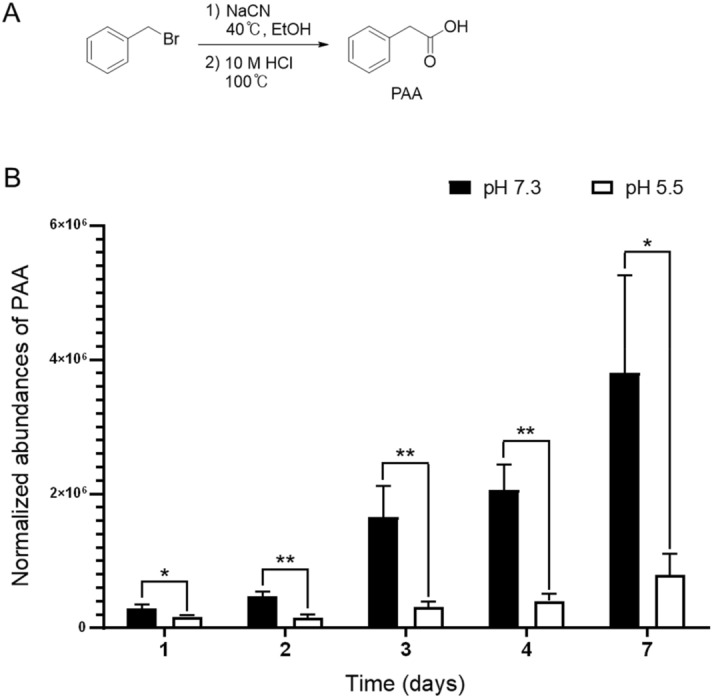
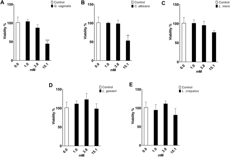
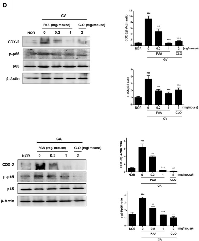
The human microbiome contains genetic information that regulates metabolic processes in response to host health and disease. While acidic vaginal pH is maintained in normal conditions, the pH level increases in infectious vaginitis. We propose that this change in the vaginal environment triggers the biosynthesis of anti-vaginitis metabolites. Gene expression levels of Chryseobacterium gleum, a vaginal symbiotic bacterium, were found to be affected by pH changes. The distinctive difference in the metabolic profiles between two C. gleum cultures incubated under acidic and neutral pH conditions was suggested to be an anti-vaginitis molecule, which was identified as phenylacetic acid (PAA) by spectroscopic data analysis. The antimicrobial activity of PAA was evaluated in vitro, showing greater toxicity toward Gardnerella vaginalis and Candida albicans, two major vaginal pathogens, relative to commensal Lactobacillus spp. The activation of myeloperoxidase, prostaglandin E2, and nuclear factor-κB, and the expression of cyclooxygenase-2 were reduced by an intravaginal administration of PAA in the vaginitis mouse model. In addition, PAA displayed the downregulation of mast cell activation. Therefore, PAA was suggested to be a messenger molecule that mediates interactions between the human microbiome and vaginal health.

Keywords: Chryseobacterium gleum; Human microbiome; Phenylacetic acid; Vaginitis.


Conflict of interest statement

The authors declare no competing interests.

Figures

Figure 1
Figure 1
Chemical and biological synthesis of phenylacetic acid (PAA). (A) Synthetic scheme and chemical structure of PAA. (B) Relative abundance of PAA production by C. gleum cultured at pH 7.3 and 5.5. The MS peak areas of PAA were normalized by the corresponding OD600 values, and data were collected from three independent cultures at each time point and pH level (*P < 0.05, **P < 0.01).
Figure 2
Figure 2
Relative viability of microorganisms upon treatment with PAA. The OD600 values of (A) Gardnerella vaginalis, (B) Candida albicans, (C) Lactobacillus iners, (D) Lactobacillus gasseri, and (E) Lactobacillus crispatus after treatment with PAA at concentrations of 1.0, 3.8, and 15.1 mM, converted to a percentage of the control. Data were collected from three independent cultures at each concentration (**P < 0.01, ***P < 0.001).
Figure 3
Figure 3
Effect of PAA on Gardnerella vaginalis (GV)- and/or Candida albicans (CA)-induced vaginitis and the expression of inflammatory markers in female mice. (A) Effect on GV- and/or CA-inflamed vagina and uterus. (B) Effect on myeloperoxidase (MPO) activity in vaginal tissues. (C) Production of PGE2. (D) Effect on COX-2 expression and NF-κB activation analyzed by Western blotting. β-Actin was used as an internal control. Female mouse vaginas were infected with GV and CA (both at 1 × 108 CFU/mouse), except in the normal control group (NOR, normal group treated with vehicle alone). PAA and clotrimazole (CLO) were intravaginally administered once a day for 14 days. On day 15 post-infection, the mice were euthanized. The expression of COX-2 and activation of MPO, PGE2, and NF-kB were measured in vaginal tissues. Western blots from three independent results were quantified by densitometry using ImageJ. All values are shown as the mean ± SD of five replicate mice (n = 5). ###P < 0.001 vs. normal control group. **P < 0.01 and ***P < 0.001 vs. GV- and CA-treated control group.
Figure 3
Figure 3
Effect of PAA on Gardnerella vaginalis (GV)- and/or Candida albicans (CA)-induced vaginitis and the expression of inflammatory markers in female mice. (A) Effect on GV- and/or CA-inflamed vagina and uterus. (B) Effect on myeloperoxidase (MPO) activity in vaginal tissues. (C) Production of PGE2. (D) Effect on COX-2 expression and NF-κB activation analyzed by Western blotting. β-Actin was used as an internal control. Female mouse vaginas were infected with GV and CA (both at 1 × 108 CFU/mouse), except in the normal control group (NOR, normal group treated with vehicle alone). PAA and clotrimazole (CLO) were intravaginally administered once a day for 14 days. On day 15 post-infection, the mice were euthanized. The expression of COX-2 and activation of MPO, PGE2, and NF-kB were measured in vaginal tissues. Western blots from three independent results were quantified by densitometry using ImageJ. All values are shown as the mean ± SD of five replicate mice (n = 5). ###P < 0.001 vs. normal control group. **P < 0.01 and ***P < 0.001 vs. GV- and CA-treated control group.
Figure 4
Figure 4
Effect of PAA on compound 48/80-stimulated mast cell activation. (A) HMC-1 cells were treated with PAA (0, 5, 10, and 50 μM) for 24 h. Cell viability was measured by the MTT assay. (B–E) HMC-1 cells were treated with different concentrations (0, 5, 10, and 50 μM) (B–D) or 50 μM (E) of PAA for 1 h, followed by stimulation with 30 μg/mL of compound 48/80 for 40 min. Degranulation (B) and secretion of LTC4 (C) and PGD2 (D) were measured. All values are shown as the mean ± SD from three independent experiments (*P < 0.05, **P < 0.01, and ***P < 0.001 vs. control; #P < 0.05, ##P < 0.01, and ###P < 0.001 vs. compound 48/80 alone). Cell lysates were subjected to Western blotting with the indicated antibodies (E) and are representative of three independent experiments. GAPDH was used as an internal control.

References

    1. Cho I, Blaser MJ. The human microbiome: At the interface of health and disease. Nat. Rev. Genet. 2012;13:260–270. doi: 10.1038/nrg3182. - DOI - PMC - PubMed
    1. Wang B, Yao M, Lv L, Ling Z, Li L. The human microbiota in health and disease. Engineering (Beijing) 2017;3:71–82. doi: 10.1016/J.ENG.2017.01.008. - DOI
    1. Proctor L, LoTempio J, Marquitz A, Daschner P, Xi D, Flores R, Brown L, Ranallo R, Maruvada P, Regan K, Lunsford RD, Reddy M, Caler L. A review of 10 years of human microbiome research activities at the US National Institutes of Health, Fiscal Years 2007–2016. Microbiome. 2019;7:31. doi: 10.1186/s40168-019-0620-y. - DOI - PMC - PubMed
    1. Donia MS, Cimermancic P, Schulze CJ, Brown LCW, Martin J, Mitreva M, Clardy J, Linington RG, Fischbach MA. A systematic analysis of biosynthetic gene clusters in the human microbiome reveals a common family of antibiotics. Cell. 2014;158:1402–1414. doi: 10.1016/j.cell.2014.08.032. - DOI - PMC - PubMed
    1. Ceccarani C, Foschi C, Parolin C, D’Antuono A, Gaspari V, Consolandi C, Laghi L, Camboni T, Vitali B, Severgnini M, Marangoni A. Diversity of vaginal microbiome and metabolome during genital infections. Sci. Rep. 2019;9:14095. doi: 10.1038/s41598-019-50410-x. - DOI - PMC - PubMed

LinkOut - more resources