

The significance of cerebrospinal fluid dynamics in adolescent idiopathic scoliosis using time-SLIP MRI
- PMID: 38806612
- PMCID: PMC11133356
- DOI: 10.1038/s41598-024-63135-3
The significance of cerebrospinal fluid dynamics in adolescent idiopathic scoliosis using time-SLIP MRI
Abstract
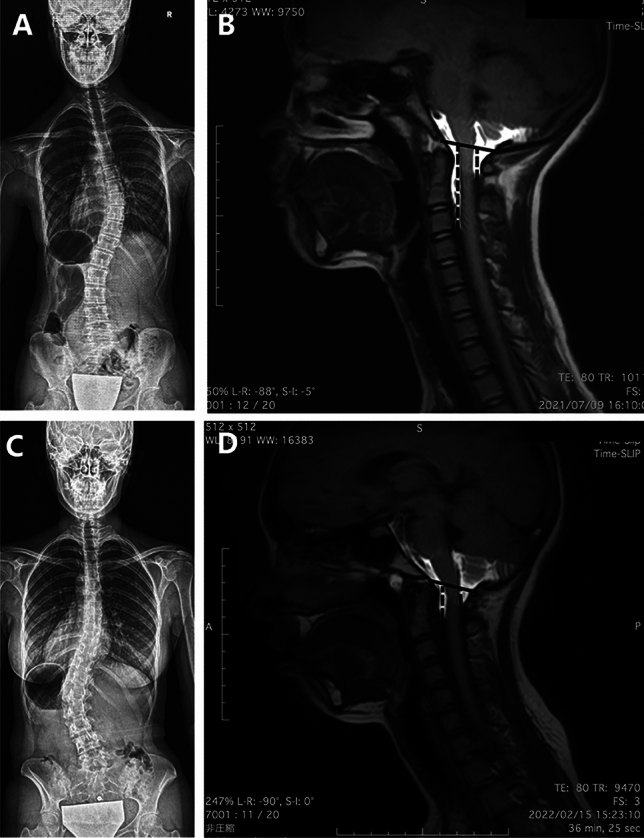
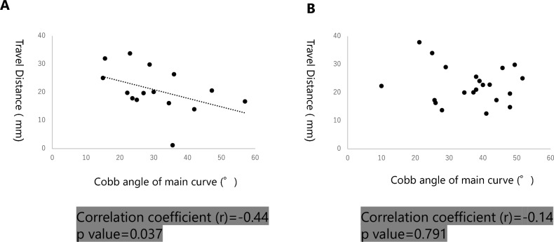
Adolescent idiopathic scoliosis (AIS) affects approximately 3% of the global population. Recent studies have drawn attention to abnormalities in the dynamics of the CSF as potential contributors. This research aims to employ the Time-Spatial Labeling Inversion Pulse (Time-SLIP) MRI to assess and analyze cerebrospinal fluid (CSF) dynamics in AIS patients. 101 AIS patients underwent Time-SLIP MRI. Images were taken at the mid-cervical and craniocervical junction regions. The sum of the maximum movement distances of CSF on the ventral and dorsal sides of the spinal canal within a single timeframe was defined and measured as Travel Distance (TD). Correlations between TD, age, Cobb angle, and Risser grade were analyzed. TD comparisons were made across Lenke classifications. TD for all patients was a weak correlation with the Cobb angle (r = - 0.16). Comparing TD between Lenke type 1 and 5, type 5 patients display significantly shorter TD (p < 0.05). In Risser5 patients with Lenke type 5 showed a significant negative correlation between Cobb angle and TD (r = - 0.44). Lenke type 5 patients had significantly shorter CSF TD compared to type1, correlating with worsening Cobb angles. Further analysis and exploration are required to understand the mechanism of onset and progression.
Keywords: Adolescent idiopathic scoliosis; Cerebrospinal fluid; Time-SLIP MRI.
© 2024. The Author(s).
Conflict of interest statement
The authors declare no competing interests.
Figures


Similar articles
-
Radiological Analysis of Cerebrospinal Fluid Dynamics at the Craniovertebral Junction Using Time-Spatial Labeling Inversion Pulse Magnetic Resonance Imaging in Patients with Cervical Spinal Canal Stenosis.World Neurosurg. 2024 Apr;184:e731-e736. doi: 10.1016/j.wneu.2024.02.020. Epub 2024 Feb 9. World Neurosurg. 2024. PMID: 38340799
-
The effectiveness of selective thoracic fusion for treating adolescent idiopathic scoliosis: a systematic review protocol.JBI Database System Rev Implement Rep. 2015 Nov;13(11):4-16. doi: 10.11124/jbisrir-2015-2338. JBI Database System Rev Implement Rep. 2015. PMID: 26657460
-
Cervical sagittal alignment in adolescent idiopathic scoliosis patients (Lenke type 1-6).J Orthop Sci. 2017 Mar;22(2):254-259. doi: 10.1016/j.jos.2016.12.006. Epub 2016 Dec 23. J Orthop Sci. 2017. PMID: 28025024
-
Immediate postoperative coronal imbalance in Lenke 5 and Lenke 6 adolescent idiopathic scoliosis: Is it predictable?Eur Spine J. 2019 Sep;28(9):2042-2052. doi: 10.1007/s00586-019-06019-9. Epub 2019 May 30. Eur Spine J. 2019. PMID: 31147844 Clinical Trial.
-
Pediatric cervical kyphosis in the MRI era (1984-2008) with long-term follow up: literature review.Childs Nerv Syst. 2022 Feb;38(2):361-377. doi: 10.1007/s00381-021-05409-z. Epub 2021 Nov 22. Childs Nerv Syst. 2022. PMID: 34806157 Review.
Cited by
-
Transmantle pressure under the influence of free breathing: non-invasive quantification of the aqueduct pressure gradient in healthy adults.Fluids Barriers CNS. 2025 Jan 3;22(1):1. doi: 10.1186/s12987-024-00612-x. Fluids Barriers CNS. 2025. PMID: 39754238 Free PMC article.
-
Interrater and intrarater reliability of aqueduct CSF flow parameters measured by phase-contrast MRI in healthy adults.Quant Imaging Med Surg. 2025 Jul 1;15(7):6399-6413. doi: 10.21037/qims-24-1589. Epub 2025 Jun 30. Quant Imaging Med Surg. 2025. PMID: 40727364 Free PMC article.
References
MeSH terms
LinkOut - more resources
Full Text Sources
Medical
