Case Reports
. 2024 Nov;32(11):1412-1418.
doi: 10.1038/s41431-024-01627-6. Epub 2024 May 28.

Utilization of automated cilia analysis to characterize novel INPP5E variants in patients with non-syndromic retinitis pigmentosa

Affiliations
Case Reports

Utilization of automated cilia analysis to characterize novel INPP5E variants in patients with non-syndromic retinitis pigmentosa

Kae R Whiting et al. Eur J Hum Genet. 2024 Nov.

Abstract

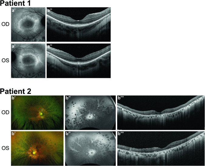
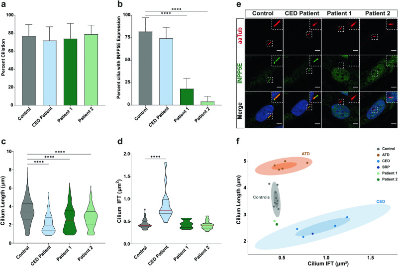
INPP5E encodes inositol polyphosphate-5-phosphatase E, an enzyme involved in regulating the phosphatidylinositol (PIP) makeup of the primary cilium membrane. Pathogenic variants in INPP5E hence cause a variety of ciliopathies: genetic disorders caused by dysfunctional cilia. While the majority of these disorders are syndromic, such as the neuronal ciliopathy Joubert syndrome, in some cases patients will present with an isolated phenotype-most commonly non-syndromic retinitis pigmentosa (RP). Here, we report two novel variants in INPP5E identified in two patients with non-syndromic RP: patient 1 with compound heterozygous variants (c.1516C > T, p.(Q506*), and c.847G > A, p.(A283T)) and patient 2 with a homozygous variant (c.1073C > T, p.(P358L)). To determine whether these variants were causative for the phenotype in the patients, automated ciliary phenotyping of patient-derived dermal fibroblasts was performed for percent ciliation, cilium length, retrograde IFT trafficking, and INPP5E localization. In both patients, a decrease in ciliary length and loss of INPP5E localization in the primary cilia were seen. With these molecular findings, we can confirm functionally that the novel variants in INPP5E are causative for the RP phenotypes seen in both patients. Additionally, this study demonstrates the usefulness of utilizing ciliary phenotyping as an assistant in ciliopathy diagnosis and phenotyping.


Conflict of interest statement

Competing interests The authors declare no competing interests. Ethics approval Institutional ethics approval was not required. All individuals or their legal guardians provided informed written consent for genetic testing and publication of clinical details and images.

Figures

Fig. 1
Fig. 1. Clinical phenotypes of patients.
Patient 1. a’ Fundus autofluorescence images showing a typical hyperautofluorescent ring in the macula and hypo-autofluorescent spots outside the macula. a” Optical coherence tomography (OCT) images of the macula, showing typical thinning of the outer retinal layers (ellipsoid zone and external limiting membrane) outside the central macula 1. Patient 2. b’ Wide-field fundus photograph showing extensive hyperpigmentations in the context of retinitis pigmentosa. b” Fundus autofluorescence showing a hyperautofluorescent ring in the macula, and extensive areas of hypo-autofluorescent atrophy of the retinal pigment epithelium. b”’ OCT for showing extensive outer retinal atrophy.
Fig. 2
Fig. 2. Morphology of the primary cilium is affected in patient-derived fibroblasts.
a Ciliogenesis of fibroblasts, as a percentage of cells with cilia. There was little variation between control and patient ciliation. Control cells had a ciliation of 77 ± 1.3%, the CED ciliopathy patient 72% ± 2.7%, and the INPP5E patients 74% ± 3.0% and 79% ± 1.8%, respectively. b INPP5E localization, as the percent of cilia with INPP5E expression. 82.0 ± 2.1% of control cilia and 74.4 ± 3.1% of CED patient cilia had INPP5E expressed. Both INPP5E patients had a significant loss of INPP5E in the cilia, with 18.3 ± 2.6% and 4.0 ± 1.3% of cilia expressing INPP5E, respectively. c Cilium length of control and patient fibroblasts, measured using ALPACA. Both patients showed shorter cilia with a mean of 2.65 ± 0.10 µm and 2.77 ± 0.08 µm when compared to control fibroblasts (3.44 ± 0.06 µm), however, this is more moderate than what is seen in the CED ciliopathy patient (2.16 ± 0.06 µm). d Retrograde trafficking was measured using accumulation of IFT88 along the cilium. The mean of control cells was 0.44 ± 0.1 µm2, with the INPP5E patients showing no change in accumulation size (0.44 ± 0.1 µm2 and 0.42 ± 0.1 µm2, respectively). The CED patient showed an increase in accumulation size (0.86 ± 0.3 µm2), as observed previously. e Representative images of control, CED patient, Patient 1 and Patient 2 fibroblasts stained for α-acetylated tubulin (aaTub, red), INPP5E (green), and DAPI (blue) (Scale bar = 5 μm). f Subgrouping of patient and control fibroblasts based on data from Doornbos et al. [30]. Confidence intervals of 0.5 and 0.9 are indicated for each identified cluster—control, CED, and ATD. Patients 1 and 2 from this study group were between the control cluster and CED cluster. (**** denotes p < 0.0001) (N = 3).
Fig. 3
Fig. 3. Current knowledge of pathogenic INPP5E variants.
a Location of variants along INPP5E. The specific colors represent the clinical outcome of each mutation (Red—Joubert Syndrome; Green—Retinitis Pigmentosa; Blue—Other/not described). In-depth descriptions of each known sequence variant can be found in supplemental table 1. Most variants cluster around the phosphatase catalytic domain, however localization of the variant does not determine phenotypic outcome. Comparison of diagnosis (b), patient sex (c), and type of variant (d) as reported in literature. e Age and sex comparison of patients. The majority of male patients show symptoms at an early age (0–10 years), while the majority of female patients show symptoms during late teenage years (15–20 years). f Prevalence of common ciliopathy phenotypes in patients. Retinal dystrophy, intellectual disability, the molar tooth sign, and other neurological features are most commonly described phenotypes in the cohort.

Similar articles

Cited by

References

    1. Daiger SP, Sullivan LS, Bowne SJ. RetNet. Retinal Information Network. Available from: https://sph.uth.edu/retnet/.
    1. Sangermano R, Deitch I, Peter VG, Ba-Abbad R, Place EM, Zampaglione E, et al. Broadening INPP5E phenotypic spectrum: detection of rare variants in syndromic and non-syndromic IRD. NPJ Genom Med. 2021;6:1–10. - PMC - PubMed
    1. Georgiou M, Fujinami K, Michaelides M. Inherited retinal diseases: therapeutics, clinical trials and end points—a review. Clin Exp Ophthalmol. 2021;49:270–88. - PubMed
    1. Veltel S, Gasper R, Eisenacher E, Wittinghofer A. The retinitis pigmentosa 2 gene product is a GTPase-activating protein for Arf-like 3. Nat Struct Mol Biol. 2008;15:373–80. - PubMed
    1. Hanany M, Rivolta C, Sharon D, Daiger SP. Worldwide carrier frequency and genetic prevalence of autosomal recessive inherited retinal diseases. Proc Natl Acad Sci USA. 2020;117:2710–16. - PMC - PubMed

Publication types

Substances

LinkOut - more resources