

Asbestos exposure determined 357 days after death through autopsy: a report of a multidisciplinary approach
- PMID: 38806807
- PMCID: PMC11953153
- DOI: 10.1007/s12024-024-00838-z
Asbestos exposure determined 357 days after death through autopsy: a report of a multidisciplinary approach
Abstract
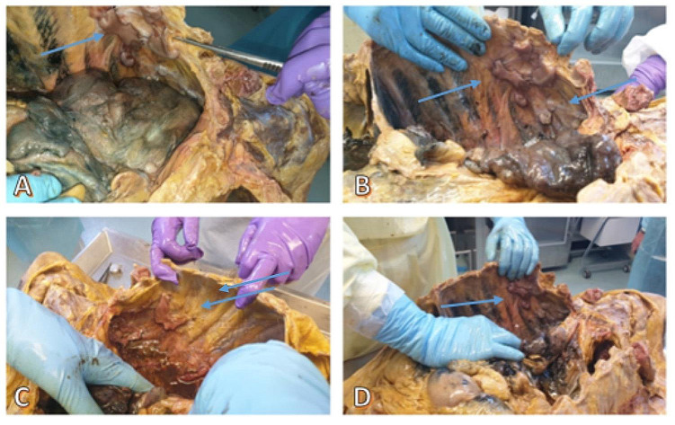
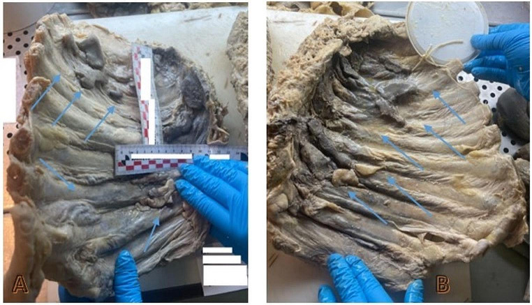
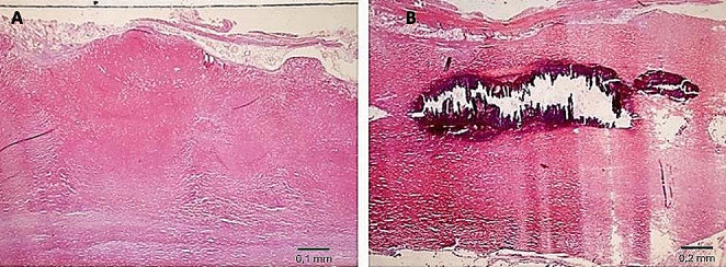
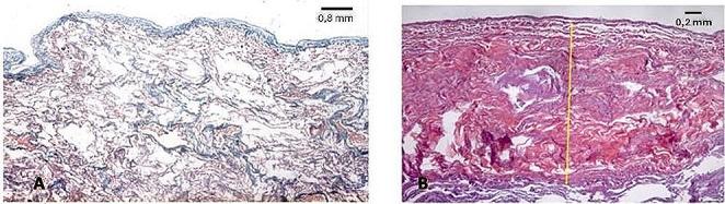
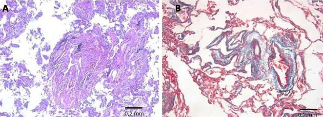
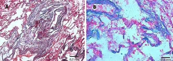
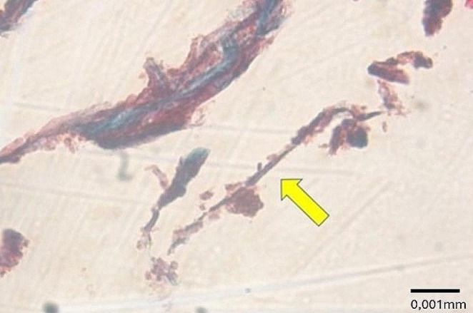
Asbestosis is an interstitial lung disease caused by the inhalation of asbestos fibers and poses a significant risk to individuals working in construction, shipping, mining, and related industries. In a forensic context, postmortem investigations are crucial for accurate diagnosis, for which the gold standard is the histopathological examination. This case report describes the autopsy and related investigations conducted on an 84-year-old man, nearly one year (357 days) after his death. After a post-mortem CT scan, an autoptic investigation was performed, followed by histopathological, immunohistochemical, and scanning electron microscopy examinations. The integration of the evidence from these examinations with previously available personal and clinical information conclusively confirmed the diagnosis of asbestosis. We demonstrated the efficacy and reliability of our diagnostic protocol in detecting asbestosis and asbestos fibers and excluding mesothelioma even in decomposed tissues. According to our findings autopsy remains the diagnostic gold standard in cases of suspected asbestosis within a forensic context, even 1 year after death, therefore it is always highly recommended, even in cases where the body has decomposed.
Keywords: Asbestos fibers; Asbestosis; Autopsy; Exhumation; Multidisciplinary approach; Post-mortem CT (PMCT); Scanning electron microscopy.
© 2024. The Author(s).
Conflict of interest statement
Declarations. Ethics approval: Our investigations were carried out following the rules of the Declaration of Helsinki of 1975, revised in 2013. According to Italian legislation, ethical approval for a single case is not required, as long as the data are kept anonymous and the investigations performed do not imply genetic results. Consent to participate: The current Italian legislation requires neither the family’s consent nor ethical approval for a single case, as long as the data are strictly kept anonymous. Because summoning the parents was not possible, as it would badly interfere with the grieving process, the parents’ consent was completely waived, according to the Italian Authority of Privacy and Data Protection (“Garante della Privacy”: GDPR nr 679/2016; 9/2016 and recent law addition number 424/19 July 2018; http://www.garanteprivacy.it ). Conflict of interest: The authors declare no conflict of interest as there’s no financial/personal interest or belief that could affect their objectivity.
Figures


Similar articles
-
Clinical, radiological, and pathological investigation of asbestosis.Int J Environ Res Public Health. 2011 Mar;8(3):899-912. doi: 10.3390/ijerph8030899. Epub 2011 Mar 22. Int J Environ Res Public Health. 2011. PMID: 21556185 Free PMC article.
-
[Pulmonary asbestosis: an autopsy case].Nihon Hoigaku Zasshi. 2007 Nov;61(2):129-33. Nihon Hoigaku Zasshi. 2007. PMID: 18064879 Japanese.
-
The risk of asbestos exposure in South African diamond mine workers.Ann Occup Hyg. 2011 Jul;55(6):569-77. doi: 10.1093/annhyg/mer028. Ann Occup Hyg. 2011. PMID: 21742625
-
Asbestos exposure diagnosis in pulmonary tissues.Pathologica. 2024 Aug;116(4):207-215. doi: 10.32074/1591-951X-930. Pathologica. 2024. PMID: 39377502 Free PMC article. Review.
-
The role of analytical techniques in the diagnosis of asbestos-associated disease.Crit Rev Clin Lab Sci. 1985;22(1):1-42. doi: 10.1080/10408368509176814. Crit Rev Clin Lab Sci. 1985. PMID: 3891228 Review.
References
-
- Bhandari J, Thada PK, Sedhai YR. Asbestosis. 2020. - PubMed
-
- Attanoos RL, Gibbs AR. Asbestos-related deaths. Curr Diagn Pathol. 2002;8(6):373–83. 10.1054/cdip.2002.0141.
-
- INAIL. Il registro nazionale dei mesoteliomi - Settimo rapporto. 2021.
-
- Nadesan K. The importance of the medico-legal autopsy. Malays J Pathol. 1997;19(2):105–9. - PubMed
Publication types
MeSH terms
Substances
LinkOut - more resources
Full Text Sources
Medical
