. 2024 May 28;22(1):294.
doi: 10.1186/s12951-024-02558-6.

MnO2 and roflumilast-loaded probiotic membrane vesicles mitigate experimental colitis by synergistically augmenting cAMP in macrophage

Affiliations

MnO2 and roflumilast-loaded probiotic membrane vesicles mitigate experimental colitis by synergistically augmenting cAMP in macrophage

Chengjun Song et al. J Nanobiotechnology. .

Abstract

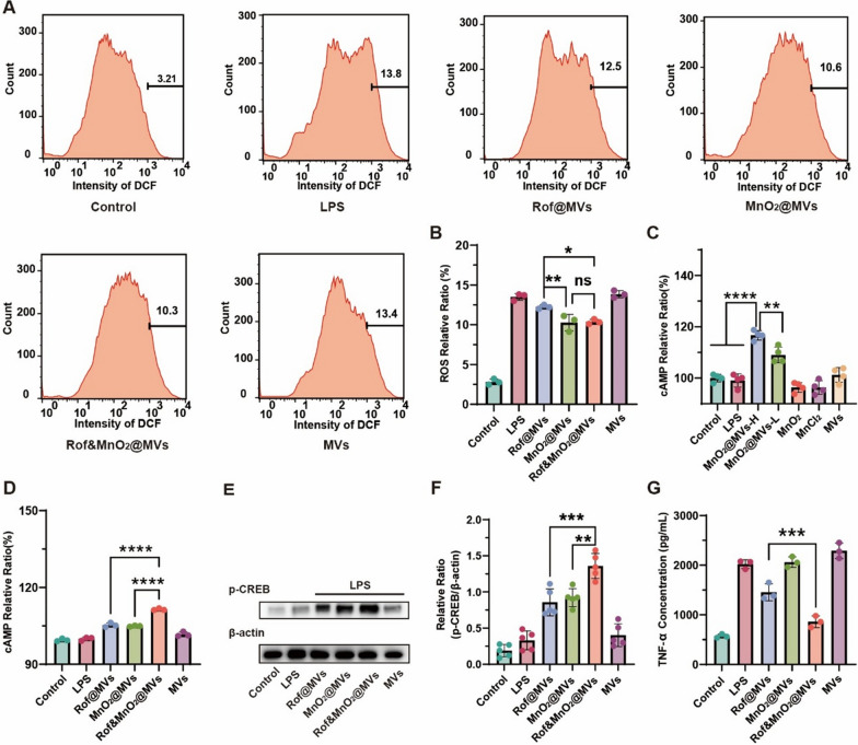
Background: Ulcerative colitis (UC) is one chronic and relapsing inflammatory bowel disease. Macrophage has been reputed as one trigger for UC. Recently, phosphodiesterase 4 (PDE4) inhibitors, for instance roflumilast, have been regarded as one latent approach to modulating macrophage in UC treatment. Roflumilast can decelerate cyclic adenosine monophosphate (cAMP) degradation, which impedes TNF-α synthesis in macrophage. However, roflumilast is devoid of macrophage-target and consequently causes some unavoidable adverse reactions, which restrict the utilization in UC.

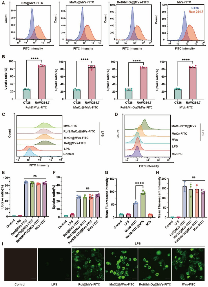
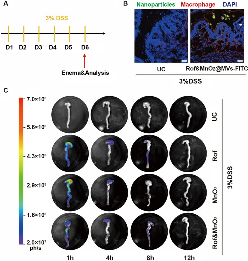
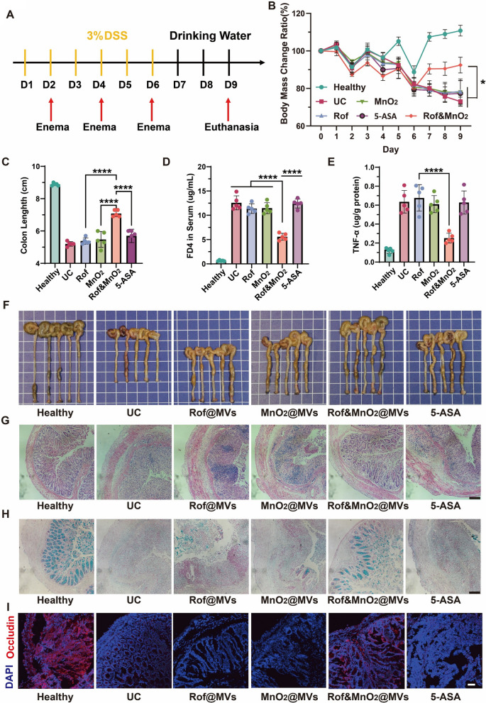
Results: Membrane vesicles (MVs) from probiotic Escherichia coli Nissle 1917 (EcN 1917) served as a drug delivery platform for targeting macrophage. As model drugs, roflumilast and MnO2 were encapsulated in MVs (Rof&MnO2@MVs). Roflumilast inhibited cAMP degradation via PDE4 deactivation and MnO2 boosted cAMP generation by activating adenylate cyclase (AC). Compared with roflumilast, co-delivery of roflumilast and MnO2 apparently produced more cAMP and less TNF-α in macrophage. Besides, Rof&MnO2@MVs could ameliorate colitis in mouse model and regulate gut microbe such as mitigating pathogenic Escherichia-Shigella and elevating probiotic Akkermansia.

Conclusions: A probiotic-based nanoparticle was prepared for precise codelivery of roflumilast and MnO2 into macrophage. This biomimetic nanoparticle could synergistically modulate cAMP in macrophage and ameliorate experimental colitis.

Keywords: Macrophage; Membrane vesicles; MnO2; Roflumilast; Ulcerative colitis; cAMP.


Conflict of interest statement

The authors declare that they have no competing interests.

Figures

Scheme 1
Scheme 1
The schematic procedure of various nanoparticles preparation and the potential mechanism of UC treatment. A Rof@MVs, MnO2@MVs and Rof&MnO2@MVs were synthesized by sonication. Gelatin NPs, Gelatin nanoparticles. EcN, Escherichia coli Nissle 1917. B After enema, Rof&MnO2@MVs passed through the defective epithelial layer in the inflammatory mucosa and were swallowed by macrophage in the lamina propria. MnO2 was reduced into Mn2+ by H2O2 and Mn2+ bonded with adenylate cyclase (AC). Besides, roflumilast inhibited the activity of PDE4. Then roflumilast and MnO2 increased the concentration of cAMP, in terms of elimination and production respectively. As a second messenger, cAMP could bind with protein kinase A (PKA) to phosphorylate cAMP-response element binding protein (CREB). Phosphorylated CREB (p-CREB) entered the cell nucleus and downregulated TNF-α expression
Fig. 1
Fig. 1
Characterization of MVs-based nanoparticles. A Dynamic light scattering diameter of MnO2. B X-ray photoelectron spectroscopy of MnO2. C The higher-resolution manganese spectra. D Dynamic light scattering diameter of Rof@MVs (Rof, 20 μg/mL). E Dynamic light scattering diameter of MnO2@MVs (MnO2, 80 μg/mL). F Dynamic light scattering diameter of Rof&MnO2@MVs (Rof, 20 μg/mL; MnO2, 80 μg/mL). G ζ potential of Rof@MVs (Rof, 20 μg/mL), MnO2@MVs (MnO2, 80 μg/mL) and Rof&MnO2@MVs (Rof, 20 μg/mL; MnO2, 80 μg/mL). H Scanning electron microscopy of Rof&MnO2@MVs (white scale bar 100 nm, black scale bar 1 μm). I Stability of MVs-based nanoparticles in simulated colon fluid (SCF, pH 7.2–7.8) at 37 °C
Fig. 2
Fig. 2
Macrophage-target of MVs-based nanoparticles. A Comparison of nanoparticle uptake in CT26 (blue) and RAW264.7 (orange) via flow cytometer. B Quantification of uptake ratio of the same nanoparticle between CT26 and RAW264.7 cells with signal intensity exceeding 100 were concluded (n = 5). C The uptake of various MVs-based nanoparticles in RAW264.7 via flow cytometer. D MVs could prompt the cargo MnO2 to accumulate in RAW264.7. E Quantification of uptake ratio of different nanoparticles in RAW264.7 (n = 5). F Quantification of uptake ratio of various nanoparticles in CT26 (n = 5). G Analysis of MnO2 uptake in RAW264.7 with or without MVs (n = 5). H Mean fluorescent intensity of FITC-labelled nanoparticles in RAW264.7 when observed under CLSM. (n = 3). I Representative fluorescent graph of FITC-labelled nanoparticles in RAW264.7 (scale bar 20 μm). These data were manifested as mean ± SD. *p < 0.05, **p < 0.01, ***p < 0.001, ****p < 0.0001, ns (none significance). Data was statistically analyzed via two-tail Student’s t test or one-way ANOVA multiple comparisons tests (Tukey’s test was used for comparison of multiple groups)
Fig. 3
Fig. 3
The underlying biological mechanism of roflumilast and MnO2. A The intracellular decomposition of MnO2 by H2O2, illustrated via flow cytometer. B Quantifying ROS relative ratio of different nanoparticles in RAW264.7. Cells with signal intensity surpassing 1000 were analyzed (n = 3). C The effect of diverse formulations of Mn on the cytosolic cAMP. MnO2@MVs-H means high concentration (5 μg/mL MnO2). Similarly, MnO2@MVs-L represents low concentration (0.5 μg/mL MnO2). The Mn concentration of both MnO2 and MnCl2 was 5 μg/mL. To obtain the relative ratio, the concentrations of cAMP in groups were divided by the control (n = 4). D The effect of MVs-based nanoparticles on the cytosolic cAMP. (n = 3). E The phosphorylation of CREB in various MVs-based nanoparticles in Western Blot. F The relative quantification of p-CREB in Western Blot (n = 5). G The synergistical efficacy of roflumilast and MnO2 in inhibiting TNF-α secretion from RAW264.7 (n = 3). Rof@MVs (Rof, 1.25 μg/mL), MnO2@MVs (MnO2, 5 μg/mL), Rof&MnO2@MVs (Rof, 1.25 μg/mL; MnO2, 5 μg/mL) and MVs (62.5 μg/mL). The concentration ratio of roflumilast and MnO2 is 1:4. These data were manifested as mean ± SD. *p < 0.05, **p < 0.01, ***p < 0.001, ****p < 0.0001, ns (none significance). Data was statistically analyzed via one-way ANOVA multiple comparisons tests (Tukey’s test was used for comparison of multiple groups)
Fig. 4
Fig. 4
The biodistribution of nanoparticles in colon after enema. A Experiment scheme. 3% (wt %) DSS was used to establish colitis via free drinking from day 1 to day 6. Mice were given enema with FITC-labelled nanoparticles and analyzed on day 6. B Representative fluorescent imaging of nanoparticles uptake in macrophage in Vivo (scale bar 20 μm). C The detention time of FITC-labelled nanoparticles in colon after enema via in vivo imaging system. Abbreviations: Rof, Rof@MVs-FITC; MnO2, MnO2@ MVs-FITC; Rof&MnO2, Rof&MnO2@ MVs-FITC
Fig. 5
Fig. 5
The efficacy of MVs-based nanoparticles in ameliorating DSS-induced colitis. A The scheme of experiment. Mice were firstly raised 7 days for acclimation before colitis establishment. Then, 3% (wt %) DSS was used to establish colitis via free drinking from day 1 to day 6. During this process, enema was performed on day 2, 4 and 6 with Rof (Rof@MVs, roflumilast 1 mg/kg), MnO2 (MnO2@MVs MnO2, 4 mg/kg), Rof&MnO2 (Rof&MnO2@MVs, roflumilast, 1 mg/kg and MnO2, 4 mg/kg) and 5-ASA (1.25 mg/kg). From day 7, DSS was substituted with drinking water until euthanasia. On day 9, mice were sacrificed and colon was collected for further analysis. B Dynamic body weight mass in different groups during the experiment (n = 5). C Measurement of colon length in various groups (n = 5). D Analysis of intestinal permeability with FD4 (n = 5). E TNF-α level in colon tissue (n = 5). F Photograph of colon from mice (n = 5). The grid behind the specimen is 1 cm × 1 cm. G Representative images of H&E stain of colon in each group (scale bar 100 μm). H Representative graphs of goblet cells in each group by Alcian blue staining (scale bar 100 μm). I Immunofluorescence examination in Occludin of colon from each group (scale bar 20 μm). These data were manifested as mean ± SD. *p < 0.05, **p < 0.01, ***p < 0.001, ****p < 0.0001. Data was statistically analyzed via two-way or one-way ANOVA multiple comparisons tests (Tukey’s test was used for comparison of multiple groups)
Fig. 6
Fig. 6
The efficacy of MVs-based nanoparticles in gut microbes. A The Chao1 of different groups (n = 5). These data were manifested as mean ± SD. B The observed operation taxonomy units (OTUs) of different groups (n = 5). These data were manifested as mean ± SD. C The Simpson diversity index of various groups (n = 5). These data were manifested as mean ± SD. D Principal components analysis (PCA) of intestinal microorganism. E Representative top 30 bacteria in genus level. F The relative abundance of Escherichia–Shigella in genus (n = 5). G The relative abundance of Akkermansia in genus (n = 5). Rof: Rof@MVs; MnO2: MnO2@MVs; Rof&MnO2: Rof&MnO2@MVs. *p < 0.05, **p < 0.01, ***p < 0.001, ****p < 0.0001. Data in A, B was statistically analyzed via one-way ANOVA multiple comparisons tests (Tukey’s test was used for comparison of multiple groups). Data in C, F, G was statistically analyzed via nonparametric tests (Mann–Whitney test was used for comparison of two groups)

References

    1. Kobayashi T, Siegmund B, Le Berre C, Wei SC, Ferrante M, Shen B, Bernstein CN, Danese S, Peyrin-Biroulet L, Hibi T. Ulcerative colitis. Nat Rev Dis Prim. 2020;6:74. doi: 10.1038/s41572-020-0205-x. - DOI - PubMed
    1. Ungaro R, Mehandru S, Allen PB, Peyrin-Biroulet L, Colombel J-F. Ulcerative colitis. Lancet. 2017;389:1756–1770. doi: 10.1016/S0140-6736(16)32126-2. - DOI - PMC - PubMed
    1. Zhang M, Li X, Zhang Q, Yang J, Liu G. Roles of macrophages on ulcerative colitis and colitis-associated colorectal cancer. Front Immunol. 2023;14:1103617. doi: 10.3389/fimmu.2023.1103617. - DOI - PMC - PubMed
    1. Allison MC, Cornwall S, Poulter LW, Dhillon AP, Pounder RE. Macrophage heterogeneity in normal colonic mucosa and in inflammatory bowel disease. Gut. 1988;29:1531. doi: 10.1136/gut.29.11.1531. - DOI - PMC - PubMed
    1. Na YR, Stakenborg M, Seok SH, Matteoli G. Macrophages in intestinal inflammation and resolution: a potential therapeutic target in IBD. Nat Rev Gastroenterol Hepatol. 2019;16:531–543. doi: 10.1038/s41575-019-0172-4. - DOI - PubMed

MeSH terms