. 2024 May 29;28(1):185.
doi: 10.1186/s13054-024-04956-6.

Biological effects of corticosteroids on pneumococcal pneumonia in Mice-translational significance

Affiliations

Biological effects of corticosteroids on pneumococcal pneumonia in Mice-translational significance

Hiroki Taenaka et al. Crit Care. .

Abstract

Background: Streptococcus pneumoniae is the most common bacterial cause of community acquired pneumonia and the acute respiratory distress syndrome (ARDS). Some clinical trials have demonstrated a beneficial effect of corticosteroid therapy in community acquired pneumonia, COVID-19, and ARDS, but the mechanisms of this benefit remain unclear. The primary objective of this study was to investigate the effects of corticosteroids on the pulmonary biology of pneumococcal pneumonia in a mouse model. A secondary objective was to identify shared transcriptomic features of pneumococcal pneumonia and steroid treatment in the mouse model and clinical samples.

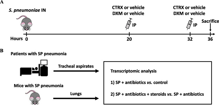
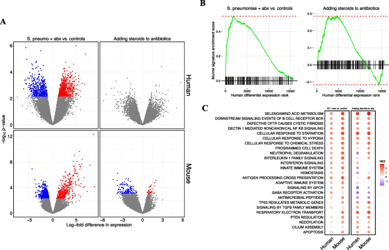
Methods: We carried out comprehensive physiologic, biochemical, and histological analyses in mice to identify the mechanisms of lung injury in Streptococcus pneumoniae with and without adjunctive steroid therapy. We also studied lower respiratory tract gene expression from a cohort of 15 mechanically ventilated patients (10 with Streptococcus pneumoniae and 5 controls) to compare with the transcriptional studies in the mice.

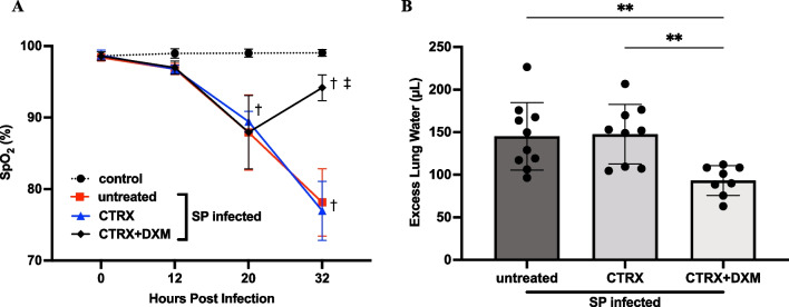
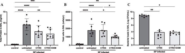
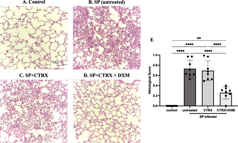
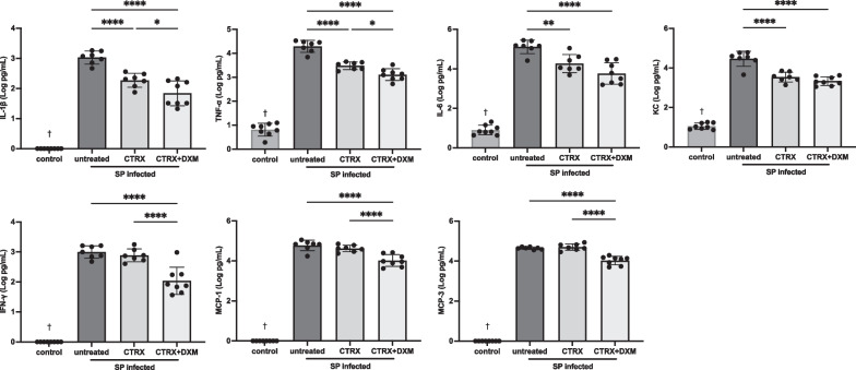
Results: In mice with pneumonia, dexamethasone in combination with ceftriaxone reduced (1) pulmonary edema formation, (2) alveolar protein permeability, (3) proinflammatory cytokine release, (4) histopathologic lung injury score, and (5) hypoxemia but did not increase bacterial burden. Transcriptomic analyses identified effects of steroid therapy in mice that were also observed in the clinical samples.

Conclusions: In combination with appropriate antibiotic therapy in mice, treatment of pneumococcal pneumonia with steroid therapy reduced hypoxemia, pulmonary edema, lung permeability, and histologic criteria of lung injury, and also altered inflammatory responses at the protein and gene expression level. The transcriptional studies in patients suggest that the mouse model replicates some of the features of pneumonia in patients with Streptococcus pneumoniae and steroid treatment. Overall, these studies provide evidence for the mechanisms that may explain the beneficial effects of glucocorticoid therapy in patients with community acquired pneumonia from Streptococcus Pneumoniae.

Keywords: Acute respiratory distress syndrome; Glucocorticoids; Pneumonia; Streptococcal infections.


Conflict of interest statement

The authors declare that they have no competing interests.

Figures

Fig. 1
Fig. 1
Study design. A Mice were inoculated intranasally (IN) with 108 colony-forming units of Streptococcus pneumoniae (SP) and received 10 mg/kg of dexamethasone or vehicle control in conjunction with 150 mg/kg of ceftriaxone or vehicle control intraperitoneally (IP) 20 and 32 h after bacterial inoculation. Mice were sacrificed 36 h after infection. B Tracheal aspirate samples were collected from an observational cohort of mechanically ventilated adults within 72 h of admission to the ICU. Patients who had SP in a respiratory tract culture or met criteria for SP infection using an established metagenomic sequencing classifier model. Lungs were collected from mouse model of SP pneumonia. Abbreviations: IN intranasal injection; IP intraperitoneal injection; CTRX ceftriaxone; DXM dexamethasone; SP streptococcus pneumoniae
Fig. 2
Fig. 2
Dexamethasone prevents the progression of hypoxemia and pulmonary edema in mice with pneumococcal pneumonia. A Arterial oxygen saturation (SpO2) was measured 12, 20, and 32 h after bacterial inoculation. SpO2 in the infected mice declined 20 h after infection. Dexamethasone-treated mice had higher SpO2 32 h after infection than mice without dexamethasone. n = 5/group. B Excess lung water, a measure of edema in the interstitial and alveolar spaces, were reduced with combination treatment of dexamethasone and ceftriaxone when compared to untreated mice and ceftriaxone alone. n = 7–10/group. All data are shown as means ± SD. Statistical differences between groups were calculated with (A) two-way repeated ANOVA followed by Tukey’s multiple comparison test, or B one-way ANOVA followed by Tukey’s multiple comparison test. P < 0.05 compared with control. P < 0.05 compared with groups without dexamethasone. **P < 0.01. Abbreviations: CTRX ceftriaxone; DXM dexamethasone; SpO2; arterial oxygen saturation; SP streptococcus pneumoniae
Fig. 3
Fig. 3
Dexamethasone reduces both alveolar protein permeability and cell infiltration. A Total protein concentration in bronchoalveolar lavage (BAL) fluid were elevated in the infected mice, indicating alveolar-capillary barrier disruption. The combination of dexamethasone and ceftriaxone reduced the BAL protein concentration, whereas ceftriaxone alone did not. n = 7–8/group. B Total cell number in BAL fluid was elevated in infected mice. The combination of dexamethasone and ceftriaxone reduced the total cell number compared with untreated and ceftriaxone alone. n = 7–8/group. C Ceftriaxone substantially reduced the bacterial loads 36 h after infection. The addition of dexamethasone to appropriate antibiotic treatment with ceftriaxone did not increase bacterial loads. n = 7–8/group. All data are shown as means ± SD. Statistical differences between groups were calculated with one-way ANOVA followed by Tukey’s multiple comparison test or Kruskal–Wallis followed by Dunn’s multiple comparison. *P < 0.05, **P < 0.01, ***P < 0.001, ****P < 0.0001. Abbreviations: CFU colony forming unit; BAL bronchoalveolar lavage; SP streptococcus pneumoniae; CTRX ceftriaxone; DXM dexamethasone
Fig. 4
Fig. 4
Dexamethasone improves histopathology in mice with pneumococcal pneumonia. AD Representative images of lung histology; E Histological scores for each group. Compared with normal healthy control (A), the lungs of mice inoculated with Streptococcus pneumoniae (SP) had severe inflammation including the infiltration of neutrophils into the airspace, alveolar septal thickening, and alveolar edema fluid at 36 h after infection (B). Treatment with ceftriaxone had little effect on the inflammation despite of its capacity to eliminate the bacteria (C). The combination of dexamethasone and ceftriaxone further reduced the inflammation (D) and significantly improved the histopathological score comparable to healthy control (E). n = 4/group. Data are shown as means ± SD. Statistical differences between groups were calculated with with one-way ANOVA followed by Tukey’s multiple comparison test. **P < 0.01, ****P < 0.0001. Abbreviations: SP streptococcus pneumoniae; CTRX ceftriaxone; DXM dexamethasone.
Fig. 5
Fig. 5
Dexamethasone attenuates the inflammatory response in mice with pneumococcal pneumonia. Inflammatory biomarkers including IL-1β, tumor necrosis factor-α (TNF-α), IL-6, keratinocyte chemoattractant (KC), interferon-γ (IFN-γ), monocyte chemoattractant protein-1 (MCP-1) and MCP-3 were significantly higher in infected mice compared with normal healthy control. Ceftriaxone reduced IL-1β, TNF-α, IL-6, and KC, but did not impact the levels of IFN-γ, MCP-1, and MCP-3. The combination of ceftriaxone and dexamethasone significantly attenuated all of these inflammatory cytokine and chemokine accumulation in the air spaces compared with untreated controls and significantly reduced IL-1β, TNF-α, IFN-γ, MCP-1, and MCP-3 as compared to ceftriaxone alone. n = 7–8/group. Data are shown as means ± SD. Statistical differences between groups were calculated with one-way ANOVA followed by Tukey’s multiple comparison test. *P < 0.05, **P < 0.01, ***P < 0.001, ****P < 0.0001. P < 0.05 compared with infected groups. Abbreviations: SP streptococcus pneumoniae; CTRX ceftriaxone; DXM dexamethasone; TNF-α tumor necrosis factor-α; KC keratinocyte chemoattractant; IFN-γ interferon-γ; MCP monocyte chemoattractant protein
Fig. 6
Fig. 6
RNA sequencing identifies reproducible effects of pneumococcal pneumonia and steroid treatment in humans and mice. A Volcano plots of differential gene expression. Each point represents an individual gene. The log2-fold difference in gene expression estimated by limma for each comparison is on the x-axis, and the p-value for each comparison is on the y axis. A positive difference in gene expression indicates the gene is more highly expressed in humans or mice with S. pneumoniae treated with antibiotics in the first column, and more highly expressed in humans or mice who received steroids in the second column. Grey points were not statistically significant (FDR < 0.1) after adjusting for multiple hypothesis testing. B Enrichment plots for mouse gene signatures in human differential expression. Genes were ranked along the x-axis by the estimated log-fold difference in gene expression between groups in human tracheal aspirate transcriptomes. A vertical line along the x-axis identifies the genes that are present in the mouse gene signature. The green line represents the running GSEA enrichment score at that position on the ranked gene list. C Dot plot of GSEA net enrichment scores (NES) for the differential expression data in panel B. Red indicates genes in selected Reactome pathways that are relatively more highly expressed in humans or mice with S. pneumoniae treated with antibiotics in first and second columns, and more highly expressed in humans or mice who received steroids in the third and fourth columns. A solid circle indicates GSEA FDR < 0.1. Abbreviations: SP streptococcus pneumoniae; NES net enrichment score

Update of

Similar articles

Cited by

References

    1. Matthay MA, Zemans RL, Zimmerman GA, Arabi YM, Beitler JR, Mercat A, Herridge M, Randolph AG, Calfee CS. Acute respiratory distress syndrome. Nat Rev Dis Primers. 2019;5(1):18. doi: 10.1038/s41572-019-0069-0. - DOI - PMC - PubMed
    1. Troeger C, Blacker B, Khalil IA, Rao PC, Cao J, Zimsen SRM, Albertson SB, Deshpande A, Farag T, Abebe Z, et al. Estimates of the global, regional, and national morbidity, mortality, and aetiologies of lower respiratory infections in 195 countries, 1990–2016: a systematic analysis for the Global Burden of Disease Study 2016. Lancet Infect Dis. 2018;18(11):1191–1210. doi: 10.1016/S1473-3099(18)30310-4. - DOI - PMC - PubMed
    1. Ortqvist A, Hedlund J, Kalin M. Streptococcus pneumoniae: epidemiology, risk factors, and clinical features. Semin Respir Crit Care Med. 2005;26(6):563–574. doi: 10.1055/s-2005-925523. - DOI - PubMed
    1. Weiser JN, Ferreira DM, Paton JC. Streptococcus pneumoniae: transmission, colonization and invasion. Nat Rev Microbiol. 2018;16(6):355–367. doi: 10.1038/s41579-018-0001-8. - DOI - PMC - PubMed
    1. File TM, Ramirez JA. Community-acquired pneumonia. N Engl J Med. 2023;389(7):632–641. doi: 10.1056/NEJMcp2303286. - DOI - PubMed