

Intrauterine inflammation and postnatal intravenous dopamine alter the neurovascular unit in preterm newborn lambs
- PMID: 38807204
- PMCID: PMC11134744
- DOI: 10.1186/s12974-024-03137-0
Intrauterine inflammation and postnatal intravenous dopamine alter the neurovascular unit in preterm newborn lambs
Abstract
Background: Intrauterine inflammation is considered a major cause of brain injury in preterm infants, leading to long-term neurodevelopmental deficits. A potential contributor to this brain injury is dysregulation of neurovascular coupling. We have shown that intrauterine inflammation induced by intra-amniotic lipopolysaccharide (LPS) in preterm lambs, and postnatal dopamine administration, disrupts neurovascular coupling and the functional cerebral haemodynamic responses, potentially leading to impaired brain development. In this study, we aimed to characterise the structural changes of the neurovascular unit following intrauterine LPS exposure and postnatal dopamine administration in the brain of preterm lambs using cellular and molecular analyses.
Methods: At 119-120 days of gestation (term = 147 days), LPS was administered into the amniotic sac in pregnant ewes. At 126-7 days of gestation, the LPS-exposed lambs were delivered, ventilated and given either a continuous intravenous infusion of dopamine at 10 µg/kg/min or isovolumetric vehicle solution for 90 min (LPS, n = 6; LPSDA, n = 6). Control preterm lambs not exposed to LPS were also administered vehicle or dopamine (CTL, n = 9; CTLDA, n = 7). Post-mortem brain tissue was collected 3-4 h after birth for immunohistochemistry and RT-qPCR analysis of components of the neurovascular unit.
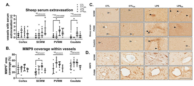
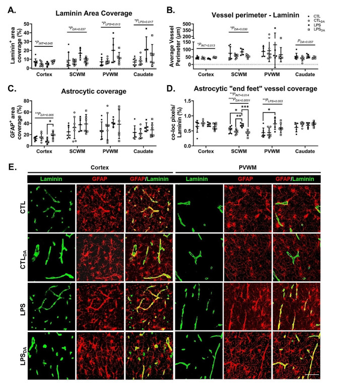
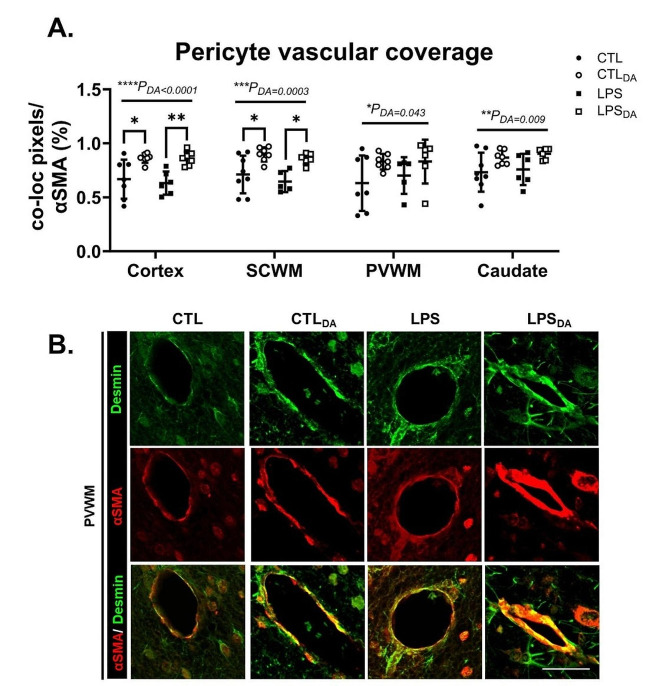
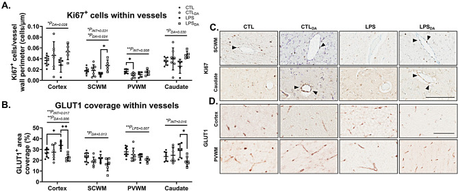
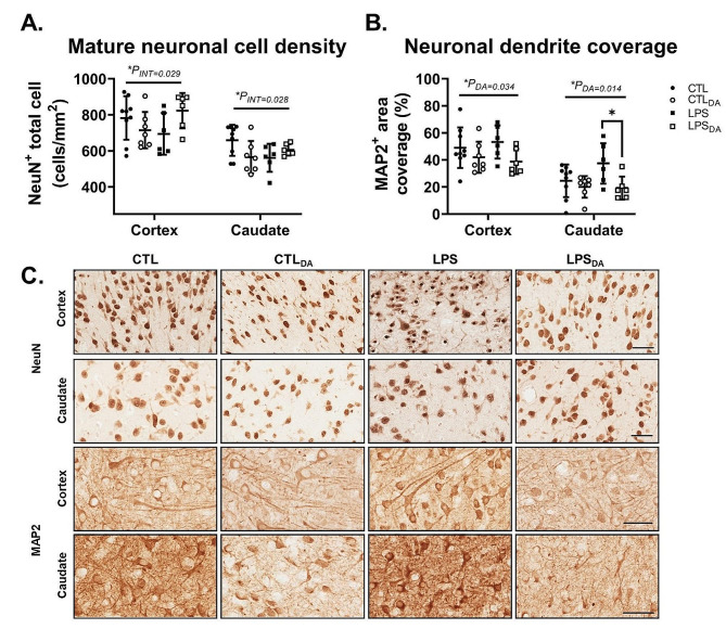
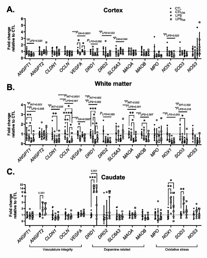
Results: LPS exposure increased vascular leakage in the presence of increased vascular density and remodelling with increased astrocyte "end feet" vessel coverage, together with downregulated mRNA levels of the tight junction proteins Claudin-1 and Occludin. Dopamine administration decreased vessel density and size, decreased endothelial glucose transporter, reduced neuronal dendritic coverage, increased cell proliferation within vessel walls, and increased pericyte vascular coverage particularly within the cortical and deep grey matter. Dopamine also downregulated VEGFA and Occludin tight junction mRNA, and upregulated dopamine receptor DRD1 and oxidative protein (NOX1, SOD3) mRNA levels. Dopamine administration following LPS exposure did not exacerbate any effects induced by LPS.
Conclusion: LPS exposure and dopamine administration independently alters the neurovascular unit in the preterm brain. Alterations to the neurovascular unit may predispose the developing brain to further injury.
Keywords: Chorioamnionitis; Dopamine; Intrauterine inflammation; Neurovascular coupling; Neurovascular unit; Preterm brain.
© 2024. The Author(s).
Conflict of interest statement
The authors declare no competing interests.
Figures
Similar articles
-
The cerebral haemodynamic response to somatosensory stimulation in preterm newborn lambs is reduced following intrauterine inflammation and dopamine infusion.Exp Neurol. 2022 Jun;352:114049. doi: 10.1016/j.expneurol.2022.114049. Epub 2022 Mar 17. Exp Neurol. 2022. PMID: 35305987
-
Lipopolysaccharide-induced changes in the neurovascular unit in the preterm fetal sheep brain.J Neuroinflammation. 2020 May 28;17(1):167. doi: 10.1186/s12974-020-01852-y. J Neuroinflammation. 2020. PMID: 32466771 Free PMC article.
-
Intrauterine inflammation, insufficient to induce parturition, still evokes fetal and neonatal brain injury.Int J Dev Neurosci. 2011 Oct;29(6):663-71. doi: 10.1016/j.ijdevneu.2011.02.011. Epub 2011 Mar 4. Int J Dev Neurosci. 2011. PMID: 21382466 Free PMC article.
-
Inflammation in utero exacerbates ventilation-induced brain injury in preterm lambs.J Appl Physiol (1985). 2012 Feb;112(3):481-9. doi: 10.1152/japplphysiol.00995.2011. Epub 2011 Nov 3. J Appl Physiol (1985). 2012. PMID: 22052871
-
Intrauterine inflammation alters cardiopulmonary and cerebral haemodynamics at birth in preterm lambs.J Physiol. 2013 Apr 15;591(8):2127-37. doi: 10.1113/jphysiol.2012.249680. Epub 2013 Feb 18. J Physiol. 2013. PMID: 23420658 Free PMC article.
References
-
- Leviton A, Joseph RM, Allred EN, Fichorova RN, O’Shea TM, Kuban KKC, et al. The risk of neurodevelopmental disorders at age 10 years associated with blood concentrations of interleukins 4 and 10 during the first postnatal month of children born extremely preterm. Cytokine. 2018;110:181–8. doi: 10.1016/j.cyto.2018.05.004. - DOI - PMC - PubMed
-
- Feng SY, Samarasinghe T, Phillips DJ, Alexiou T, Hollis JH, Yu VY et al. Acute and Chronic Effects of Endotoxin on Cerebral Circulation in Lambs. American journal of physiology Regulatory, integrative and comparative physiology. 2010. - PubMed
MeSH terms
Substances
Grants and funding
LinkOut - more resources
Full Text Sources
Miscellaneous
