. 2024 May 28;22(1):297.
doi: 10.1186/s12964-024-01666-y.

SIRT1 regulates hepatic vldlr levels

Affiliations

SIRT1 regulates hepatic vldlr levels

Mona Peyman et al. Cell Commun Signal. .

Abstract

Background: Endoplasmic reticulum (ER) stress-mediated increases in the hepatic levels of the very low-density lipoprotein (VLDL) receptor (VLDLR) promote hepatic steatosis by increasing the delivery of triglyceride-rich lipoproteins to the liver. Here, we examined whether the NAD(+)-dependent deacetylase sirtuin 1 (SIRT1) regulates hepatic lipid accumulation by modulating VLDLR levels and the subsequent uptake of triglyceride-rich lipoproteins.

Methods: Rats fed with fructose in drinking water, Sirt1-/- mice, mice treated with the ER stressor tunicamycin with or without a SIRT1 activator, and human Huh-7 hepatoma cells transfected with siRNA or exposed to tunicamycin or different inhibitors were used.

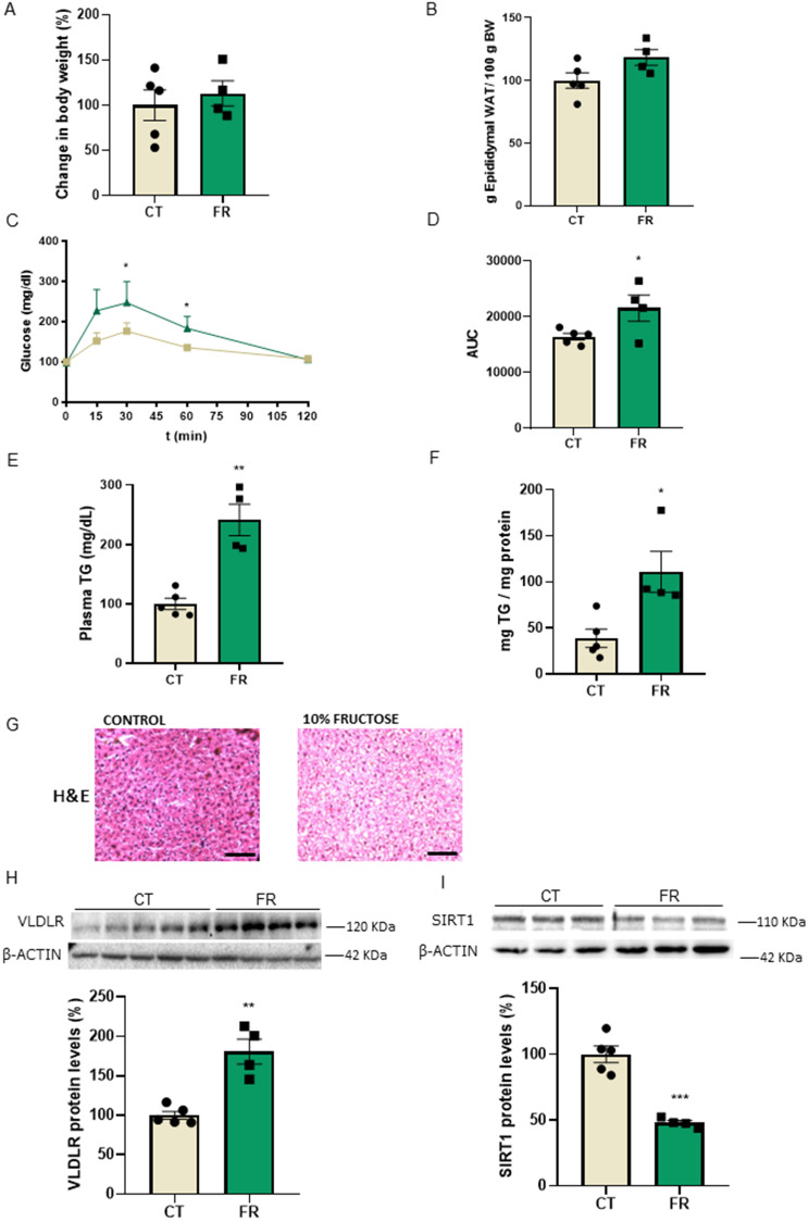
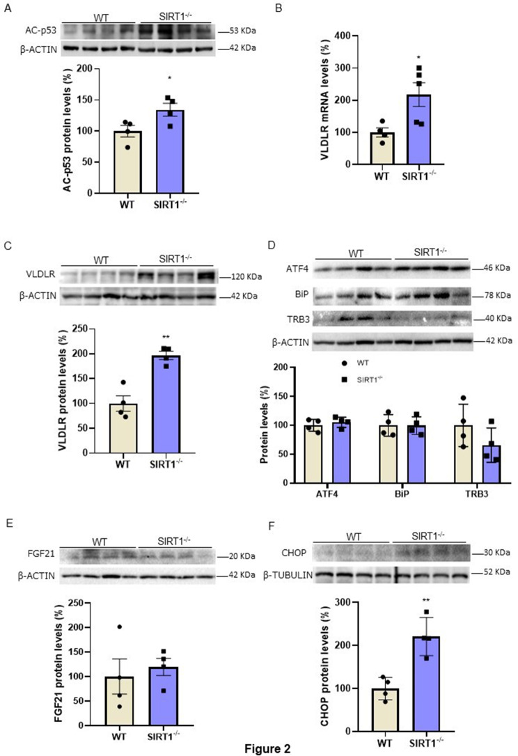
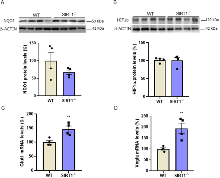
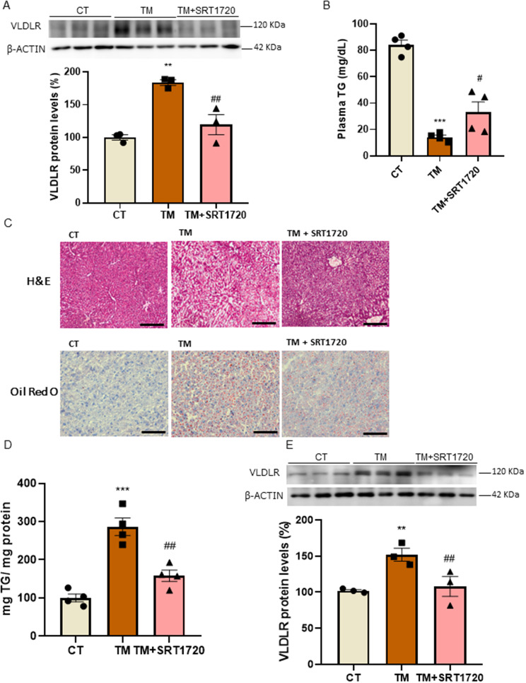
Results: Hepatic SIRT1 protein levels were reduced, while those of VLDLR were upregulated in the rat model of metabolic dysfunction-associated steatotic liver disease (MASLD) induced by fructose-drinking water. Moreover, Sirt1-/- mice displayed increased hepatic VLDLR levels that were not associated with ER stress, but were accompanied by an increased expression of hypoxia-inducible factor 1α (HIF-1α)-target genes. The pharmacological inhibition or gene knockdown of SIRT1 upregulated VLDLR protein levels in the human Huh-7 hepatoma cell line, with this increase abolished by the pharmacological inhibition of HIF-1α. Finally, SIRT1 activation prevented the increase in hepatic VLDLR protein levels in mice treated with the ER stressor tunicamycin.

Conclusions: Overall, these findings suggest that SIRT1 attenuates fatty liver development by modulating hepatic VLDLR levels.

Keywords: ER stress; HIF-1α; MASLD; SIRT1; VLDLR.


Conflict of interest statement

The authors declare no competing interests.

Figures

Fig. 1
Fig. 1
Hepatic steatosis induced by liquid fructose in rats results in an increase in VLDLR levels and a reduction in SIRT1 protein levels. (A) Changes in body weight in rats with free access to plain tap water (control, CT) or to a 10% (w/v)-fructose (FR) solution for 3 weeks. (B) Epididymal adipose tissue. (C) Glucose tolerance test (GTT) and (D) area under the curve (AUC) in CT and FR rats. (E) Plasma triglyceride (TG) levels. (F) Hepatic TG levels. (G) Representative images of liver sections with hematoxylin–eosin (H&E) staining in CT and FR rats. Scale bar: 100 μm. Immunoblot analysis of (H) VLDLR and (I) SIRT1 in the livers of CT and FR rats. Data are presented as the mean ± SEM. Significant differences were established by Student’s t-test. *p < 0.05 and **p < 0.01 vs. CT. n = 4 or 5 per group
Fig. 2
Fig. 2
VLDLR levels are increased in the livers ofSirt1−/−mice. (A) Immunoblot analysis of acetylated (Ac)-p53 in the livers of WT and Sirt1−/− mice. (B) mRNA levels of Vldlr. Immunoblot analysis of (C) VLDLR, (D) ATF4, BiP/GRP78, TRB3, (E) FGF21 and (F) CHOP in the livers of WT and Sirt1−/− mice. Data are presented as the mean ± SEM. Significant differences were established by Student’s t-test. *p < 0.05 and **p < 0.01 vs. CT. n = 4 or 5 per group
Fig. 3
Fig. 3
The expression of HIF-1α-target genes is increased in the livers ofSirt1−/−mice. Immunoblot analysis of (A) NQO1 and (B) HIF-1α in the livers of WT and Sirt1−/− mice. mRNA levels of (C) Glut1 and (D) Vegfa in the livers of WT and Sirt1−/− mice. Data are presented as the mean ± SEM. Significant differences were established by Student’s t-test. *p < 0.05 and **p < 0.01 vs. CT. n = 4 per group
Fig. 4
Fig. 4
SIRT1 inhibition increases VLDLR levels and VLDL uptake in human Huh-7 cells. (A) mRNA and (B) immunoblot analysis of VLDLR in human Huh-7 cells in the absence (control, CT) or presence of 10 µM EX-527 for 24 h. Immunoblot analysis of (C) VLDLR and (D) VLDL uptake in human Huh-7 cells in the absence (control, CT) or presence of 10 µM EX-527, or in the presence of both 10 µM EX-527 and 20 µM PX-478 for 24 h. (E) Immunoblot analysis of SIRT1 and VLDLR in Huh-7 cells transfected with control siRNA or SIRT1 siRNA in the absence or presence of 20 µM PX-478. Data are presented as the mean ± SEM. Significant differences were established by Student’s t-test or one-way ANOVA with Tukey’s post-hoc test. *p < 0.05 and **p < 0.01 vs. CT. #p < 0.05, ##p < 0.01, and ###p < 0.001 vs. EX-527 or SIRT1 siRNA. n = 3 or 4 per group
Fig. 5
Fig. 5
SIRT1 activation prevents the increase in VLDLR levels caused by the ER stressor tunicamycin. (A) Immunoblot analysis of VLDLR in human Huh-7 cells in the absence (control, CT) or presence of tunicamycin or in the presence of tunicamycin plus SRT1720 for 24 h (n = 3). (B) Plasma triglyceride (TG) levels in mice treated with the SIRT1 activator SRT1720 for 5 days and vehicle or tunicamycin for the last 24 h (n = 4 animals). (C) Representative images of liver sections with hematoxylin-eosin (H&E) and Oil Red O (ORO) staining. Scale bar: 100 μm. (D) Hepatic TG levels. (E) Immunoblot analysis of VLDLR in the livers of mice. Data are presented as the mean ± SEM. Significant differences were established by one-way ANOVA with Tukey’s post-hoc test. **p < 0.01 and ***p < 0.001 vs. CT. #p < 0.05 and ##p < 0.01 vs. tunicamycin

References

    1. Polyzos SA, et al. Obesity and nonalcoholic fatty liver disease: from pathophysiology to therapeutics. Metabolism. 2019;92:82–97. doi: 10.1016/j.metabol.2018.11.014. - DOI - PubMed
    1. Badmus OO, et al. Molecular mechanisms of metabolic associated fatty liver disease (MAFLD): functional analysis of lipid metabolism pathways. Clin Sci (Lond) 2022;136:1347–66. doi: 10.1042/CS20220572. - DOI - PMC - PubMed
    1. Jo H, et al. Endoplasmic reticulum stress induces hepatic steatosis via increased expression of the hepatic very low-density lipoprotein receptor. Hepatology. 2013;57:1366–77. doi: 10.1002/hep.26126. - DOI - PubMed
    1. Webb JC, et al. Characterization and tissue-specific expression of the human ‘very low density lipoprotein (VLDL) receptor’ mRNA. Hum Mol Genet. 1994;3:531–7. doi: 10.1093/hmg/3.4.531. - DOI - PubMed
    1. Oka K, et al. Mouse very-low-density-lipoprotein receptor (VLDLR) cDNA cloning, tissue-specific expression and evolutionary relationship with the low-density-lipoprotein receptor. Eur J Biochem. 1994;224:975–82. doi: 10.1111/j.1432-1033.1994.00975.x. - DOI - PubMed

Publication types

MeSH terms

LinkOut - more resources