

Microstructural changes of the white matter in systemic lupus erythematosus patients without neuropsychiatric symptoms: a multi-shell diffusion imaging study
- PMID: 38807248
- PMCID: PMC11134659
- DOI: 10.1186/s13075-024-03344-3
Microstructural changes of the white matter in systemic lupus erythematosus patients without neuropsychiatric symptoms: a multi-shell diffusion imaging study
Abstract
Background: Diffusion kurtosis imaging (DKI) and neurite orientation dispersion and density imaging (NODDI) provide more comprehensive and informative perspective on microstructural alterations of cerebral white matter (WM) than single-shell diffusion tensor imaging (DTI), especially in the detection of crossing fiber. However, studies on systemic lupus erythematosus patients without neuropsychiatric symptoms (non-NPSLE patients) using multi-shell diffusion imaging remain scarce.
Methods: Totally 49 non-NPSLE patients and 41 age-, sex-, and education-matched healthy controls underwent multi-shell diffusion magnetic resonance imaging. Totally 10 diffusion metrics based on DKI (fractional anisotropy, mean diffusivity, axial diffusivity, radial diffusivity, mean kurtosis, axial kurtosis and radial kurtosis) and NODDI (neurite density index, orientation dispersion index and volume fraction of the isotropic diffusion compartment) were evaluated. Tract-based spatial statistics (TBSS) and atlas-based region-of-interest (ROI) analyses were performed to determine group differences in brain WM microstructure. The associations of multi-shell diffusion metrics with clinical indicators were determined for further investigation.
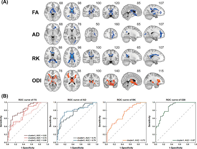
Results: TBSS analysis revealed reduced FA, AD and RK and increased ODI in the WM of non-NPSLE patients (P < 0.05, family-wise error corrected), and ODI showed the best discriminative ability. Atlas-based ROI analysis found increased ODI values in anterior thalamic radiation (ATR), inferior frontal-occipital fasciculus (IFOF), forceps major (F_major), forceps minor (F_minor) and uncinate fasciculus (UF) in non-NPSLE patients, and the right ATR showed the best discriminative ability. ODI in the F_major was positively correlated to C3.
Conclusion: This study suggested that DKI and NODDI metrics can complementarily detect WM abnormalities in non-NPSLE patients and revealed ODI as a more sensitive and specific biomarker than DKI, guiding further understanding of the pathophysiological mechanism of normal-appearing WM injury in SLE.
Keywords: Atlas-based region-of-interest (ROI) analysis; Diffusion kurtosis imaging; Neurite orientation dispersion and density imaging; Systemic lupus erythematosus; Tract-based spatial statistics.
© 2024. The Author(s).
Conflict of interest statement
The authors declare no competing interests.
Figures


References
MeSH terms
Grants and funding
LinkOut - more resources
Full Text Sources
Medical
Miscellaneous
