. 2024 May 14:15:1393485.
doi: 10.3389/fimmu.2024.1393485. eCollection 2024.

Protein kinase CK2α is overexpressed in classical hodgkin lymphoma, regulates key signaling pathways, PD-L1 and may represent a new target for therapy

Affiliations

Protein kinase CK2α is overexpressed in classical hodgkin lymphoma, regulates key signaling pathways, PD-L1 and may represent a new target for therapy

Edoardo Ruggeri et al. Front Immunol. .

Abstract

Introduction: In classical Hodgkin lymphoma (cHL), the survival of neoplastic cells is mediated by the activation of NF-κB, JAK/STAT and PI3K/Akt signaling pathways. CK2 is a highly conserved serine/threonine kinase, consisting of two catalytic (α) and two regulatory (β) subunits, which is involved in several cellular processes and both subunits were found overexpressed in solid tumors and hematologic malignancies.

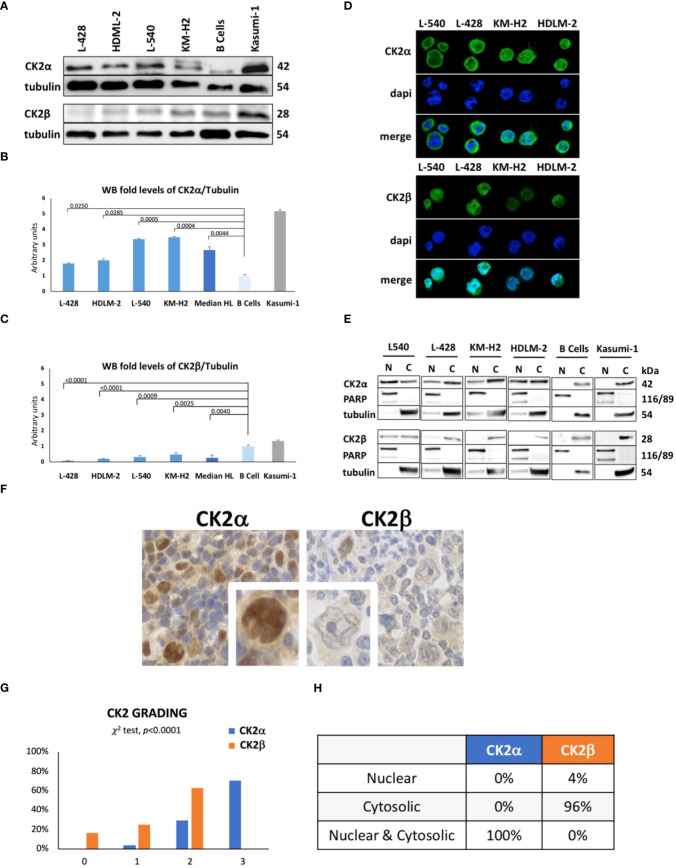
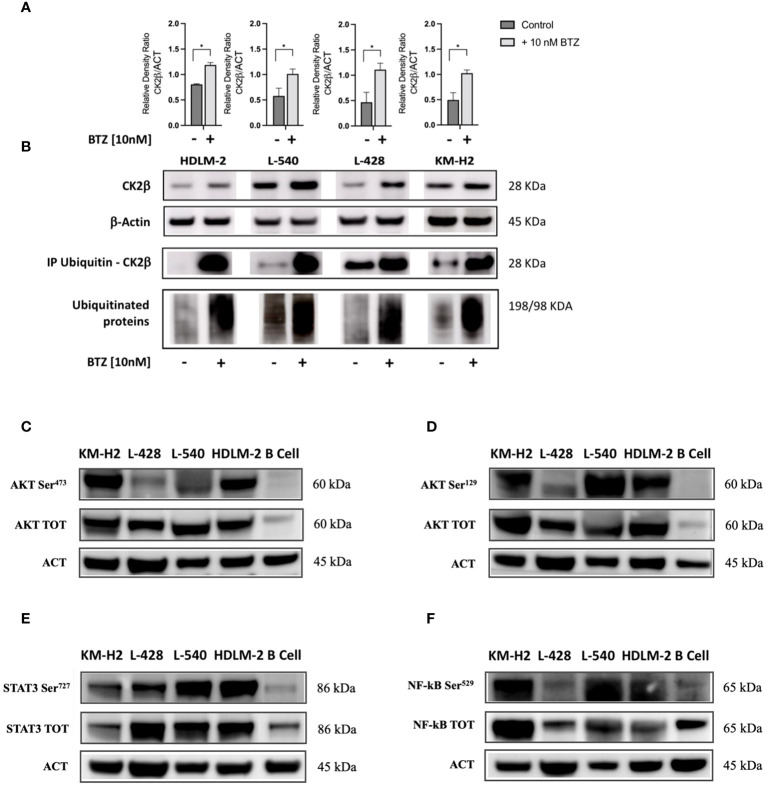
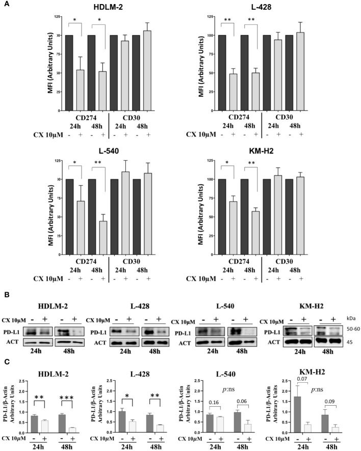
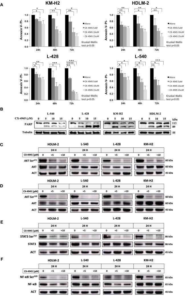
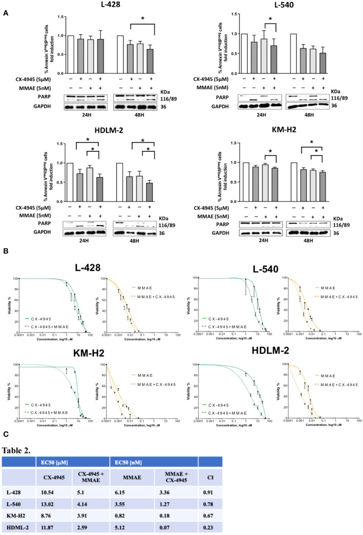
Methods and results: Biochemical analyses and in vitro assays showed an impaired expression of CK2 subunits in cHL, with CK2α being overexpressed and a decreased expression of CK2β compared to normal B lymphocytes. Mechanistically, CK2β was found to be ubiquitinated in all HL cell lines and consequently degraded by the proteasome pathway. Furthermore, at basal condition STAT3, NF-kB and AKT are phosphorylated in CK2-related targets, resulting in constitutive pathways activation. The inhibition of CK2 with CX-4945/silmitasertib triggered the de-phosphorylation of NF-κB-S529, STAT3-S727, AKT-S129 and -S473, leading to cHL cell lines apoptosis. Moreover, CX-4945/silmitasertib was able to decrease the expression of the immuno-checkpoint CD274/PD-L1 but not of CD30, and to synergize with monomethyl auristatin E (MMAE), the microtubule inhibitor of brentuximab vedotin.

Conclusions: Our data point out a pivotal role of CK2 in the survival and the activation of key signaling pathways in cHL. The skewed expression between CK2α and CK2β has never been reported in other lymphomas and might be specific for cHL. The effects of CK2 inhibition on PD-L1 expression and the synergistic combination of CX-4945/silmitasertib with MMAE pinpoints CK2 as a high-impact target for the development of new therapies for cHL.

Keywords: CK2; MMAE; PD-L1; anti-CD30; classical hodgkin lymphoma.


Conflict of interest statement

AV and LT participated to scientific board organized and received travel grant by Takeda. The remaining authors declare that the research was conducted in the absence of any commercial or financial relationships that could be construed as a potential conflict of interest. The author(s) declared that they were an editorial board member of Frontiers, at the time of submission. This had no impact on the peer review process and the final decision.

Figures

Figure 1
Figure 1
Protein expression levels of CK2 alpha (α) and beta (β) subunits in HL cell lines. Representative WB analysis (A), and the corresponding densitometric values (B, C) are reported as mean ± SD of n= 3 independent experiments. α-Tubulin was used as the loading control. (L-428, HDLM-2, L-540, KM-H2 are HL cell lines; B cell are healthy lymphocytes purified from three different buffy coats; Kasumi-1, a cell line model of acute myeloid leukemia, was used ad positive control). Above the histogram bars (B, C), the p-value is reported. In the upper-right part of the figure confocal microscopy is shown (D). The experiment was performed in different preparations for each HL cell line; the α and β subunits of CK2 were detected using green fluorescence (Alexa Fluor 488), nuclei were visualized through blue staining (DAPI), and the merge for each CK2 subunit is shown. Images were collected with 40X magnification, Scale bars = 15μm. Cell Fractionation was performed on the four HL cell lines, healthy B cells (normal control), and Kasumi-1 cell line as positive controls. WB reports α and β subunits of CK2, PARP (Poly (ADP-ribose) polymerase) used as nuclear marker, α-Tubulin as cytosolic marker. The image reports a representative case of three independent experiments (E). Expression and localization of CK2 α and β subunits assessed by tissue microarray performed in neoplastic lymph nodes from 25 patients and 5 reactive adenopathy (F). Representative case of CK2 expression in patient-derived Hodgkin and Reed-Sternberg (HRS) cells. CK2α appears to be localized in both the nucleus and cytoplasm of HRS cells. Expression of CK2 subunits according to the grading level (G), and description of their cellular localization (H). Positivity was graded as: 0 = negative; 1 = positive <30% of HRS; 2 = positive 30-60% HRS or week-moderate intensity; 3 = positive >60% HRS or strong diffuse intensity.
Figure 2
Figure 2
Western blotting analysis of ubiquitinated CK2β and phosphorylation levels of different CK2 targets. (A). Relative densitometry of CK2β subunit with/without treatment of Bortezomib (BTZ) 10nM for 36h. (B) Representative WB. The proteins were immunoprecipitated, and all polyubiquitinated proteins were purified using a Ubiquitin detection kit. The immunocomplexes were then loaded onto an SDS-PAGE gel and subsequently analyzed using an anti-CK2β antibody. The results of proteasome inhibition were evaluated using the anti-poly ubiquitinated proteins (poly-Ubi) as positive control. For each condition, total cell lysates were loaded in SDS-PAGE and probed with anti-CK2β. * indicates p<0.05 (Mann-Whitney test), ACT: β-Actin; BTZ: Bortezomib. Representative WB of the main signaling pathways overexpressed in HL cell lines sustained by CK2 activity compared to the normal B lymphocytes (C–F). The images depict phosphorylation levels at the Serine residue targeted by CK2α, and their corresponding total proteins for: AKT-S473 (C), AKT-S129 (D), STAT3-S727 (E), and NF-kB (p65) S529 (F). Densitometry for phosphorylated/total protein, total/β-Actin protein, and phosphorylated/β-Actin protein ratios are shown alongside each representative WB picture. β-Actin was used as the loading control. Analysis reports the mean ± SD of n = 3 independent experiments. * indicates p< 0.05 (unpaired t test).
Figure 3
Figure 3
CD30 and CD274/PD-L1 expression assessment. In panel (A), histograms depict the mean fluorescence intensity (MFI) of CD274 (PD-L1) and CD30 expression in the four HL cell lines, with or without 10μM CX-4945 treatment for 24 and 48 hours (three independent experiments, Student t test) CX: CX-4945. Only the viable cells were gated in the MFI analyses of PD-L1 or CD30 and normalized to their untreated condition. (B) Representative WB and (C) WB analysis of PD-L1 expression levels. HL cell lines were treated with or without 10μM CX-4945, for 24 and 48 hours. Densitometric values are reported as mean ± SD of n= 3 independent experiments, Unpaired t test. CX: CX-4945, ACT: β-Actin. *p<0.05, **p<0.01, ***p<0.001.
Figure 4
Figure 4
Apoptotic Effect of CX-4945 in HL Cell Lines. Apoptosis was detected through Annexin V propidium iodide (PI) assay by flow cytometry. Histograms illustrate the percentage of viable cells (Annexin Vneg/PIneg) for each HL cell line after treatment with 5μM, 10μM, and 15μM CX-4945 at different time points (24, 48, and 72 hours). *p<0.05, **p<0.01, ***p<0.001, and ****p<0.0001 compared to the untreated cell population, Kruskal-Wallis’s test (A). Qualitative WB was performed using anti-PARP to highlight apoptosis induction in all four HL cell lines. Protein lysates, obtained after 48 hours of treatment with 0, 5, 10, and 15μM CX-4945, are presented. α-Tubulin was used as the loading control (experimental duplicate) (B). WB illustrate the impact of CK2 inhibition with CX-4945 on CK2 substrates in the four HL cell lines. WBs depict phosphorylation levels on CK2 Serine targets, with and without CX-4945 treatment, along with corresponding total protein levels. The panels illustrate phosphorylation levels on AKT S473 (C), AKT S129 (D), STAT3 S727 (E), and NF-κB at S529 (F) at 0, 5, and 10μM CX-4945 after 24 hours of treatment (experimental duplicate). ACT: β-Actin.
Figure 5
Figure 5
Apoptotic effect of CX-4945 alone or in combination with MMAE in Hodgkin lymphoma cell lines. HL cells lines were treated for 24 and 48 hours with CX-4945 and/or MMAE or the combination of both drugs. Apoptosis was detected through Annexin V propidium iodide (PI) assay by flow cytometry. Histograms shows alive non-apoptotic cells (Annexin Vneg/PIneg). Wilcoxon matched-pairs signed rank test was used to compared paired data (A). A qualitative WB (n=2), placed beneath each histogram, shows the cleavage of PARP protein expression in response to the heightened levels of apoptosis induced by the pharmacological treatments. In the middle panel (B) dose response curves of L-428, L-540, KM-H2, and HDLM-2 cell lines incubated for 72h with increasing concentrations of CX-4945 (green curves), and MMAE (orange curves) or their combination (dotted curves) are reported. Cell viability was assessed by trypan-blue exclusion assay. The curves were generated by maintaining a constant 1:1 ratio between the respective EC50 concentrations of CX-4945 and MMAE. In the lower panel (C), EC50 values of CX-4945 and MMAE, used alone or in combination in L-428, L-540, KM-H2, and HDLM-2 cell lines incubated as in (B). A combination index (CI) < 1 means a synergistic effect. Experiments were performed in triplicate. *p<0.05. Wilcoxon matched-pairs signed rank test.

References

    1. Yung L, Linch D. Hodgkin’s lymphoma. Lancet. (2003) 361:943–51. doi: 10.1016/S0140-6736(03)12777-8 - DOI - PubMed
    1. Brice P, de Kerviler E, Friedberg JW. Classical Hodgkin lymphoma. Lancet. (2021) 398:1518–27. doi: 10.1016/S0140-6736(20)32207-8 - DOI - PubMed
    1. Aldinucci D, Borghese C, Casagrande N. For of the immunosuppressive microenvironment of classic Hodgkin lymphoma and therapeutic approaches to counter it. Int J Mol Sci. (2019) 20:2416. doi: 10.3390/ijms20102416 - DOI - PMC - PubMed
    1. Mottok A, Steidl C. Biology of classical Hodgkin lymphoma: implications for prognosis and novel therapies. Blood. (2018) 131:1654–65. doi: 10.1182/blood-2017-09-772632 - DOI - PubMed
    1. Cellini A, Scarmozzino F, Angotzi F, Ruggeri E, Dei Tos AP, Trentin L, et al. . Tackling the dysregulated immune-checkpoints in classical Hodgkin lymphoma: bidirectional regulations between the microenvironment and Hodgkin/Reed-Sternberg cells. Front Oncol. (2023) 13:1203470. doi: 10.3389/fonc.2023.1203470 - DOI - PMC - PubMed