Case Reports
. 2024 May 17;28(1):329.
doi: 10.3892/ol.2024.14462. eCollection 2024 Jul.

Synchronous liver metastasis at initial diagnosis of adrenal pheochromocytoma by CT: A case report

Affiliations
Case Reports

Synchronous liver metastasis at initial diagnosis of adrenal pheochromocytoma by CT: A case report

Zhuo Zhang et al. Oncol Lett. .

Abstract

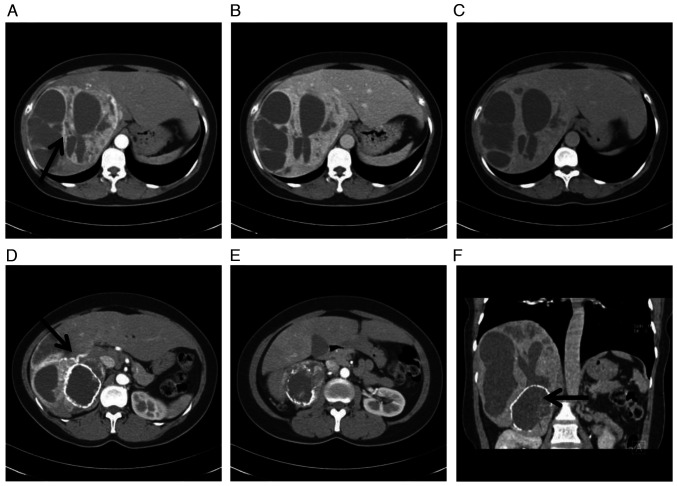
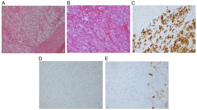
Pheochromocytoma is a tumor of the sympathetic nervous system, characterized by atypical symptoms and signs. Pheochromocytoma metastases can be found in various tissues and organs. However, synchronous metastasis at the initial diagnosis of pheochromocytoma is rare. The present study described a case with synchronous liver metastasis at the initial diagnosis of adrenal pheochromocytoma based on imaging findings. A 41-year-old woman presented with liver pain and fatigue for 1 month. Physical examination showed increased blood pressure and heart rate with sinus tachycardia. Laboratory examination revealed normal levels of liver tumor markers and increased levels of serum or urine epinephrine and norepinephrine. CT examination revealed a large cystic solid mass in the right lobe of the liver and right adrenal gland, and the solid part of the mass was enhanced after enhancement. The pathological diagnosis was pheochromocytoma of the right adrenal gland with liver metastasis. The patient underwent right hepatectomy and right adrenal tumor resection. During the postoperative follow-up, the patient's blood pressure and catecholamine levels were within the normal range. Three years after surgery, the CT examination revealed multiple liver metastases. Chemotherapy was administered to the patient. A year later, re-examination revealed an increase and enlargement of the metastases, and the mass of the right adrenal gland remained similar to the previous one. After 6 months of follow-up, the patient succumbed to recurrence and metastasis. Preoperative diagnosis of metastatic pheochromocytoma is challenging. This case mainly emphasizes that imaging findings can help the clinical diagnosis of metastatic pheochromocytoma.

Keywords: adrenal gland metastasis; liver metastasis; metastatic pheochromocytoma.


Conflict of interest statement

The authors declare that they have no competing interests.

Figures

Figure 1.
Figure 1.
CT enhanced images of the upper abdomen at diagnosis. (A) CT image of arterial phase. A huge cystic solid mass was observed in the right lobe of the liver, with an obvious enhancement of solid parts and separations but no enhancement in the cystic area (black arrow). (B) CT image of portal venous phase. There was continuous enhancement of the mass. (C) CT image of delayed phase. The density of the mass was similar to that of liver tissue. (D) The right hepatic artery was enlarged, providing blood to the mass (black arrow). (E) The density of the mass in the right adrenal gland was uneven, with annular calcification. After enhancement, the solid part was obviously enhanced, but the cystic area was not enhanced. (F) The coronal image clearly showed the positional relationship between the liver and adrenal masses and the adrenal tumors on the right side were surrounded by circular calcifications (black arrow).
Figure 2.
Figure 2.
Postoperative pathological analysis. (A) HE staining results of adrenal mass. (B) HE staining results of liver mass. (C-E) Immunohistochemistry of liver mass: (C) Chromogranin A (+) and synaptophysin (+); (D) α-fetoprotein (−); and (E) cytokeratin (−) (magnification, ×400). Blue staining indicates negative expression, whereas brown staining indicates positive expression.
Figure 3.
Figure 3.
CT enhanced images of the upper abdomen at three years after surgery. (A) Hepatic arterial stage: The right lobe of the liver was absent. The left lobe of the liver increased in volume and there were multiple nodular (black arrow) and annular (white arrow) enhancements in the liver. (B) A mass in the right adrenal gland area was unevenly enhanced after enhancement (white arrow).
Figure 4.
Figure 4.
CT enhanced images of the upper abdomen at four years after surgery. (A) There was a progressive enlargement of the liver, obvious increase, enlargement of intrahepatic metastases and uneven density. (B) The mass in the right adrenal area was similar to that before and there were spotted calcifications.

Similar articles

Cited by

References

    1. Gruber M, Darr R, Eisenhofer G. Pheochromocytoma: Update on diagnosis and therapy. Dtsch Med Wochenschr. 2014;139:486–490. doi: 10.1055/s-0033-1360082. (In German) - DOI - PubMed
    1. Reisch N, Peczkowska M, Januszewicz A, Neumann HP. Pheochromocytoma: Presentation, diagnosis and treatment. J Hypertens. 2006;24:2331–2339. doi: 10.1097/01.hjh.0000251887.01885.54. - DOI - PubMed
    1. Nölting S, Grossman A, Pacak K. Metastatic phaeochromocytoma: Spinning towards more promising treatment options. Exp Clin Endocrinol Diabetes. 2019;127:117–128. doi: 10.1055/a-0715-1888. - DOI - PMC - PubMed
    1. Soltani A, Pourian M, Davani BM. does this patient have pheochromocytoma? a systematic review of clinical signs and symptoms. J Diabetes Metab Disord. 2015;15:6. doi: 10.1186/s40200-016-0226-x. - DOI - PMC - PubMed
    1. Hori T, Yamagiwa K, Hayashi T, Yagi S, Iida T, Taniguchi K, Kawarada Y, Uemoto S. Malignant pheochromocytoma: Hepatectomy for liver metastases. World J Gastrointest Surg. 2013;5:309–313. doi: 10.4240/wjgs.v5.i11.309. - DOI - PMC - PubMed

Publication types