

A Systematic Review and Case Illustrations of Misdiagnosing Intracranial Aneurysms
- PMID: 38807799
- PMCID: PMC11130603
- DOI: 10.7759/cureus.59185
A Systematic Review and Case Illustrations of Misdiagnosing Intracranial Aneurysms
Abstract
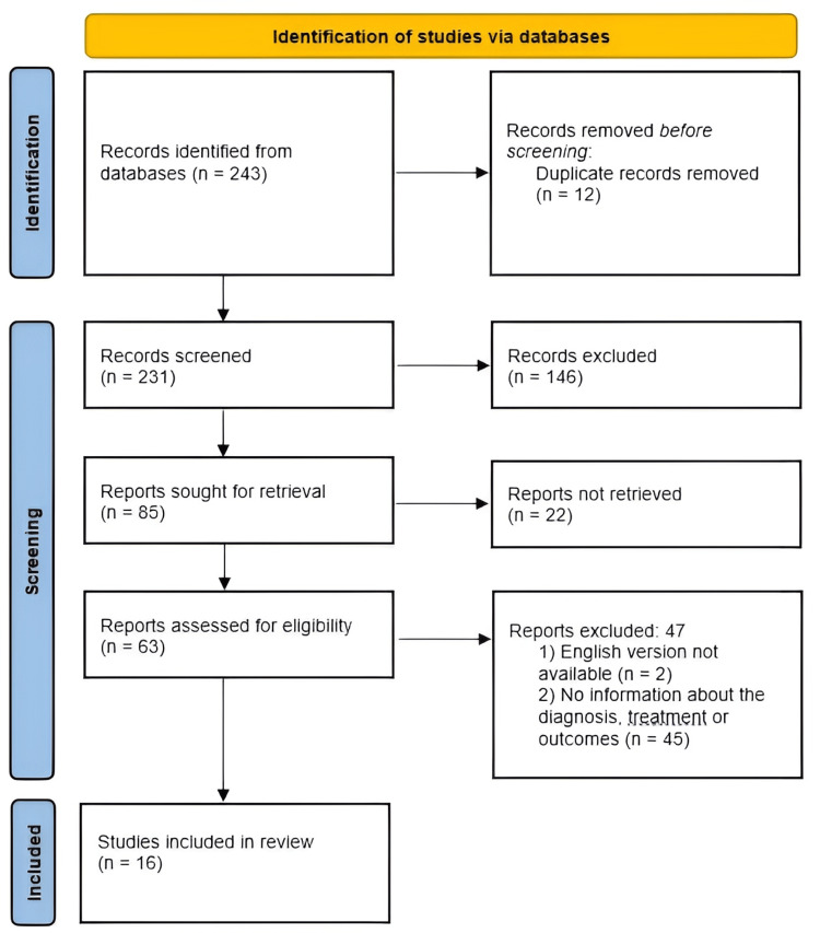
Modern neuroimaging methods do not completely rule out false diagnoses of intracranial aneurysms which can lead to an unwarranted operation associated with risks of complications. However, surgical interventions for falsely diagnosed aneurysms are quite rare. The purpose of this study is to demonstrate two clinical cases of false-positive aneurysms and a systematic review of the literature dedicated to the incidence and etiology of false-positive aneurysms, identifying risk factors associated with false-positive aneurysms. A literature search in two databases (PubMed and Web of Science) using keywords "mimicking an intracranial aneurysm", "presenting as an intracranial aneurysm", "false positive intracranial aneurysms", and "neurosurgery" was conducted. A total of 243 papers were found in the initial search in two databases. Sixteen papers (including 20 patients) were included in the final analysis. There were 10 women and 10 men. The most common location of false-positive aneurysms was the bifurcation of the middle cerebral artery (MCA). In the posterior circulation, false-positive aneurysms were identified either on the basilar artery, or at the vertebro-basilar junction. The main causes of false intracranial aneurysm diagnosis included artery occlusion with vascular stump formation, infundibular widening, fenestration, arterial dissection, contrast extravasation, and venous varix. In conclusion, summarizing the results of our analysis, we can say that surgical interventions for false-positive aneurysms are an underestimated problem in vascular neurosurgery. Despite extremely rare published clinical observations, the actual frequency of erroneous surgical interventions for false-positive aneurysms is unknown.
Keywords: clipping; false-positive diagnosis; intracranial aneurysm; masquerading; mimicking.
Copyright © 2024, Konovalov et al.
Conflict of interest statement
The authors have declared that no competing interests exist.
Figures
References
-
- Can noninvasive imaging accurately depict intracranial aneurysms? A systematic review. White PM, Wardlaw JM, Easton V. Radiology. 2000;217:361–370. - PubMed
-
- Detection and characterization of unruptured intracranial aneurysms: comparison of 3T MRA and DSA. Mine B, Pezzullo M, Roque G, David P, Metens T, Lubicz B. J Neuroradiol. 2015;42:162–168. - PubMed
-
- Screening for intracranial aneurysms in individuals with a positive first-degree family history: a systematic review. Van Hoe W, van Loon J, Demeestere J, Lemmens R, Peluso J, De Vleeschouwer S. World Neurosurg. 2021;151:235–248. - PubMed
-
- A register-based SAH study in Japan: high incidence rate and recent decline trend based on lifestyle. Ikawa F, Morita A, Nakayama T, et al. J Neurosurg. 2020;134:983–991. - PubMed
Publication types
LinkOut - more resources
Full Text Sources