

Three-dimensional Hemifacial Microsomia Classification with New Subtypes Based on the Pruzansky and Kaban Classification
- PMID: 38808145
- PMCID: PMC11132385
- DOI: 10.1097/GOX.0000000000005810
Three-dimensional Hemifacial Microsomia Classification with New Subtypes Based on the Pruzansky and Kaban Classification
Abstract
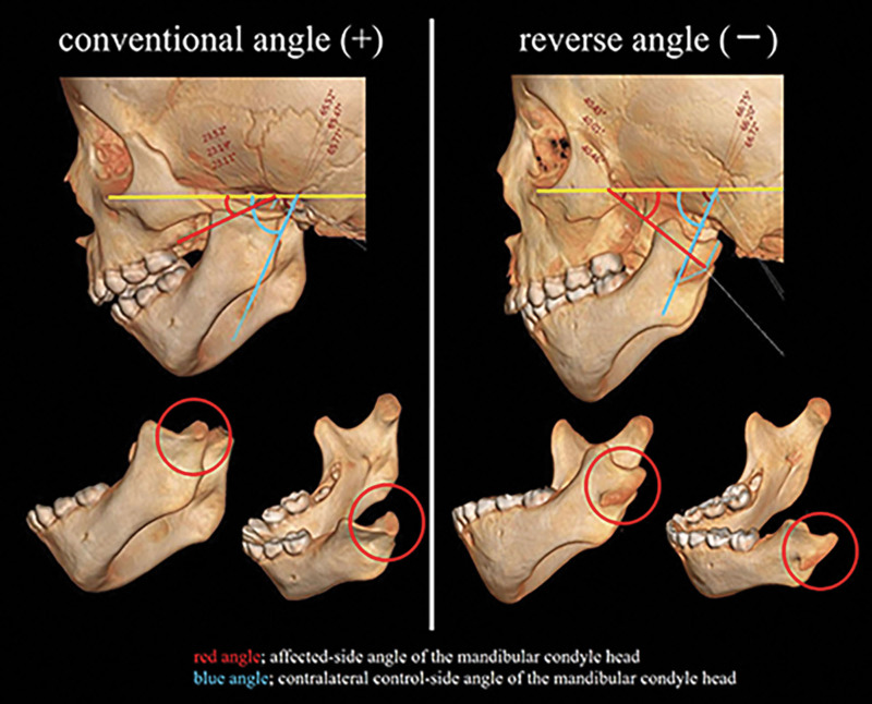
Background: Various classifications of hemifacial microsomia (HFM) have been described previously. Although some of these classifications are used widely, others use external outlines of reference organs, even in three-dimensional (3D) images. The purpose of this study was to investigate the 3D characteristics of the mandibular condyle in HFM and to update the Pruzansky and Kaban classification as a 3D classification.
Methods: Fifty-three patients with HFM were classified according to the Pruzansky and Kaban classification (type I, IIA, IIB, and III) using computed tomographic scan images. 3D images of the mandible were isolated, and the 3D characteristics were observed; furthermore, the angle of inclination of the mandibular condyle was measured in 3D.
Results: Subtypes of retroflexed mandibular condyle in 3D were observed in the Pruzansky and Kaban classification type IIA and IIB, termed as type IIAβ (33.3% in type IIA) and type IIBβ (100% in type IIB). Although some differences were observed in the inclination of the mandibular condyle between the control and the affected sides in type I and IIAα, multiple differences were observed in type IIAβ and IIBβ.
Conclusions: To the best of our knowledge, this is the first report that identified the retroflexed mandibular condyle as subtypes type IIAβ and IIBβ. Notably, this could not be identified in the two-dimensional images (lateral cephalogram) yet. We proposed to update the Pruzansky and Kaban classification as a 3D classification with a new 3D subtype. The angle of the retroflexed mandibular condyle may predict mandibular growth in HFM.
Copyright © 2024 The Authors. Published by Wolters Kluwer Health, Inc. on behalf of The American Society of Plastic Surgeons.
Conflict of interest statement
The authors have no financial interests to declare in relation to the content of this article.
Figures


Similar articles
-
Effects of mandibular distraction osteogenesis on anesthetic implications in children with hemifacial microsomia.Acta Anaesthesiol Scand. 2022 Aug;66(7):823-832. doi: 10.1111/aas.14073. Epub 2022 Apr 24. Acta Anaesthesiol Scand. 2022. PMID: 35416276
-
The mandibular deformity in hemifacial microsomia: a reassessment of the Pruzansky and Kaban classification.Plast Reconstr Surg. 2014 Feb;133(2):174e-181e. doi: 10.1097/01.prs.0000436858.63021.14. Plast Reconstr Surg. 2014. PMID: 24469188
-
Three-dimensional longitudinal changes in craniofacial growth in untreated hemifacial microsomia patients with cone-beam computed tomography.Am J Orthod Dentofacial Orthop. 2014 May;145(5):579-94. doi: 10.1016/j.ajodo.2013.09.015. Am J Orthod Dentofacial Orthop. 2014. PMID: 24785922
-
Surgical correction of the midface in craniofacial microsomia. Part 1: A systematic review.J Craniomaxillofac Surg. 2018 Sep;46(9):1427-1435. doi: 10.1016/j.jcms.2018.05.043. Epub 2018 May 25. J Craniomaxillofac Surg. 2018. PMID: 29907434
-
Modern Mandibular Distraction Applications in Hemifacial Microsomia.Clin Plast Surg. 2021 Jul;48(3):375-389. doi: 10.1016/j.cps.2021.02.001. Epub 2021 May 8. Clin Plast Surg. 2021. PMID: 34051892 Review.
References
-
- Poswillo D. The pathogenesis of the first and second branchial arch syndrome. Oral Surg Oral Med Oral Pathol. 1973;35:302–328. - PubMed
-
- Grabb WC. The first and second branchial arch syndrome. Plast Reconstr Surg. 1965;36:485–508. - PubMed
-
- Pruzansky S. Not all dwarfed mandibles are alike. Birth Defects. 1969;5:120–129.
-
- Kaban LB, Moses MH, Mulliken JB. Surgical correction of hemifacial microsomia in the growing child. Plast Reconstr Surg. 1988;82:9–19. - PubMed
-
- Kaban LB, Mulliken JB, Murray JE. Three-dimensional approach to analysis and treatment of hemifacial microsomia. Cleft Palate J. 1981;18:90–99. - PubMed
LinkOut - more resources
Full Text Sources